Protein filament

Last updated
Developing wood cells in poplar showing microfilaments (in green) and cell nuclei (in red) Microfilaments.jpg
Developing wood cells in poplar showing microfilaments (in green) and cell nuclei (in red)

In biology, a protein filament is a long chain of protein monomers, such as those found in hair, muscle, or in flagella. [1] Protein filaments form together to make the cytoskeleton of the cell. They are often bundled together to provide support, strength, and rigidity to the cell. When the filaments are packed up together, they are able to form three different cellular parts. The three major classes of protein filaments that make up the cytoskeleton include: actin filaments, microtubules and intermediate filaments.

Contents

Cellular types

Microfilaments

Compared to the other parts of the cytoskeletons, the microfilaments contain the thinnest filaments, with a diameter of approximately 7 nm. Microfilaments are part of the cytoskeleton that are composed of protein called actin. Two strands of actin intertwined together form a filamentous structure allowing for the movement of motor proteins. Microfilaments can either occur in the monomeric G-actin or filamentous F-actin. [2] Microfilaments are important when it comes to the overall organization of the plasma membrane. Actin filaments are considered to be both helical and flexible. They are composed of several actin monomers chained together which add to their flexibility. They are found in several places in the body including the microvilli, contractile rings, stress fibers, cellular cortex, etc. In a contractile ring, actin have the ability to help with cellular division while in the cellular cortex they can help with the structural integrity of the cell.      

Microfilament Polymerization

Microfilament polymerization is divided into three steps. The nucleation step is the first step, and it is the rate limiting and slowest step of the process. Elongation is the next step in this process, and it is the rapid addition of actin monomers at both the plus and minus end of the microfilament. The final step is the steady state. At this state the addition of monomers will equal the subtraction of monomers causing the microfilament to no longer grow. This is known as the critical concentration of actin. There are several toxins that have been known to limit the polymerization of actin. Cytochalasin is a toxin that will bind to the actin polymer, so it can no longer bind to the incoming actin monomers. Actin originally attached in the polymer is still leaving the microfilament causing depolymerization. Phalloidin is a toxin that will bind to actin locking the filament in place. Monomers are neither adding or leaving this polymer which causes the stabilization of the molecule. Latrunculin is similar to cytochalasin, but it is a toxin which will bind to the actin monomers preventing it from adding onto the actin polymer. This will cause the depolymerization of the actin polymer in the cell.[ citation needed ]

Actin Based Motor Protein- Myosin
This is a representation of a microfilament breakdown in a muscle fiber. It shows the different zones and disks within the sarcolemma of this muscle fiber. 1002 Organization of Muscle Fiber.jpg
This is a representation of a microfilament breakdown in a muscle fiber. It shows the different zones and disks within the sarcolemma of this muscle fiber.

There are several different proteins that interact with actin in the body. However, one of the most famous types of motor proteins is myosin. Myosin will bind to these actins causing the movement of actin. This movement of myosin along the microfilament can cause muscle contraction, membrane association, endocytosis, and organelle transport. The actin microfilament is composed of three bands and one disk. The A band is the part of the actin that will bind to the myosin during muscle contraction. The I band is the part of the actin that is not bound to the myosin, but it will still move during muscle contraction. The H zone is the space in between two adjacent actin that will shrink when the muscle begins to contract. The Z disk is the part of the microfilament that characterizes the overall end of each side of the sarcomere, a structural unit of a myofibril.[ citation needed ]

Proteins Limiting Microfilaments

These microfilaments have the potential to be limited by several factors or proteins. Tropomodulin is a protein that will cap the ends of the actin filaments causing the overall stability of the structure. Nebulin is another protein that can bind to the sides of the actin preventing the attachment of myosin to them. This causes stabilization of the actin limiting muscle contraction. Titin is another protein, but it binds to the myosin rather than the actin microfilament. Titin will help stabilize the contraction and myosin-actin structure.[ citation needed ]

Microtubules

A human cell showing the tubulin component of the cytoskeleton in green and the nucleus in red. The blue staining is a single cytoplasmic protein. Microtubule.png
A human cell showing the tubulin component of the cytoskeleton in green and the nucleus in red. The blue staining is a single cytoplasmic protein.

Microtubules are the largest type of filament, with a diameter of 25 nm wide, in the cytoskeleton. [3] A single microtubule consists of 13 linear microfilaments. Unlike microfilaments, microtubules are composed of a protein called tubulin. The tubulin consists of dimers, named either "αβ-tubulin" or "tubulin dimers", which polymerize to form the microtubules. [3] These microtubules are structurally quantified into three main groups: singlets, doublets, and triplets. Singlets are microtubule structures that are known to be found in the cytoplasm. Doublets are structures found in the cilia and flagella. Triplets are found in the basal bodies and centrioles. There are two main populations of these microtubules. There are unstable short-lived microtubules that will assemble and disassemble rapidly. The other population are stable long-lived microtubules. These microtubules will remain polymerized for longer periods of time and can be found in flagella, red blood cells, and nerve cells.  Microtubules have the ability to play a significant role in the organization of organelles and vesicles, beating of cilia and flagella, nerve and red blood cell structure, and alignment/ separation of chromosomes during mitosis and meiosis.[ citation needed ]

Orientation in Cells

When a cell is in the interphase process, microtubules tend to all orient the same way. Their negatively charged end will be close to the nucleus of the cell, while their positively end will be oriented away from the cell body. The basal body found within the cell helps the microtubule to orient in this specific fashion. In mitotic cells, they will see similar orientation as the positively charged end will be orientated away from the cell while the negatively charged end will be towards the Microtubule Organizing Center (MTOC). The positive end of these microtubules will attach to the kinetochore on the chromosome allowing for cellular division when applicable. Nerve cells tend to be a different from these other two forms of orientation. In an axon nerve cell, microtubules will arrange with their negatively charged end toward the cell body and positively charged end away from the cell body. However, in dendrites, microtubules can have a different orientation. In dendrites, microtubules can have their positively charged end toward the cell body, but their negatively charged end will likely be away from the cell body.[ citation needed ]

Drugs Disrupting Microtubules

Colchicine is an example of a drug that has been known to be used as a microtubule inhibitor. It binds to both the α and β tubulin on dimers in microtubules. At low concentrations this can cause stabilization of microtubules, but at high concentrations it can lead to depolymerization of microtubules. Taxol is another drug often times used to help treat breast cancer through targeting microtubules. Taxol binds to the side of a tubule and can lead to disruption in cell division.[ citation needed ]

Role in Cellular Division
This helps depict the role that microtubules play in cellular division. In this fluorescent image, the microtubules are highlighted green and can be seen helping to pull the cells apart. Dividing Cell Fluorescence-uk.jpg
This helps depict the role that microtubules play in cellular division. In this fluorescent image, the microtubules are highlighted green and can be seen helping to pull the cells apart.

There are three main type of microtubules involved with cellular division. Astral microtubules are those extending out of the centrosome toward the cell cortex. They can connect to the plasma membrane via cortical landmark deposits. These deposits are determined via polarity cues, growth and differentiation factors, or adhesion contacts. Polar microtubules will extend toward the middle of the cell and overlap the equator where the cell is dividing. Kinetochore microtubules will extend and bind to the kinetochore on the chromosomes assisting in the division of a cell. These microtubules will attach to the kinetochore at their positive end. NDC80 is a protein found at this binding point that will help with the stabilization of this interaction during cellular division. During the cellular division process, the overall microtubule length will not change. It will however produce a tread-milling effect that can cause the separation of these chromosomes.[ citation needed ]

Intermediate filaments

Human neural stem cells stained for Sox2, in green, and vimentin, in red. Vimentin is a type III intermediate filament (IF) protein. Intermediate filament.png
Human neural stem cells stained for Sox2, in green, and vimentin, in red. Vimentin is a type III intermediate filament (IF) protein.

Intermediate filaments are part of the cytoskeleton structure found in most eukaryotic cells. An example of an intermediate filament is a Neurofilament. They provide support for the structure of the axon and are a major part of the cytoskeleton. Intermediate filaments contain an average diameter of 10 nm, which is smaller than that of microtubules, but larger than that of microfilaments. [4] These 10 nm filaments are made up of polypeptide chains, which belong to the same family as intermediate filaments. Intermediate filaments are not involved with the direct movement of cells unlike microtubules and microfilaments. Intermediate filaments can play a role in cell communication in a process known as crosstalk. This cross talk has the potential to help with the mechanosensing. This mechanosensing can help protect the cell during cellular migration within the body. They can also help with the linkage of actin and microtubules to the cytoskeleton which will lead to the eventual movement and division of cells. Lastly these intermediate filaments have the ability to help with vascular permeability through organizing continuous adherens junctions through plectin cross-linking. [5]


Classification of Intermediate Filaments

Intermediate filaments are composed of several proteins unlike microfilaments and microtubules which are composed of primarily actin and tubulin. These proteins have been classified into 6 major categories based on their similar characteristics. Type 1 and 2 intermediate filaments are those that are composed of keratins, and they are mainly found in epithelial cells. Type 3 intermediate filaments contain vimentin. They can be found in a variety of cells which include smooth muscle cells, fibroblasts, and white blood cells. Type 4 intermediate filaments are the neurofilaments found in neurons. They can be found in many different motor axons supporting these cells. Type 5 intermediate filaments are composed of nuclear lamins which can be found in the nuclear envelope of many eukaryotic cells. They will help to assemble an orthogonal network in these cells in the nuclear membrane. Type 6 intermediate filaments are involved with nestin that interact with the stem cells of central nervous system. [6]

Related Research Articles

<span class="mw-page-title-main">Microtubule</span> Polymer of tubulin that forms part of the cytoskeleton

Microtubules are polymers of tubulin that form part of the cytoskeleton and provide structure and shape to eukaryotic cells. Microtubules can be as long as 50 micrometres, as wide as 23 to 27 nm and have an inner diameter between 11 and 15 nm. They are formed by the polymerization of a dimer of two globular proteins, alpha and beta tubulin into protofilaments that can then associate laterally to form a hollow tube, the microtubule. The most common form of a microtubule consists of 13 protofilaments in the tubular arrangement.

<span class="mw-page-title-main">Pseudopodia</span> False leg found on slime molds, archaea, protozoans, leukocytes and certain bacteria

A pseudopod or pseudopodium is a temporary arm-like projection of a eukaryotic cell membrane that is emerged in the direction of movement. Filled with cytoplasm, pseudopodia primarily consist of actin filaments and may also contain microtubules and intermediate filaments. Pseudopods are used for motility and ingestion. They are often found in amoebas.

<span class="mw-page-title-main">Cytoskeleton</span> Network of filamentous proteins that forms the internal framework of cells

The cytoskeleton is a complex, dynamic network of interlinking protein filaments present in the cytoplasm of all cells, including those of bacteria and archaea. In eukaryotes, it extends from the cell nucleus to the cell membrane and is composed of similar proteins in the various organisms. It is composed of three main components:microfilaments, intermediate filaments, and microtubules, and these are all capable of rapid growth or disassembly depending on the cell's requirements.

<span class="mw-page-title-main">Cytokinesis</span> Part of the cell division process

Cytokinesis is the part of the cell division process during which the cytoplasm of a single eukaryotic cell divides into two daughter cells. Cytoplasmic division begins during or after the late stages of nuclear division in mitosis and meiosis. During cytokinesis the spindle apparatus partitions and transports duplicated chromatids into the cytoplasm of the separating daughter cells. It thereby ensures that chromosome number and complement are maintained from one generation to the next and that, except in special cases, the daughter cells will be functional copies of the parent cell. After the completion of the telophase and cytokinesis, each daughter cell enters the interphase of the cell cycle.

<span class="mw-page-title-main">Smooth muscle</span> Involuntary non-striated muscle

Smooth muscle is an involuntary non-striated muscle, so-called because it has no sarcomeres and therefore no striations. It is divided into two subgroups, single-unit and multiunit smooth muscle. Within single-unit muscle, the whole bundle or sheet of smooth muscle cells contracts as a syncytium.

<span class="mw-page-title-main">Microfilament</span> Filament in the cytoplasm of eukaryotic cells

Microfilaments, also called actin filaments, are protein filaments in the cytoplasm of eukaryotic cells that form part of the cytoskeleton. They are primarily composed of polymers of actin, but are modified by and interact with numerous other proteins in the cell. Microfilaments are usually about 7 nm in diameter and made up of two strands of actin. Microfilament functions include cytokinesis, amoeboid movement, cell motility, changes in cell shape, endocytosis and exocytosis, cell contractility, and mechanical stability. Microfilaments are flexible and relatively strong, resisting buckling by multi-piconewton compressive forces and filament fracture by nanonewton tensile forces. In inducing cell motility, one end of the actin filament elongates while the other end contracts, presumably by myosin II molecular motors. Additionally, they function as part of actomyosin-driven contractile molecular motors, wherein the thin filaments serve as tensile platforms for myosin's ATP-dependent pulling action in muscle contraction and pseudopod advancement. Microfilaments have a tough, flexible framework which helps the cell in movement.

<span class="mw-page-title-main">Cleavage furrow</span> Plasma membrane invagination at the cell division site

In cell biology, the cleavage furrow is the indentation of the cell's surface that begins the progression of cleavage, by which animal and some algal cells undergo cytokinesis, the final splitting of the membrane, in the process of cell division. The same proteins responsible for muscle contraction, actin and myosin, begin the process of forming the cleavage furrow, creating an actomyosin ring. Other cytoskeletal proteins and actin binding proteins are involved in the procedure.

<span class="mw-page-title-main">Actin</span> Family of proteins

Actin is a family of globular multi-functional proteins that form microfilaments in the cytoskeleton, and the thin filaments in muscle fibrils. It is found in essentially all eukaryotic cells, where it may be present at a concentration of over 100 μM; its mass is roughly 42 kDa, with a diameter of 4 to 7 nm.

<span class="mw-page-title-main">Intermediate filament</span> Cytoskeletal structure

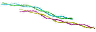
Intermediate filaments (IFs) are cytoskeletal structural components found in the cells of vertebrates, and many invertebrates. Homologues of the IF protein have been noted in an invertebrate, the cephalochordate Branchiostoma.

<span class="mw-page-title-main">Tropomyosin</span> Protein

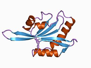
Tropomyosin is a two-stranded alpha-helical, coiled coil protein found in many animal and fungal cells. In animals, it is an important component of the muscular system which works in conjunction with troponin to regulate muscle contraction. It is present in smooth and striated muscle tissues, which can be found in various organs and body systems, including the heart, blood vessels, respiratory system, and digestive system. In fungi, tropomyosin is found in cell walls and helps maintain the structural integrity of cells.

In cell biology, microtubule-associated proteins (MAPs) are proteins that interact with the microtubules of the cellular cytoskeleton. MAPs are integral to the stability of the cell and its internal structures and the transport of components within the cell.

<span class="mw-page-title-main">Motor protein</span> Class of molecular proteins

Motor proteins are a class of molecular motors that can move along the cytoplasm of cells. They convert chemical energy into mechanical work by the hydrolysis of ATP. Flagellar rotation, however, is powered by a proton pump.

<span class="mw-page-title-main">ADF/Cofilin family</span>

ADF/cofilin is a family of actin-binding proteins associated with the rapid depolymerization of actin microfilaments that give actin its characteristic dynamic instability. This dynamic instability is central to actin's role in muscle contraction, cell motility and transcription regulation.

<span class="mw-page-title-main">Treadmilling</span> Simultaneous growth and breakdown on opposite ends of a protein filament

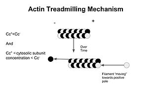
In molecular biology, treadmilling is a phenomenon observed within protein filaments of the cytoskeletons of many cells, especially in actin filaments and microtubules. It occurs when one end of a filament grows in length while the other end shrinks, resulting in a section of filament seemingly "moving" across a stratum or the cytosol. This is due to the constant removal of the protein subunits from these filaments at one end of the filament, while protein subunits are constantly added at the other end. Treadmilling was discovered by Wegner, who defined the thermodynamic and kinetic constraints. Wegner recognized that: “The equilibrium constant (K) for association of a monomer with a polymer is the same at both ends, since the addition of a monomer to each end leads to the same polymer.”; a simple reversible polymer can’t treadmill; ATP hydrolysis is required. GTP is hydrolyzed for microtubule treadmilling.

<span class="mw-page-title-main">Prokaryotic cytoskeleton</span> Structural filaments in prokaryotes

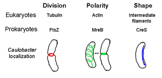
The prokaryotic cytoskeleton is the collective name for all structural filaments in prokaryotes. It was once thought that prokaryotic cells did not possess cytoskeletons, but advances in visualization technology and structure determination led to the discovery of filaments in these cells in the early 1990s. Not only have analogues for all major cytoskeletal proteins in eukaryotes been found in prokaryotes, cytoskeletal proteins with no known eukaryotic homologues have also been discovered. Cytoskeletal elements play essential roles in cell division, protection, shape determination, and polarity determination in various prokaryotes.

<span class="mw-page-title-main">Stress fiber</span> Contractile actin bundles found in non-muscle cells

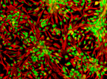
Stress fibers are contractile actin bundles found in non-muscle cells. They are composed of actin (microfilaments) and non-muscle myosin II (NMMII), and also contain various crosslinking proteins, such as α-actinin, to form a highly regulated actomyosin structure within non-muscle cells. Stress fibers have been shown to play an important role in cellular contractility, providing force for a number of functions such as cell adhesion, migration and morphogenesis.

Actin remodeling is the biochemical process that allows for the dynamic alterations of cellular organization. The remodeling of actin filaments occurs in a cyclic pattern on cell surfaces and exists as a fundamental aspect to cellular life. During the remodeling process, actin monomers polymerize in response to signaling cascades that stem from environmental cues. The cell's signaling pathways cause actin to affect intracellular organization of the cytoskeleton and often consequently, the cell membrane. Again triggered by environmental conditions, actin filaments break back down into monomers and the cycle is completed. Actin-binding proteins (ABPs) aid in the transformation of actin filaments throughout the actin remodeling process. These proteins account for the diverse structure and changes in shape of Eukaryotic cells. Despite its complexity, actin remodeling may result in complete cytoskeletal reorganization in under a minute.

<span class="mw-page-title-main">Cytoskeletal drugs</span> Substances or medications that interact with actin or tubulin

Cytoskeletal drugs are small molecules that interact with actin or tubulin. These drugs can act on the cytoskeletal components within a cell in three main ways. Some cytoskeletal drugs stabilize a component of the cytoskeleton, such as taxol, which stabilizes microtubules, or Phalloidin, which stabilizes actin filaments. Others, such as Cytochalasin D, bind to actin monomers and prevent them from polymerizing into filaments. Drugs such as demecolcine act by enhancing the depolymerisation of already formed microtubules. Some of these drugs have multiple effects on the cytoskeleton: for example, Latrunculin both prevents actin polymerization as well as enhancing its rate of depolymerization. Typically the microtubule targeting drugs can be found in the clinic where they are used therapeutically in the treatment of some forms of cancer. As a result of the lack of specificity for specific type of actin, the use of these drugs in animals results in unacceptable off-target effects. Despite this, the actin targeting compounds are still useful tools that can be used on a cellular level to help further our understanding of how this complex part of the cells' internal machinery operates. For example, Phalloidin that has been conjugated with a fluorescent probe can be used for visualizing the filamentous actin in fixed samples.

<span class="mw-page-title-main">Microtubule plus-end tracking protein</span>

Microtubule plus-end/positive-end tracking proteins or +TIPs are a type of microtubule associated protein (MAP) which accumulate at the plus ends of microtubules. +TIPs are arranged in diverse groups which are classified based on their structural components; however, all classifications are distinguished by their specific accumulation at the plus end of microtubules and their ability to maintain interactions between themselves and other +TIPs regardless of type. +TIPs can be either membrane bound or cytoplasmic, depending on the type of +TIPs. Most +TIPs track the ends of extending microtubules in a non-autonomous manner.

Edwin W. Taylor is an adjunct professor of cell and developmental biology at Northwestern University. He was elected to the National Academy of Sciences in 2001. Taylor received a BA in physics and chemistry from the University of Toronto in 1952; an MSc in physical chemistry from McMaster University in 1955, and a PhD in biophysics from the University of Chicago in 1957. In 2001 Taylor was elected to the National Academy of Scineces in Cellular and Developmental Biology and Biochemistry.

References

  1. Speer B, Waggoner B (13 August 1995). "Filament". UCMP Glossary: Cell biology. Berkeley, CA: Museum of Paleontology, University of California. Retrieved November 2, 2011.
  2. Hohmann T, Dehghani F (April 2019). "The Cytoskeleton-A Complex Interacting Meshwork". Cells. 8 (4): 362. doi: 10.3390/cells8040362 . PMC   6523135 . PMID   31003495.
  3. 1 2 Goodson HV, Jonasson EM (June 2018). "Microtubules and Microtubule-Associated Proteins". Cold Spring Harbor Perspectives in Biology. 10 (6): a022608. doi:10.1101/cshperspect.a022608. PMC   5983186 . PMID   29858272.
  4. Herrmann H, Aebi U (November 2016). "Intermediate Filaments: Structure and Assembly". Cold Spring Harbor Perspectives in Biology. 8 (11): a018242. doi:10.1101/cshperspect.a018242. PMC   5088526 . PMID   27803112.
  5. Ndiaye, Anne-Betty; Koenderink, Gijsje H.; Shemesh, Michal (2022). "Intermediate Filaments in Cellular Mechanoresponsiveness: Mediating Cytoskeletal Crosstalk From Membrane to Nucleus and Back". Frontiers in Cell and Developmental Biology. 10: 882037. doi: 10.3389/fcell.2022.882037 . ISSN   2296-634X. PMC   9035595 . PMID   35478961.
  6. Cooper, Geoffrey M. (2000). "Intermediate Filaments". The Cell: A Molecular Approach. 2nd Edition.