Radiation-induced cognitive decline

Last updated

Radiation-induced cognitive decline describes the possible correlation between radiation therapy and cognitive impairment. Radiation therapy is used mainly in the treatment of cancer. Radiation therapy can be used to cure care or shrink tumors that are interfering with quality of life. Sometimes radiation therapy is used alone; other times it is used in conjunction with chemotherapy and surgery. For people with brain tumors, radiation can be an effective treatment because chemotherapy is often less effective due to the blood–brain barrier.[ citation needed ] Unfortunately for some patients, as time passes, people who received radiation therapy may begin experiencing deficits in their learning, memory, and spatial information processing abilities. The learning, memory, and spatial information processing abilities are dependent on proper hippocampus functionality. Therefore, any hippocampus dysfunction will result in deficits in learning, memory, and spatial information processing ability.

Contents

The hippocampus is one of two structures of the central nervous system where neurogenesis continues after birth. The other structure that undergoes neurogenesis is the olfactory bulb. Therefore, it has been proposed that neurogenesis plays some role in the proper functionality of the hippocampus and the olfactory bulb. [1] To test this proposal, a group of rats with normal hippocampal neurogenesis (control) were subjected to a placement recognition exercise that required proper hippocampus function to complete. Afterwards a second group of rats (experimental) were subjected to the same exercise but in that trial their neurogenesis in the hippocampus was arrested. It was found that the experimental group was not able to distinguish between its familiar and unexplored territory. The experimental group spent more time exploring the familiar territory, while the control group spent more time exploring the new territory. The results indicate that neurogenesis in the hippocampus is important for memory and proper hippocampal functionality. [2] Therefore, if radiation therapy inhibits neurogenesis in the hippocampus it would lead to the cognitive decline observed in patients who have received this radiation therapy.

In animal studies discussed by Monje and Palmer in "Radiation Injury and Neurogenesis", it has been proven that radiation does indeed decrease or arrest neurogenesis altogether in the hippocampus. This decrease in neurogenesis is due to apoptosis of the neurons which usually occurs after irradiation. However it has not been proven whether the apoptosis is a direct result of the radiation itself or if there are other factors that cause neuronal apoptosis, namely changes in the hippocampus micro-environment or damage to the precursor pool. [3] Determining the exact cause of the cell apoptosis is important because then it may be possible to inhibit the apoptosis and reverse the effects of the arrested neurogenesis.

Radiation therapy

Ionizing radiation is classified as a neurotoxicant. [4] A 2004 cohort study concluded that irradiation of the brain with dose levels overlapping those imparted by computed tomography can, in at least some instances, adversely affect intellectual development. [5] [6]

Radiation therapy at doses around "23.4 Gy" was found to cause cognitive decline that was especially apparent in young children who underwent the treatment for cranial tumors, between the ages of 5 and 11. Studies found, for example, that the IQ of 5-year-old children declined each year after treatment by additional several IQ points, thereby the child's IQ decreased and decreased while growing older though may plateau at adulthood. [7]

Radiation of 100 mGy to the head at infancy resulted in the beginning appearance of statistically significant cognitive-deficits in one Swedish/radiation-therapy follow-up study. [5] Radiation of 1300-1500mGy to the head at childhood was similarly found to be roughly the threshold dose for the beginning increase in statistically significant rates of schizophrenia. [8]

From soliciting for participants in a study and then examination of the prenatally exposed at Hiroshima & Nagasaki, those who experienced the prompt burst of ionizing radiation at the 8-15 and 16–25 week periods after gestation were to, especially in the closest survivors, have a higher rate of severe mental retardation as well as variation in intelligence quotient (IQ) and school performance. It is uncertain, if there exists a threshold dose, under which one or more of these effects, of prenatal exposure to ionizing radiation, do not exist, though from analysis of the limited data, "0.1" Gy is suggested for both. [9] [8]

Warfare

Adult humans receiving an acute whole body incapacitating dose (30 Gy) have their performance degraded almost immediately and become ineffective within several hours. A dose of 5.3 Gy to 8.3 Gy is considered lethal within months to half of male adults but not immediately incapacitating. Personnel exposed to this amount of radiation have their cognitive performance degraded in two to three hours. [10] [11] Depending on how physically demanding the tasks they must perform are, and remain in this disabled state at least two days. However, at that point they experience a recovery period and can perform non-demanding tasks for about six days, after which they relapse for about four weeks. At this time they begin exhibiting symptoms of radiation poisoning of sufficient severity to render them totally ineffective. Death follows for about half of males at approximately six weeks after exposure.

Nausea and vomiting generally occur within 24–48 hours after exposure to mild (1–2  Gy) doses of radiation. Headache, fatigue, and weakness are also seen with mild exposure. [12]

Exposure of adults to 150−500 mSv results in the beginning observance of cerebrovascular pathology, and exposure to 300 mSv results in the beginning of the observance of neuropsychiatric and neurophysiological dose-related effects. [8] Cumulative equivalent doses above 500 mSv of ionizing radiation to the head, were proven with epidemiological evidences to cause cerebrovascular atherosclerotic damage, thus increasing the chances of stroke in later life. [13] The equivalent dose of 0.5 Gy (500 mGy) x-rays is 500 mSv. [14]

Acute ablation of precursor cells

Recent studies have shown that there is a decrease in neurogenesis in the hippocampus after irradiation therapy. The decrease in neurogenesis is the result of a reduction in the stem cell pool due to apoptosis. However, the question remains whether radiation therapy results in a complete ablation of the stem cell pool in the hippocampus or whether some stem cells survive. Animal studies have been performed by Monje and Palmer to determine if there is an acute ablation of the stem cell pool. In the study, rats were subjected to 10 Gy dosage of radiation. The 10 Gy radiation dosage is comparable to that used in irradiation therapy in humans. One month after the reception of the dosage, living precursor cells from these rats’ hippocampus were successfully isolated and cultured. Therefore, a complete ablation of the precursor cell pool by irradiation does not occur. [3]

Precursor cell integrity

Precursor cells may be damaged by radiation. This damage of the cells may prevent the precursor cells from differentiating into neurons and result in decreased neurogenesis. To determine whether the precursor cells are impaired in their ability to differentiate, two cultures were prepared by Fike et al. One of these cultures contained precursor cells from an irradiated rat's hippocampus and the second culture contained non-irradiated precursor cells from a rat hippocampus. The precursor cells were then observed while they continued to develop. The results indicated that the irradiated culture contained a higher number of differentiated neuron and glial cells in comparison to the control. It was also found that the ratios of glial cells to neurons in both cultures were similar. [15] These results suggest that the radiation did not impair the precursor cells ability to differentiate into neurons and therefore neurogenesis is still possible.

Alterations in hippocampus microenvironment

The microenvironment is an important component to consider for precursor survival and differentiation. It is the microenvironment that provides the signals to the precursor cells that help it survive, proliferate, and differentiate. To determine if the microenvironment is altered as a result of radiation, an animal study was performed by Fike et al. where highly enriched, BrdU labeled, non-irradiated stem cells from a rat hippocampus were implanted into a hippocampus that was irradiated one month prior. The stem cells were allowed to remain in the live rat for 3–4 weeks. Afterwards, the rat was killed and the stem cells were observed using immunohistochemistry and confocal microscopy. The results show that stem cell survival was similar to that found in a control subject (normal rat hippocampus); however, the number of neurons generated was decreased by 81%. Therefore, alterations of the microenvironment post radiation can lead to a decrease in neurogenesis. [15]

In addition, studies mentioned by Fike et al. found that there are two main differences between the hippocampus of an irradiated rat and a non-irradiated rat that are part of the microenvironment. There was a significantly larger number of activated microglia cells in the hippocampus of irradiated rats in comparison to non-irradiated rats. [16] The presence of microglia cells is characteristic of the inflammatory response which is most likely due to radiation exposure. Also the expected clustering of stem cells around the vasculature of the hippocampus was disrupted. [15] Therefore, focusing on the microglial activation, inflammatory response, and microvasculature may produce a direct link to the decrease in neurogenesis post irradiation.

Inflammatory response affects neurogenesis

Radiation therapy usually results in chronic inflammation, and in the brain this inflammatory response comes in the form of activated microglia cells. Once activated, these microglia cells start to release stress hormones and various pro-inflammatory cytokines. [16] [17] Some of what is released by the activated microglia cells, like the glucocorticoid stress hormone, may result in a decrease in neurogenesis. To investigate this concept, an animal study was performed by Monje et al. in order to determine the specific cytokines or stress hormones that were released by activated microglial cells that decrease neurogenesis in an irradiated hippocampus. In this study, microglia cells were exposed to bacterial lipopolysaccharide to elicit an inflammatory response, thus activating the microglia cells. These activated microglia were then co-cultured with normal hippocampal neural stem cells. Also, as a control, non-activated microglia cells were co-cultured with normal hippocampal neural stem cells. In comparing the two co-cultures, it was determined that neurogenesis in the activated microglia cell culture was 50% less than in the control. A second study was also performed to ensure that the decrease in neurogenesis was the result of released cytokines and not cell-to-cell contact of microglia and stem cells. In this study, neural stem cells were cultured on preconditioned media from activated microglia cells and a comparison was made with a neural stem cells cultured on plain media. The results of this study indicated that neurogenesis also showed a similar decrease in the preconditioned media culture versus the control. [17]

When microglia cells are activated, they release the pro-inflammatory cytokine IL-1β, TNF-α, INF-γ, and IL-6. In order to identify the cytokines that decreased neurogenesis, Monje et al. allowed progenitor cells to differentiate while exposed to each cytokine. The results of the study showed that only the recombinant IL-6 and TNF-α exposure significantly reduced neurogenesis. Then the IL-6 was inhibited and neurogenesis was restored. This implicates IL-6 as the main cytokine responsible for the decrease of neurogenesis in the hippocampus. [17]

Microvasculature and neurogenesis

The microvasculature of the subgranular zone, located in dentate gyrus of hippocampus, plays an important role in neurogenesis. As precursor cells develop in the subgranular zone, they form clusters. These clusters usually contain dozens of cells. The clusters are made up of endothelial cells and neuronal precursor cells that have the ability to differentiate into either neurons or glia cells. With time, these clusters eventually migrate towards microvessels in the subgranular zone. As the clusters get closer to the vessels, some of the precursor cells differentiate in glia cells and eventually the remaining precursor cells will differentiate into neurons. Upon investigation of the close association between the vessels and clusters, it is apparent that the actual migration of the precursor cells to these vessels is not random. [18] Since endothelial cells forming the vessel wall do secrete brain-derived neurotrophic factor, it is plausible that the neuronal precursor cells migrate to those regions in order to grow, survive, and differentiate. [19] Also, since the clusters do contain endothelial cells, they might be attracted to the vascular endothelial growth factor that is released in the area of vessels to promote endothelial survival and angiogenesis. [19] However, as noted previously, clustering along the capillaries in the subgranular zone does decrease when the brain is subject to radiation. [15] The exact reasoning for this disruption of the close association between cluster and vessels remains unknown. It is possible that any signaling that would normally attract the clusters to the region, for example the bone-derived growth factor and the vascular endothelial growth factor, may be suppressed.

Reversal

Blocking inflammatory cascade

Neurogenesis in the hippocampus usually decreases after exposure to radiation and usually leads to a cognitive decline in patients undergoing radiation therapy. As discussed above, the decrease in neurogenesis is heavily influenced by changes in the microenvironment of the hippocampus upon exposure to radiation. Specifically, disruption of the cluster/vessel association in the subgranular zone of the dentate gyrus and cytokines released by activated microglia as part of the inflammatory response do impair neurogenesis in the irradiated hippocampus. Thus several studies have used this knowledge to reverse the reduction in neurogenesis in the irradiated hippocampus. In one study, indomethacin treatment was given to the irradiated rat during and after irradiation treatment. It was found that the indomethacin treatment caused a 35% decrease in the number of activated microglia per dentate gyrus in comparison to microglia activation in irradiated rats without indomethacin treatment. This decrease in microglia activation reduces the amount of cytokines and stress-hormone release, thus reducing the effect of the inflammatory response. When the number of precursor cells adopting a neuronal fate was quantified, it was determined that the ratio of neurons to glia cells increased. This increase in neurogenesis was only 20-25% of that observed in control animals. However, in this study the inflammatory response was not eliminated entirely, and some cytokines or stress hormones continued to be secreted by the remaining activated microglia cells causing the reduction in neurogenesis. [17] In a second study, the inflammatory cascade was also blocked at another stage. This study focused mainly on the c-Jun NH2 – terminal kinase pathway which when activated results in the apoptosis of neurons. This pathway was chosen because, upon irradiation, it is the only mitogen-activated protein kinase that is activated. The mitogen-activated protein kinases are important for regulation of migration, proliferation, differentiation, and apoptosis. The JNK pathway is activated by cytokines released by activated microglia cells, and blocking this pathway significantly reduces neuronal apoptosis. In the study, the JNK was inhibited using 5 μM SP600125 dosage, and this resulted in a decrease of neural stem cells apoptosis. This decrease in apoptosis results in increased neuronal recovery. [20]

Environmental enrichment

In previous work, environmental enrichment has been used to determine its effect on brain activity. In these studies, the environmental enrichment has positively impacted the brain functionality in both normal, healthy animals and animals that had suffered severe brain injury. It has already been shown by Elodie Bruel-Jungerman et al. that subjecting animals to learning exercises that are heavily dependent on the hippocampus results in increased neurogenesis. [1] Therefore, the question of whether environmental enrichment can enhance neurogenesis in an irradiated hippocampus is raised. In a study performed by Fan et al., the effects of environmental enrichment on gerbils were tested. There were four groups of gerbils used for this experiment, where group one consisted on non-irradiated animals that lived in a standard environment, group two were non-irradiated animals that lived in an enriched environment, group three were irradiated animals that lived in a standard environment, and group four were irradiated animals that lived in an enriched environment. After two months of maintaining the gerbils in the required environments, they were killed and hippocampal tissue was removed for analysis. It was found that the number of precursor neurons that were differentiated into neurons from group four (irradiated and enriched environment) was significantly more than group three (irradiated and standard environment). Similarly, the number of neuron precursor cells was more in group two (non-irradiated and enriched environment), in comparison to group one (non-irradiated and standard environment). The results indicate that neurogenesis was increased in the animals that were exposed to the enriched environment, in comparison to animals in the standard environment. This outcome indicates that environmental enrichment can indeed increase neurogenesis and reverse the cognitive decline. [21]

See also

Related Research Articles

<span class="mw-page-title-main">Dentate gyrus</span> Region of the hippocampus in the brain

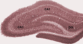
The dentate gyrus (DG) is part of the hippocampal formation in the temporal lobe of the brain, which also includes the hippocampus and the subiculum. The dentate gyrus is part of the hippocampal trisynaptic circuit and is thought to contribute to the formation of new episodic memories, the spontaneous exploration of novel environments and other functions.

<span class="mw-page-title-main">Adult neurogenesis</span> Generating of neurons from neural stem cells in adults

Adult neurogenesis is the process in which neurons are generated from neural stem cells in the adult. This process differs from prenatal neurogenesis.

<span class="mw-page-title-main">Rostral migratory stream</span> One path neural stem cells take to reach the olfactory bulb


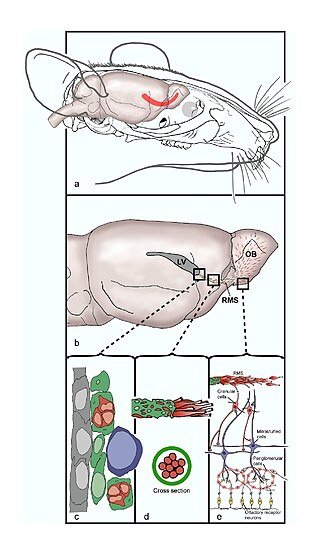
The rostral migratory stream (RMS) is a specialized migratory route found in the brain of some animals along which neuronal precursors that originated in the subventricular zone (SVZ) of the brain migrate to reach the main olfactory bulb (OB). The importance of the RMS lies in its ability to refine and even change an animal's sensitivity to smells, which explains its importance and larger size in the rodent brain as compared to the human brain, as our olfactory sense is not as developed. This pathway has been studied in the rodent, rabbit, and both the squirrel monkey and rhesus monkey. When the neurons reach the OB they differentiate into GABAergic interneurons as they are integrated into either the granule cell layer or periglomerular layer.

HIV-associated neurocognitive disorders (HAND) are neurological disorders associated with HIV infection and AIDS. It is a syndrome of progressive deterioration of memory, cognition, behavior, and motor function in HIV-infected individuals during the late stages of the disease, when immunodeficiency is severe. HAND may include neurological disorders of various severity. HIV-associated neurocognitive disorders are associated with a metabolic encephalopathy induced by HIV infection and fueled by immune activation of macrophages and microglia. These cells are actively infected with HIV and secrete neurotoxins of both host and viral origin. The essential features of HIV-associated dementia (HAD) are disabling cognitive impairment accompanied by motor dysfunction, speech problems and behavioral change. Cognitive impairment is characterised by mental slowness, trouble with memory and poor concentration. Motor symptoms include a loss of fine motor control leading to clumsiness, poor balance and tremors. Behavioral changes may include apathy, lethargy and diminished emotional responses and spontaneity. Histopathologically, it is identified by the infiltration of monocytes and macrophages into the central nervous system (CNS), gliosis, pallor of myelin sheaths, abnormalities of dendritic processes and neuronal loss.

<span class="mw-page-title-main">Neuroimmune system</span>

The neuroimmune system is a system of structures and processes involving the biochemical and electrophysiological interactions between the nervous system and immune system which protect neurons from pathogens. It serves to protect neurons against disease by maintaining selectively permeable barriers, mediating neuroinflammation and wound healing in damaged neurons, and mobilizing host defenses against pathogens.

Elizabeth Gould is an American neuroscientist and the Dorman T. Warren Professor of Psychology at Princeton University. She was an early investigator of adult neurogenesis in the hippocampus, a research area that continues to be controversial. In November 2002, Discover magazine listed her as one of the 50 most important women scientists.

Neuroepithelial cells, or neuroectodermal cells, form the wall of the closed neural tube in early embryonic development. The neuroepithelial cells span the thickness of the tube's wall, connecting with the pial surface and with the ventricular or lumenal surface. They are joined at the lumen of the tube by junctional complexes, where they form a pseudostratified layer of epithelium called neuroepithelium.

Neural stem cells (NSCs) are self-renewing, multipotent cells that firstly generate the radial glial progenitor cells that generate the neurons and glia of the nervous system of all animals during embryonic development. Some neural progenitor stem cells persist in highly restricted regions in the adult vertebrate brain and continue to produce neurons throughout life. Differences in the size of the central nervous system are among the most important distinctions between the species and thus mutations in the genes that regulate the size of the neural stem cell compartment are among the most important drivers of vertebrate evolution.

<span class="mw-page-title-main">Subventricular zone</span> Region outside each lateral ventricle of the brain

The subventricular zone (SVZ) is a region situated on the outside wall of each lateral ventricle of the vertebrate brain. It is present in both the embryonic and adult brain. In embryonic life, the SVZ refers to a secondary proliferative zone containing neural progenitor cells, which divide to produce neurons in the process of neurogenesis. The primary neural stem cells of the brain and spinal cord, termed radial glial cells, instead reside in the ventricular zone (VZ).

<span class="mw-page-title-main">Subgranular zone</span>

The subgranular zone (SGZ) is a brain region in the hippocampus where adult neurogenesis occurs. The other major site of adult neurogenesis is the subventricular zone (SVZ) in the brain.

<span class="mw-page-title-main">Colony stimulating factor 1 receptor</span> Protein found in humans

Colony stimulating factor 1 receptor (CSF1R), also known as macrophage colony-stimulating factor receptor (M-CSFR), and CD115, is a cell-surface protein encoded by the human CSF1R gene. CSF1R is a receptor that can be activated by two ligands: colony stimulating factor 1 (CSF-1) and interleukin-34 (IL-34). CSF1R is highly expressed in myeloid cells, and CSF1R signaling is necessary for the survival, proliferation, and differentiation of many myeloid cell types in vivo and in vitro. CSF1R signaling is involved in many diseases and is targeted in therapies for cancer, neurodegeneration, and inflammatory bone diseases.

<span class="mw-page-title-main">Environmental enrichment</span> Brain stimulation through physical and social surroundings

Environmental enrichment is the stimulation of the brain by its physical and social surroundings. Brains in richer, more stimulating environments have higher rates of synaptogenesis and more complex dendrite arbors, leading to increased brain activity. This effect takes place primarily during neurodevelopment, but also during adulthood to a lesser degree. With extra synapses there is also increased synapse activity, leading to an increased size and number of glial energy-support cells. Environmental enrichment also enhances capillary vasculation, providing the neurons and glial cells with extra energy. The neuropil expands, thickening the cortex. Research on rodent brains suggests that environmental enrichment may also lead to an increased rate of neurogenesis.

Erythropoietin in neuroprotection is the use of the glycoprotein erythropoietin (Epo) for neuroprotection. Epo controls erythropoiesis, or red blood cell production.

Endogenous regeneration in the brain is the ability of cells to engage in the repair and regeneration process. While the brain has a limited capacity for regeneration, endogenous neural stem cells, as well as numerous pro-regenerative molecules, can participate in replacing and repairing damaged or diseased neurons and glial cells. Another benefit that can be achieved by using endogenous regeneration could be avoiding an immune response from the host.

While researchers have found that moderate alcohol consumption in older adults is associated with better cognition and well-being than abstinence, excessive alcohol consumption is associated with widespread and significant brain lesions. Other data – including investigated brain-scans of 36,678 UK Biobank participants – suggest that even "light" or "moderate" consumption of alcohol by itself harms the brain, such as by reducing brain grey matter volume. This may imply that alternatives and generally aiming for lowest possible consumption could usually be the advisable approach.

Travel outside the Earth's protective atmosphere, magnetosphere, and in free fall can harm human health, and understanding such harm is essential for successful crewed spaceflight. Potential effects on the central nervous system (CNS) are particularly important. A vigorous ground-based cellular and animal model research program will help quantify the risk to the CNS from space radiation exposure on future long distance space missions and promote the development of optimized countermeasures.

Neuroinflammation is inflammation of the nervous tissue. It may be initiated in response to a variety of cues, including infection, traumatic brain injury, toxic metabolites, or autoimmunity. In the central nervous system (CNS), including the brain and spinal cord, microglia are the resident innate immune cells that are activated in response to these cues. The CNS is typically an immunologically privileged site because peripheral immune cells are generally blocked by the blood–brain barrier (BBB), a specialized structure composed of astrocytes and endothelial cells. However, circulating peripheral immune cells may surpass a compromised BBB and encounter neurons and glial cells expressing major histocompatibility complex molecules, perpetuating the immune response. Although the response is initiated to protect the central nervous system from the infectious agent, the effect may be toxic and widespread inflammation as well as further migration of leukocytes through the blood–brain barrier may occur.

<span class="mw-page-title-main">Raz Yirmiya</span>

Raz Yirmiya is an Israeli behavioral neuroscientist and director of the Laboratory for Psychoneuroimmunology at the Hebrew University of Jerusalem in Israel. He is best known for providing the first experimental evidence for the role of immune system activation in depression, for discovering that disturbances in brain microglia cells underlie some forms of depression, and for elucidating the involvement of inflammatory cytokines in regulation of cognitive and emotional processes.

Neurogenesis is the process by which nervous system cells, the neurons, are produced by neural stem cells (NSCs). In short, it is brain growth in relation to its organization. This occurs in all species of animals except the porifera (sponges) and placozoans. Types of NSCs include neuroepithelial cells (NECs), radial glial cells (RGCs), basal progenitors (BPs), intermediate neuronal precursors (INPs), subventricular zone astrocytes, and subgranular zone radial astrocytes, among others.

<span class="mw-page-title-main">Erin M. Gibson</span> Glial and circadian biologist

Erin M. Gibson is a glial and circadian biologist as well as an assistant professor in the Department of Psychiatry and Behavioral Sciences and the Stanford Center for Sleep Sciences and Medicine at Stanford University. Gibson investigates the role of glial cells in sculpting neural circuits and mechanistically probes how the circadian rhythm modulates glial biology.

References

  1. 1 2 Bruel-Jungerman, Elodie; Rampon, Claire; Laroche, Serge (2007). "Adult Hippocampal Neurogenesis, Synaptic Plasticity and Memory: Facts and Hypotheses". Reviews in the Neurosciences. 18 (2): 93–114. doi:10.1515/REVNEURO.2007.18.2.93. PMID   17593874. S2CID   21585753.
  2. Madsen, T.M; Kristjansen, P.E.G; Bolwig, T.G; Wörtwein, G (2003). "Arrested neuronal proliferation and impaired hippocampal function following fractionated brain irradiation in the adult rat". Neuroscience. 119 (3): 635–42. doi:10.1016/S0306-4522(03)00199-4. PMID   12809684. S2CID   25715150.
  3. 1 2 Monje, Michelle L.; Palmer, Theo (2003). "Radiation injury and neurogenesis". Current Opinion in Neurology. 16 (2): 129–34. doi:10.1097/00019052-200304000-00002. PMID   12644738.
  4. Mendola, Pauline; Selevan, Sherry G.; Gutter, Suzanne; Rice, Deborah (2002). "Environmental factors associated with a spectrum of neurodevelopmental deficits". Mental Retardation and Developmental Disabilities Research Reviews. 8 (3): 188–97. doi:10.1002/mrdd.10033. PMID   12216063.
  5. 1 2 Hall, P.; Adami, HO; Trichopoulos, D; Pedersen, NL; Lagiou, P; Ekbom, A; Ingvar, M; Lundell, M; Granath, F (2004). "Effect of low doses of ionising radiation in infancy on cognitive function in adulthood: Swedish population based cohort study". BMJ. 328 (7430): 19. doi:10.1136/bmj.328.7430.19. PMC   313898 . PMID   14703539.
  6. Asteriadis, I (2004). "Low radiation doses; are they harmful in infancy?". Hellenic Journal of Nuclear Medicine. 7 (1): 2–4. PMID   16868634.
  7. "Late Effects of Treatment for Childhood Cancer". National Cancer Institute. 12 April 2012. Retrieved 7 June 2012.
  8. 1 2 3 Loganovsky, K (2009). "Do Low Doses of Ionizing Radiation Affect the Human Brain?". Data Science Journal. 8: BR13–35. doi: 10.2481/dsj.BR-04 .
  9. Otake, M.; Schull, WJ (1998). "Review: Radiation-related brain damage and growth retardation among the prenatally exposed atomic bomb survivors". International Journal of Radiation Biology. 74 (2): 159–71. doi:10.1080/095530098141555. PMID   9712546.
  10. NUCLEAR EVENTS AND THEIR CONSEQUENCES by the Borden institute. Chapter 1
  11. NUCLEAR EVENTS AND THEIR CONSEQUENCES by the Borden institute. Chapter 7 BEHAVIORAL AND NEUROPHYSIOLOGICAL CHANGES WITH EXPOSURE TO IONIZING RADIATION
  12. Mayo Clinic Staff (May 9, 2008). "Symptoms". Radiation sickness. Mayo Clinic. Retrieved 2011-12-24.
  13. Picano, Eugenio; Vano, Eliseo; Domenici, Luciano; Bottai, Matteo; Thierry-Chef, Isabelle (2012). "Cancer and non-cancer brain and eye effects of chronic low-dose ionizing radiation exposure". BMC Cancer. 12 (1): 157. doi: 10.1186/1471-2407-12-157 . PMC   3495891 . PMID   22540409.
  14. Jain, Prasoon; Mehta, Atul C. (2011). "Radiation terminology". In Wang, Ko-Pen; Mehta, Atul C.; Turner, J. Francis (eds.). Flexible Bronchoscopy. John Wiley & Sons. p. 21. ISBN   978-1-4443-4640-4. For X rays, the radiation-weighting factor is equal to one; so the equivalent dose in Sv units is equal to the absorbed dose in Gy.
  15. 1 2 3 4 Monje, Michelle L.; Mizumatsu, Shinichiro; Fike, John R.; Palmer, Theo D. (2002). "Irradiation induces neural precursor-cell dysfunction". Nature Medicine. 8 (9): 955–62. doi:10.1038/nm749. PMID   12161748. S2CID   10347561.
  16. 1 2 Mizumatsu, S; Monje, ML; Morhardt, DR; Rola, R; Palmer, TD; Fike, JR (2003). "Extreme sensitivity of adult neurogenesis to low doses of X-irradiation". Cancer Research. 63 (14): 4021–7. PMID   12874001.
  17. 1 2 3 4 Monje, M. L.; Toda, H; Palmer, TD (2003). "Inflammatory Blockade Restores Adult Hippocampal Neurogenesis". Science. 302 (5651): 1760–5. Bibcode:2003Sci...302.1760M. doi: 10.1126/science.1088417 . PMID   14615545. S2CID   36806485.
  18. Palmer, Theo D.; Willhoite, Andrew R.; Gage, Fred H. (2000). "Vascular niche for adult hippocampal neurogenesis". The Journal of Comparative Neurology. 425 (4): 479–94. doi:10.1002/1096-9861(20001002)425:4<479::AID-CNE2>3.0.CO;2-3. PMID   10975875. S2CID   46440541.
  19. 1 2 Louissaint Jr., Abner; Rao, Sudha; Leventhal, Caroline; Goldman, Steven A. (2002). "Coordinated Interaction of Neurogenesis and Angiogenesis in the Adult Songbird Brain". Neuron. 34 (6): 945–60. doi: 10.1016/S0896-6273(02)00722-5 . PMID   12086642.
  20. Kanzawa, T; Iwado, E; Aoki, H; Iwamaru, A; Hollingsworth, E F; Sawaya, R; Kondo, S; Kondo, Y (2006). "Ionizing radiation induces apoptosis and inhibits neuronal differentiation in rat neural stem cells via the c-Jun NH2-terminal kinase (JNK) pathway". Oncogene. 25 (26): 3638–48. doi: 10.1038/sj.onc.1209414 . PMID   16491125.
  21. Fan, Yang; Liu, Zhengyan; Weinstein, Philip R.; Fike, John R.; Liu, Jialing (2007). "Environmental enrichment enhances neurogenesis and improves functional outcome after cranial irradiation". European Journal of Neuroscience. 25 (1): 38–46. doi:10.1111/j.1460-9568.2006.05269.x. PMID   17241265. S2CID   43259184.

Further reading