Roy R. Parker

Last updated
Roy Parker
Born
Roy R. Parker
TitleMember of the National Academy of Sciences
Alma mater Carnegie Mellon University (BSc)
University of California, San Francisco (PhD)
Known forDistinguished Professor of Chemistry and Biochemistry
Cech-Leinwand Endowed Chair of Biochemistry at the University of Colorado Boulder
Scientific career
Fields Biochemistry
Chemistry
Institutions University of Arizona
University of Colorado Boulder
University of California, San Francisco
University of California, San Diego
University of Massachusetts Medical School
Howard Hughes Medical Institute
National Academy of Sciences
Website www.colorado.edu/lab/parkergroup/
External videos
Nuvola apps kaboodle.svg Roy Parker, “Improving Graduate Training”, iBiology
Nuvola apps kaboodle.svg Roy Parker, “The Life of Eukaryotic mRNA: Localization, Translation, and Degradation”, iBiology

Roy R. Parker is a biochemist who has been an active investigator in science since the 1970s. He is currently a Distinguished Professor of Chemistry and Biochemistry and Cech-Leinwand Endowed Chair of Biochemistry at the University of Colorado Boulder. Throughout his life, Parker has contributed a vast degree of knowledge to research and studies of biochemistry. His current focus includes the biogenesis, function, and degradation of multiple forms of RNA in eukaryotes. Parker aims to use his research to understand how various diseases and pathologies result from abnormalities in RNA. [1] In 2012, Parker was elected to the National Academy of Sciences in Biochemistry. [2]

Contents

Education

To start his career, Parker attended Carnegie Mellon University in Pittsburgh, Pennsylvania, where he received his Bachelor of Science degree in chemistry. After graduating from Carnegie Mellon in 1979, he moved on to receive his PhD in genetics with Christine Guthrie at the University of California, San Francisco, California in 1985. From 1985-1987, Parker worked in laboratories for his postdoctoral degree at both the University of California, San Francisco, and the University of California, San Diego. [3] Later, Parker became a postdoctoral fellow with Allan Jacobson at the University of Massachusetts Medical School in Worcester, MA. [4]

Career

At the University of Arizona, Parker taught molecular and cellular biology until 2012 when he moved to be a professor at the University of Colorado Boulder. [4] He is currently the Cech-Leinwand Endowed Chair of Biochemistry as well as professor in chemistry and biochemistry courses. Parker also conducts research at the Howard Hughes Medical Institute. [1]

Research interests

The primary investigations of Parker's research include the analysis of eukaryotic RNA and the effects that flawed types of RNA have on the biochemical mechanisms of the body. [1] More technically, his lab addresses the regulation of RNA molecules, how that impacts normal physiology of eukaryotic cells, and how aberrant RNA regulation contributes to human disease. His lab uses yeast and mammalian cells to examine oligo(U) and oligo(A) tailing and detailing, tau protein in RNA biology and RNA chaperones.

Key conclusions

Important research findings first began when Parker and his lab team established the major pathways of eukaryotic mRNA turnover. These pathways have the potential to generate mRNA degradation and decapping. Next, the lab group pinpointed major nucleases that contribute to mRNA degradation, as well as the molecules that help facilitate this event. Some of these facilitators include decapping enzymes, 3' to 5' decay complexes, and deadenylases. In years following, Parker and co-workers collected enough data to conclude that some decapping and decay proteins are translational repressors. In other words, Parker determined that decapping and translation could be thought of as rivals, for their mechanisms oppose each other. Finally, most recently, the lab group has discovered several cytoplasmic mRNP granules most notably P bodies. [2]

Major contributions

In addition to publishing hundreds of research articles in scientific journals, Parker has supported the education of students around the country. Some of his talks are published online. These focus on topics including improving graduate training and "The Life of Eukaryotic mRNA: Localization, Translation, and Degradation". These video lectures are published online to educate students and other scientists. [4]

Publications

Throughout his education and career, Parker has published 165 original papers and 77 chapters and invited reviews. Some of his most-cited publications and discoveries include the following: [3]

“PEP4 Gene Function Is Required for Expression of Several Vacuolar Hydrolases in Saccharomyces Cerevisiae”(1982) Parker’s first original publication was in 1982 where he worked with two other scientists, Zubenko and Jones, to investigate the role of the PEP4 gene on yeast S. cerevisiae. [5]

A Turnover Pathway for Both Stable and Unstable mRNAs in Yeast: Evidence for a Requirement for Deadenylation”(1993) The purpose of this investigation was to trace the pathways involved in mRNA turnover in yeast. To do so, Parker and Decker conducted experimentation in four genes by analyzing the degradation of nascent transcripts. They found a connection between both deadenylation rate of mRNA and stability of oligo(A) with mRNA half-lives. More specifically, they looked at the effects of RNA secondary structures and realized that after deadenylation, those without the 5’ sequence began to aggregate. After evaluating these results, they concluded that deadenylation contributes to changes in mRNA, creating an mRNA decay pathway. [6]

“Degradation of mRNA in Eukaryotes”(1995) With this research study, Parker and Beelman investigated the various pathways of degradation to several subtypes of mRNA. They offer evidence that there is a connection between decay rate and turnover pathways of mRNA. After developing conclusions from much observation and research in this study, Parker and Beelman hoped to pinpoint the genetic products that control nucleolytic events and promote degradation pathways. [7]

“mRNA Turnover in Yeast Promoted by the MATalpha1 Instability Element”(1996) In 1996, Parker and Caponigro analyzed the decay rates of mRNA in yeast by deleting a component of the MATalpha1 instability element and observing the effects. [8]

“Recognition of Yeast mRNAs As “Nonsense Containing” Leads to Both Inhibition of mRNA Translation and mRNA Degradation: Implications for the Control of mRNA Decapping”(1999) This paper examined promoters for decapping and how the promoters impede the translation and destruction of mRNA. [9]

“Decapping and Decay of Messenger RNA Occur in Cytoplasmic Processing Bodies”(2003) In 2003, Parker and Sheth concentrated on deadenylation, decapping, and exonucleolytic decay in this publication. They studied yeast models and proposed a model that P bodies are the sites of degradation in the cell and contain intermediates of mRNA degradation. With this information, they concluded that mRNAs move between polysomes and P bodies, and this is a critical aspect of mRNA metabolism in the cytoplasm. [10]

“Cytoplasmic Degradation of Splice-Defective Pre-mRNAs and Intermediates”(2003) With this publication, Hilleren and Parker discovered that the buildup of certain intermediates in a Dbr1p-dependent pathway indicates that same pathway regulates pre-mRNA splicing. [11]

“General Translational Repression by Activators of mRNA Decapping”(2005) Coller and Parker singled out activators for decapping mechanisms in this study. These decapping activators inhibit translation and stimulate the formation of P bodies. Using staining techniques to identify the activity of decapping activators, they were successfully able to conclude that there is a mechanism which signals mRNAs for decapping and therefore restricts mRNAs from going through translation. [12]

“Movement of Eukaryotic mRNAs between Polysomes and Cytoplasmic Processing Bodies”(2005) Throughout this paper, Parker and two other scientists, Brengues and Teixeira, demonstrated that mRNAs migrate between polysomes and cytoplasmic processing bodies as a form of mRNA packing. [13]

“Endonucleolytic Cleavage of Eukaryotic mRNAs with Stalls in Translation Elongation”(2006) Doma and Parker identified the process of non-stop decay by which special proteins are able to distinguish stalled mRNA translation, and then initiate the process of endonucleolytic cleavage. [14]

“P Bodies and the Control of mRNA Translation and Degradation”(2007) This publication focuses on eukaryotes and the formation of P bodies due to aggregations of mRNAs that failed to undergo translation. P bodies are linked to maternal and neuronal mRNA granules. This link led to uncovering of the relationship between polysomes and mRNAs within P bodies with regards to cytoplasmic mRNA activity. Parker and Sheth’s prime conclusion from this publication is that translation is inhibited, and decay is promoted when mRNA-specific regulatory factors, such as miRNAs and RISC, engage with P bodies. [15]

“Eukaryotic Stress Granules: The Ins and Outs of Translation”(2009) The focus of this publication is the formation of stress granules due to the body’s stress response which prevents translation from occurring. Parker and Buchan researched that stress granules cause many problems to forms of RNA, particularly mRNAs, by making them less stable and by preventing their translation. They were able to conclude that stress granules may communicate with P bodies to create even more harmful effects to the function of mRNA. [16]

“Skill Development in Graduate Education”(2012) This unique paper does not discuss biochemical processes but rather explores the impacts of proper education in the field of biochemistry. Parker demonstrates the elevated dropout rates in students pursuing graduate degrees and analyzes the necessary steps taken and skills acquired for a student to succeed. [17]

“Analysis of Double-Stranded RNA from Microbial Communities Identifies Double-Stranded RNA Virus-Like Elements”(2014) Decker and Parker suggested from this publication that investigating double-stranded RNAs could potentially lead to a multitude of discoveries about the genetics of viruses and other microbes. [18]

“The Link between Adjacent Codon Pairs and mRNA Stability”(2017) In 2017, Harigaya and Parker found that there was a connection between the stability of mRNA and codon pairs that act to suppress certain functions in the codon-mediated gene regulation. [19]

“Transcriptome-Wide Comparison of Stress Granules and P Bodies Reveals that Translation Plays a Major Role in RNA Partitioning”(2019) Matheny, Rao, and Parker collaborated to identify that translation is an extremely important process in the formation of P bodies and stress granules, which is often suppressed during stress. [20]

Honors and awards

Parker has received many honors and awards, including the following: [3]

Related Research Articles

<span class="mw-page-title-main">Messenger RNA</span> RNA that is read by the ribosome to produce a protein

In molecular biology, messenger ribonucleic acid (mRNA) is a single-stranded molecule of RNA that corresponds to the genetic sequence of a gene, and is read by a ribosome in the process of synthesizing a protein.

Polyadenylation is the addition of a poly(A) tail to an RNA transcript, typically a messenger RNA (mRNA). The poly(A) tail consists of multiple adenosine monophosphates; in other words, it is a stretch of RNA that has only adenine bases. In eukaryotes, polyadenylation is part of the process that produces mature mRNA for translation. In many bacteria, the poly(A) tail promotes degradation of the mRNA. It, therefore, forms part of the larger process of gene expression.

In molecular biology, the five-prime cap is a specially altered nucleotide on the 5′ end of some primary transcripts such as precursor messenger RNA. This process, known as mRNA capping, is highly regulated and vital in the creation of stable and mature messenger RNA able to undergo translation during protein synthesis. Mitochondrial mRNA and chloroplastic mRNA are not capped.

<span class="mw-page-title-main">Nonsense-mediated decay</span> Elimination of mRNA with premature stop codons in eukaryotes

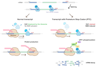
Nonsense-mediated mRNA decay (NMD) is a surveillance pathway that exists in all eukaryotes. Its main function is to reduce errors in gene expression by eliminating mRNA transcripts that contain premature stop codons. Translation of these aberrant mRNAs could, in some cases, lead to deleterious gain-of-function or dominant-negative activity of the resulting proteins.

In cellular biology, P-bodies, or processing bodies, are distinct foci formed by phase separation within the cytoplasm of a eukaryotic cell consisting of many enzymes involved in mRNA turnover. P-bodies are highly conserved structures and have been observed in somatic cells originating from vertebrates and invertebrates, plants and yeast. To date, P-bodies have been demonstrated to play fundamental roles in general mRNA decay, nonsense-mediated mRNA decay, adenylate-uridylate-rich element mediated mRNA decay, and microRNA (miRNA) induced mRNA silencing. Not all mRNAs which enter P-bodies are degraded, as it has been demonstrated that some mRNAs can exit P-bodies and re-initiate translation. Purification and sequencing of the mRNA from purified processing bodies showed that these mRNAs are largely translationally repressed upstream of translation initiation and are protected from 5' mRNA decay.

<span class="mw-page-title-main">Stress granule</span> Cytoplasmic biomolecular condensates of proteins and RNA occurring in cells under stress

In cellular biology, stress granules are biomolecular condensates in the cytosol composed of proteins and RNAs that assemble into 0.1–2 μm membraneless organelles when the cell is under stress. The mRNA molecules found in stress granules are stalled translation pre-initiation complexes associated with 40S ribosomal subunits, translation initiation factors, poly(A)+ mRNAs and RNA-binding proteins (RBPs). While they are membraneless organelles, stress granules have been proposed to be associated with the endoplasmatic reticulum. There are also nuclear stress granules. This article is about the cytosolic variety.

<span class="mw-page-title-main">Exosome complex</span> Protein complex that degrades RNA

The exosome complex is a multi-protein intracellular complex capable of degrading various types of RNA molecules. Exosome complexes are found in both eukaryotic cells and archaea, while in bacteria a simpler complex called the degradosome carries out similar functions.

<span class="mw-page-title-main">Cap binding complex</span> Formation on 5 ends of mRNAs

The 5' cap of eukaryotic messenger RNA is bound at all times by various cap-binding complexes (CBCs).

<span class="mw-page-title-main">PABPC1</span> Protein-coding gene in the species Homo sapiens

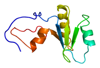
Polyadenylate-binding protein 1 is a protein that in humans is encoded by the PABPC1 gene. The protein PABP1 binds mRNA and facilitates a variety of functions such as transport into and out of the nucleus, degradation, translation, and stability. There are two separate PABP1 proteins, one which is located in the nucleus (PABPN1) and the other which is found in the cytoplasm (PABPC1). The location of PABP1 affects the role of that protein and its function with RNA.

<span class="mw-page-title-main">Poly(A)-specific ribonuclease</span> Protein-coding gene in the species Homo sapiens

Poly(A)-specific ribonuclease (PARN), also known as polyadenylate-specific ribonuclease or deadenylating nuclease (DAN), is an enzyme that in humans is encoded by the PARN gene.

<span class="mw-page-title-main">DCP2</span> Protein found in humans

mRNA-decapping enzyme 2 is a protein that in humans is encoded by the DCP2 gene.

<span class="mw-page-title-main">DCP1A</span> Protein found in humans

mRNA-decapping enzyme 1A is a protein that in humans is encoded by the DCP1A gene.

<span class="mw-page-title-main">Messenger RNA decapping</span> Removal of the 5 cap structure on mRNA

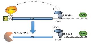
The process of messenger RNA decapping consists of hydrolysis of the 5' cap structure on the RNA exposing a 5' monophosphate. In eukaryotes, this 5' monophosphate is a substrate for the 5' exonuclease Xrn1 and the mRNA is quickly destroyed. There are many situations which may lead to the removal of the cap, some of which are discussed below.

<span class="mw-page-title-main">Decapping complex</span> Eukaryotic protein complex that removes the 5 cap on mRNA

The mRNA decapping complex is a protein complex in eukaryotic cells responsible for removal of the 5' cap. The active enzyme of the decapping complex is the bilobed Nudix family enzyme Dcp2, which hydrolyzes 5' cap and releases 7mGDP and a 5'-monophosphorylated mRNA. This decapped mRNA is inhibited for translation and will be degraded by exonucleases. The core decapping complex is conserved in eukaryotes. Dcp2 is activated by Decapping Protein 1 (Dcp1) and in higher eukaryotes joined by the scaffold protein VCS. Together with many other accessory proteins, the decapping complex assembles in P-bodies in the cytoplasm.

mRNA surveillance mechanisms are pathways utilized by organisms to ensure fidelity and quality of messenger RNA (mRNA) molecules. There are a number of surveillance mechanisms present within cells. These mechanisms function at various steps of the mRNA biogenesis pathway to detect and degrade transcripts that have not properly been processed.

Cryptic unstable transcripts (CUTs) are a subset of non-coding RNAs (ncRNAs) that are produced from intergenic and intragenic regions. CUTs were first observed in S. cerevisiae yeast models and are found in most eukaryotes. Some basic characteristics of CUTs include a length of around 200–800 base pairs, a 5' cap, poly-adenylated tail, and rapid degradation due to the combined activity of poly-adenylating polymerases and exosome complexes. CUT transcription occurs through RNA Polymerase II and initiates from nucleosome-depleted regions, often in an antisense orientation. To date, CUTs have a relatively uncharacterized function but have been implicated in a number of putative gene regulation and silencing pathways. Thousands of loci leading to the generation of CUTs have been described in the yeast genome. Additionally, stable uncharacterized transcripts, or SUTs, have also been detected in cells and bear many similarities to CUTs but are not degraded through the same pathways.

<span class="mw-page-title-main">Gephyronic acid</span> Chemical compound

Gephyronic acid is a polyketide that exists as an equilibrating mixture of structural isomers. In nature, gephyronic acid is produced by slow growing myxobacterium: Archangium gephyra strain Ar3895 and Cystobacter violaceus strain Cb vi76. It is the first antibiotic in myxobacteria that was reported to specifically inhibit eukaryotic protein synthesis.

<span class="mw-page-title-main">Sandra Wolin</span> American microbiologist and physician-scientist

Sandra Lynn Wolin is an American microbiologist and physician-scientist specialized in biogenesis, function, and turnover of non-coding RNA. She is chief of the RNA Biology Laboratory at the National Cancer Institute.

In molecular biology, the NAD+ five-prime cap refers to a molecule of nicotinamide adenine dinucleotide (NAD+), a nucleoside-containing metabolite, covalently bonded the 5’ end of cellular mRNA. While the more common methylated guanosine (m7G) cap is added to RNA by a capping complex that associates with RNA polymerase II, the NAD cap is added during transcriptional initiation by the RNA polymerase itself, acting as a non-canonical initiating nucleotide (NCIN). As such, while m7G capping can only occur in organisms possessing specialized capping complexes, because NAD capping is performed by RNAP itself, it is hypothesized to occur in most, if not all, organisms.

<span class="mw-page-title-main">CCR4-Not</span> Multiprotein complex used in gene expression

Carbon Catabolite Repression—Negative On TATA-less, or CCR4-Not, is a multiprotein complex that functions in gene expression. The complex has multiple enzymatic activities as both a poly(A) 3′-5′ exonuclease and a ubiquitin ligase. The exonuclease activity of CCR4-Not shortens the poly(A) tail found at 3' end of almost every eukaryotic mRNA. The complex is present both in the nucleus where it regulates transcription and in the cytoplasm where it associates with translating ribosomes and RNA processing bodies. In mammalian cell, it has a function in the regulation of the cell cycle, chromatin modification, activation and inhibition of transcription initiation, control of transcription elongation, RNA export, nuclear RNA surveillance, and DNA damage repair in nucleus. Ccr4–Not complex plays an important role in mRNA decay and protein quality control in the cytoplasm.

References

  1. 1 2 3 “Roy Parker, PhD”. Our Scientists. Howard Hughes Medical Institute. Retrieved 5 March 2020. https://www.hhmi.org/scientists/roy-parker
  2. 1 2 “Roy Parker”. Member Directory. National Academy of Sciences. Retrieved 4 March 2020. http://www.nasonline.org/member-directory/members/20027268.html
  3. 1 2 3 4 5 6 7 8 9 10 11 12 13 14 15 16 17 18 19 20 21 “Parker, Roy”. CU Experts. University of Colorado Boulder. Retrieved 6 March 2020. https://experts.colorado.edu/display/fisid_151440
  4. 1 2 3 “Roy Parker”. Speakers. iBiology. Retrieved 5 March 2020. https://www.ibiology.org/speakers/roy-parker/
  5. Jones, Elizabeth W.; Zubenko, George S.; Parker, Roy R (December 1982). “PEP4 Gene Function Is Required for Expression of Several Vacuolar Hydrolases in Saccharomyces Cerevisiae”. Genetics. 102 (4): 665-677.
  6. Decker, Carolyn J.; Parker, Roy (12 April 1993). “A Turnover Pathway for Both Stable and Unstable mRNAs in Yeast: Evidence for a Requirement for Deadenylation”. Genes & Development. 7: 1632-1643. doi:10.1101/gad.7.8.1632
  7. Beelman, Clare A.; Parker, Roy (21 April 1995). “Degradation of mRNA in Eukaryotes”. Cell. 81 (2): 179-183. doi:10.1016/0092-8674(95)90326-7
  8. Caponigro, Giordano; Parker, Roy (1 November 1996). “mRNA Turnover in Yeast Promoted by the MATalpha1 Instability Element”. Nucleic Acid Research. 24 (21): 4304-4312. doi:10.1093/nar/24.21.4304
  9. Muhlrad, Denise; Parker, Roy (November 1999). “Recognition of Yeast mRNAs As “Nonsense Containing” Leads to Both Inhibition of mRNA Translation and mRNA Degradation: Implications for the Control of mRNA Decapping”. Molecular Biology of the Cell. 10 (11): 3971-3978. doi:10.1091/mbc.10.11.3971
  10. Sheth, Ujwal; Parker, Roy (2 May 2003). “Decapping and Decay of Messenger RNA Occur in Cytoplasmic Processing Bodies”. Science. 300 (5620): 805-808. doi:10.1126/science.1082320
  11. Hilleren, Patricia J.; Parker, Roy (1 December 2003). “Cytoplasmic Degradation of Splice-Defective Pre-mRNAs and Intermediates”. Molecular Cell. 12 (6): 1453-1465. doi:10.1016/S1097-2765(03)00488-X
  12. Coller, Jeff; Parker, Roy (23 September 2005). “General Translational Repression by Activators of mRNA Decapping”. Cell. 122 (6): 875-886. doi:10.1016/j.cell.2005.07.012
  13. Brengues, Muriel; Teixeira, Daniela; Parker, Roy (21 October 2005). “Movement of Eukaryotic mRNAs between Polysomes and Cytoplasmic Processing Bodies”. Science. 310 (5747): 486-489. doi:10.1126/science.1115791
  14. Doma, Meenakshi K.; Parker, Roy (23 March 2006). “Endonucleolytic Cleavage of Eukaryotic mRNAs with Stalls in Translation Elongation”. Nature. 440 (7083): 561-564. doi:10.1038/nature04530
  15. Parker, Roy; Sheth, Ujwal (9 March 2007). “P Bodies and the Control of mRNA Translation and Degradation”. Molecular Cell. 25 (5): 635-646. doi:10.1016/j.molcel.2007.02.011
  16. Buchan, Ross J.; Parker, Roy (24 December 2009). “Eukaryotic Stress Granules: The Ins and Outs of Translation”. Molecular Cell. 36 (6): 932-941. doi:10.1016/j.molcel.2009.11.020
  17. Parker, Roy (25 May 2012). “Skill Development in Graduate Education”. Molecular Cell. 46 (4): 377-381. doi:10.1016/j.molcel.2012.05.003
  18. Decker, Carolyn J.; Parker, Roy (8 May 2014). “Analysis of Double-Stranded RNA from Microbial Communities Identifies Double-Stranded RNA Virus-Like Elements”. Cell Reports. 7 (3): 898-906. doi:10.1016/j.celrep.2014.03.049
  19. Harigaya, Yuriko; Parker, Roy (10 May 2017). “The Link between Adjacent Codon Pairs and mRNA Stability”. BMC Genomics. 18 (364): 704. doi:10.1186/s12864-017-3749-8
  20. Matheny, Tyler; Rao, Bhalchandra S.; Parker, Roy (25 November 2019). “Transcriptome-Wide Comparison of Stress Granules and P-Bodies Reveals that Translation Plays a Major Role in RNA Partitioning”. Molecular Cell Biology. 39 (24): 313-319. doi:10.1128/MCB.00313-19
  21. “6 CU Boulder Faculty Members Become Distinguished Professors”. CU Boulder Today. University of Colorado Boulder. Retrieved 10 April 2020. https://www.colorado.edu/today/2018/09/14/6-cu-boulder-faculty-members-become-distinguished-professors
  22. “Ada Doisy Lectures in Biochemistry”. Department of Biochemistry. University of Illinois at Urbana-Champaign. Retrieved 10 April 2020. https://mcb.illinois.edu/departments/biochemistry/doisy
  23. “RNA at the Bench and Bedside”. Investors and Media. Ionis Pharmaceuticals. Retrieved 10 April 2020. https://ir.ionispharma.com/events/event-details/ionis-pharmaceuticals-nature-structural-molecular-biology-nature-biotechnology
  24. “Seminars & Symposia”. Max Planck Institute of Biochemistry. Max-Planck-Gesellschaft. Retrieved 10 April 2020. https://www.biochem.mpg.de/en/seminars
  25. “March 30, 2018 U-M Center for RNA Biomedicine 3rd Annual Symposium”. Center for RNA Biomedicine. University of Michigan. Retrieved 10 April 2020. https://rna.umich.edu/2018-symposium/
  26. “RNA Localisation and Local Translation”. Speakers. EMBO Conference. Retrieved 10 April 2020. https://meetings.embo.org/event/17-rna-localisation
  27. “UA College of Science Chooses First Galileo Circle Fellows”. UA News. The University of Arizona. Retrieved 10 April 2020. https://uanews.arizona.edu/story/ua-college-science-chooses-first-galileo-circle-fellows
  28. “Roy R. Parker”. Awardees. Searle Scholars Program. Retrieved 10 April 2020. https://www.searlescholars.net/people/roy-r-parker