Scalp reconstruction

Last updated

Scalp reconstruction is a surgical procedure for people with scalp defects. Scalp defects may be partial or full thickness and can be congenital or acquired. Because not all layers of the scalp are elastic and the scalp has a convex shape, the use of primary closure is limited. Sometimes the easiest way of closing the wound may not be the ideal or best way. The choice for a reconstruction depends on multiple factors, such as the defect itself, the patient characteristics and surgeon preference.

Contents

History

Trepanated skull, Bronze Age.JPG
Bronze Age
Incan brain surgery.jpg
Incan brain surgery
Hieronymus Bosch-Removing the Rocks from the Head-Detail.jpg
Middle Ages
Sanjay Gupta & medical team prepare for brain surgery on USS Carl Vinson (CVN-70) 2010-01-18.jpg
Modern day
Scalp surgery through history in different times and places.

Skull and brain 'surgery' are known from the prehistoric era. There is evidence of scalp reconstructions dating back to the Egyptians in 3000 B.C., and to the Roman Empire. The word plastic surgery likely comes from the Greek πλαστικός (plastikós lit. "formable", "mouldable"). Attempts were made to reconstruct damaged skulls despite minimal knowledge of neurology, anatomy, and the brain. Skulls showing manipulation that can be interpreted as a primitive form of surgery have been found from various eras around the world. In Medieval times, people were convinced that trepanation was a remedy for various diseases.[ citation needed ]

Indications

Flowchart for use in scalp reconstruction. Flowchart for scalp reconstruction.jpg
Flowchart for use in scalp reconstruction.

Main reasons for scalp reconstruction are divided into two groups: congenital or acquired. Congenital defects may include aplasia cutis congenita, congenital nevus, congenital vascular malformations and congenital tumors. Acquired defects can be caused by burns, blunt, penetrating, or avulsion injuries, tumor invasion, infection, oncologic resection, radiation, or wound-healing difficulties. [1] Alopecia can be an aesthetic motivation for hair-bearing scalp reconstruction. [2] As the incidence of basal-cell carcinoma and squamous-cell carcinoma is rising and about 80% are located in the head and neck area, the number of scalp reconstructions will likely increase in the future. Depending on the size and nature of the defect, an appropriate reconstructive method has to be used. By using Mohs surgery the defect can be kept minimal, but nevertheless infiltrative basal cell carcinoma may have the need to remove a large part of the scalp.[ citation needed ]

On the flowchart, a simplified algorithm for scalp reconstruction is depicted. The options range from simple solutions for small skin defects to complicated reconstructions requiring multi-tissue reconstructions.

An active and severe infection must be controlled first by surgical debridement and antibiotic treatment before reconstruction is performed, as infection can cause bacteraemia and has a negative effect on wound healing.

Surgical anatomy

The five layers of the scalp, from superficial to deep, can be easily memorized by using the mnemonic SCALP. The Skin of the scalp has been scientifically examined for thickness. [3] The posterior scalp skin thickness is 1.48 mm; [3] the temporal scalp is 1.38mm; [3] and the anterior scalp thickness is 1.18 mm. [3] The scalp contains approximately 100.000 hairs.[ citation needed ] Hair lines make scalp reconstruction difficult because the hair lines must be respected to attain a satisfying aesthetic result. [4] The subCutis is a layer of fat, enclosed in compartments formed by rigid fibrous septa. Their inelasticity prevents bleeding vessels from collapsing and retracting under the skin to achieve haemostasis. All large blood vessels and nerves of the scalp are located in this layer. [5] The next layer is the galea Aponeurotica, which separates the underlying bone and the overlying layers. The large blood vessels and nerves of the scalp don’t pierce this layer. [4] Loose connective tissue between the periosteum and the aponeurosis makes these two rigid structures easily slide over each other and contribute to skin movement. Thus, if vascular and nervous anatomy is respected, the skin, subcutaneous tissue and galea aponeurotica can be lifted off the skull with minimal bleeding, nerve damage, or chance of necrosis. This method was first described by Orticochea in 1967, but has been updated to minimize scarring. [6] The fifth layer is the Periosteum of the skull, also referred to as pericranium. It can be separated from the skull, except near the sutures. The skull consists of an inner and outer table, with spongy bone in between known as diploë. [5]

Vascular supply

On both sides of the scalp, there are five large arteries that perfuse the scalp. Local flaps used for scalp reconstruction must contain at least one of these major arteries, to maintain a reliable blood supply. The scalp can be divided into four different vascular territories:

Gray557.png
Veins
Head arteries.gif
Arteries
Head nerves.gif
Nerves
Head lymph.gif
Lymphatic system
Blood vessels, nerves and lymphatic system of the scalp.

The veins anastomose frequently with each other and enter the diploic veins of the skull bones and the dural sinuses. This is an extra difficulty as the vein pattern differs. The scalp veins accompany the arteries and have similar names:

Lymphatic system

The frontal part of the scalp is drained to the parotid, submandibular, and deep cervical lymph nodes. The posterior part is drained to the posterior auricular and occipital lymph nodes. Malignancies of the scalp can metastasize to these lymph nodes. Brain tumors, however, tend to metastasize haematogenously (through the blood).

Innervation

The scalp is innervated by motor nerves and sensory nerves. The trigeminal nerve (CNV) is one of the important cranial sensory nerves which innervates the scalp. From anterior to posterior front to back the nerves are:

Non-skin reconstruction

Dura mater reconstruction

Dural lesions should be closed to avoid CSF leakage. Also, a defect acts like a port of entry for micro-organisms that can cause meningitis. If fibrin glue or primary closure is not possible, patches have to be used. These are made from cadaveric dura mater, xenografts (tachosil, duragen, durepair), or synthetic grafts materials (PTFE, neuropatch). However, (vascularised) autografts (fascia lata, muscle or omentum majus) are preferred in irradiated or severely infected defects. [8]

Bone defects

Forehead bulk defect picture 1.jpg
Pre-operative frontal defect
Forehead bulk defect picture 2.jpg
Titanium plate and fully expanded tissue expander in place
Forehead bulk defect picture 3.jpg
After removal of the tissue expander, the skin is transposed to close the defect
Forehead bulk defect picture 4.jpg
Final result
Reconstruction of a frontal skull defect and tissue expansion for coverage of the titanium plate.

Skull defects should be closed in order to protect the brain. The occipital and temporal regions bear the most pressure while sleeping and therefore need to be reconstructed. Frontal bone defects cause a contour defect; therefore, aesthetic considerations are often taken into account to reconstruct this area. Midsagittal defects are of lower importance, as they allow only penetrating trauma. When the reconstruction cannot be performed immediately, wearing a helmet is advised. Skull deformities can result in high intracranial pressure which can cause complaints ranging from headaches to epilepsy-like seizures. Small defects can be filled with morcellized bone, which will consolidate in some weeks. Because of the anatomy of the skull, the external table can be split off the internal table and then be moved over the defect. Rib grafts (whether or not accompanied with the latissimus dorsi muscle) are suitable for larger defects and can bear pressure, but do not cover the whole defect. Implants can be used as well, but are not preferred in patients who are to be irradiated or recently have had an infection or necrosis, because of the increased risk of infection and extrusion. [8] These implants can be factory-made out of metal (titanium), synthetic materials (PMMA, PEEK) or synthetic body-own material (Hydroxylapatite). In the pictures, a reconstruction using a titanium plate is shown. The skull contour has been restored.

Soft tissue defects

Robert McGee, scalped as a child by Sioux Chief Little Turtle in 1864-2.jpg
1890, Robert McGee, scalped in 1864
Occipital defect picture 1.jpg
Occipital scalp defect before closure with a regional trapezius flap
Occipital defect picture 2.jpg
After transposition of the flap and closure of all wounds
Occipital defect picture 3.jpg
Final result
Reconstruction of an occipital scalp defect using a pedicled myocutaneous trapezius flap.

If the periosteum or underlying muscles (frontalis, occipitalis, temporalis) are intact, secondary closure by granulation is possible. Before surgical intervention this was the only option available, as used to treat Robert McGee in 1864.[ citation needed ] Considering modern aesthetic standards, a better option is the use of split-thickness or full-thickness skin grafts, which is also quicker. When bulk is needed for a better contour a free flap is used, or as shown in the pictures, a regional flap. [9]

Skin reconstruction

Local reconstruction

Forehead scalp defect picture 1.jpg
Frontal scalp defect
Forehead scalp defect picture 2.jpg
Full-thickness scalp flap lifted
Forehead scalp defect picture 3.jpg
Flap rotated and wounds closed primarily
Forehead scalp defect picture 4.jpg
Final result
Frontal scalp defect closed with a rotation flap.

If the skin defect does not exceed 3 cm in diameter, it can be closed primarily. If this is not possible without tension, the surrounding loose connective tissue can be undermined to attain more mobility. [10] Different kinds of transpositions to close the defect with adjacent skin are possible: V-Y, Z, pinwheel flaps, advancement flaps, Orticochea flaps and rotation flaps. All these transpositions generate tension of the skin and may distort hair lines. A combination of the Orticochea and rotation flaps is illustrated by the pictures about the forehead defect. Another option is secondary healing, but this is aesthetically inferior to primary closure in hair bearing areas because of the resulting alopecia. If the scalp cannot be closed primarily and local reconstruction with hair bearing skin is needed because of hair lines, tissue expansion of hair bearing skin may be possible. The expansion process is uncomfortable on the short term, but long-term results are good.

Regional reconstruction

If local reconstruction is not possible due to lack of local tissue, regional reconstruction is the next rung on the reconstructive ladder. This includes pedicled flaps as the trapezius or supraclavicular flap or tissue expansion of nearby regions. Alternatively, the Crane principle, as described by Millard in 1969, can be used. A healthy part is used to resurface the defect and when this flap takes, the skin is returned to its original site leaving the subcutaneous tissue on the defect, which then needs a split skin graft. [1]

Skin grafts

If only skin is missing and underlying galea, muscle or connective tissue are intact, a skin graft can be used. A skin graft needs healthy, vascularised tissue beneath it to take; otherwise it will become necrotic.

Free flap

Free flaps are usually the best solution for reconstruction of large defects that cannot be closed locally and that have unfavourable wound conditions such as severe infection, exposed sinuses, dura or brain tissue, CSF leakage or radiation damage. This method is the most complex of the reconstructive ladder. In scalp reconstruction free flaps have the great benefit of being completely detached from their original location ("donor site") before transferral to the scalp, which makes the inset easier than for pedicled flaps. Another advantage is that free flaps provide a more robust vascular supply to the wound compared to pedicled flaps, controlling infection and radiation induced damage. In addition, muscle or myocutaneous free flaps provide additional bulk that obliterates empty spaces (e.g. exposed sinuses) and covers dura mater defects more than all other options, reducing postoperative wound infections and CSF leakages. Disadvantages are the complexity of the operation, leading to prolonged operation times, the need for specialised personnel, and the chance for total flap necrosis due to microvascular complications. Another challenge with the use of free flaps is to achieve an aesthetically pleasing result with good color and contour match, especially if the defect is deep. [1] [11]

Outcome

Temporal defect picture 1.jpg
Retroauricular squamous cell carcinoma
Temporal defect picture 2.jpg
A free ALT flap for coverage; a selective neck dissection was also performed
Temporal defect picture 3.jpg
Final result after radiation therapy, needing an ear prothesis
Reconstruction of a lateral full thickness scalp defect with a free ALT flap.

As the incidence of basal cell carcinoma and squamous-cell carcinoma rises, so does the need for reconstructions after radical excision of these skin malignancies. Reconstructive options depend on location and size. Most of the time the problem is not solved in just one operation, because small adjustments are necessary for good aesthetic results. Further postoperative treatment modalities as radiation therapy and chemotherapy may impact the final result.[ citation needed ]

Aesthetic and functional

Temporal and forehead defects offer a more difficult aesthetic challenge and are best covered with a thin flap so the aesthetic unit appears equal. Although the forehead is not a highly important aesthetic unit, color mismatch and bulkiness will draw attention quickly. Free flap reconstruction of the forehead can be bulky and color match is variable and depends on the ethnic and genetic background. Some people are not satisfied with the outcome and may experience psychological problems such as low self-confidence or even depression. Sometimes a second operation is needed to improve skin color. Overgrafting the skin with skin grafts from the scalp can improve color match.[ citation needed ]

Complications

Post-operative complications can be divided into donor-site and recipient-site problems. Donor-site complications include wound infection, hematoma, and seroma. Recipient-site complications include (total or partial) flap necrosis, wound infection, dehiscence, hematoma or skin graft failure. To avoid major bleeding or sensibility disorders, the anatomy of the scalp must be respected, such as by making incisions parallel to rather than across blood vessels. Due to the rich perfusion, scalp injuries can lead to serious bleeding, which may be difficult to stem if the cut blood vessels retract into the fat. [7]

Related Research Articles

Plastic surgery Medical surgical specialty

Plastic surgery is a surgical specialty involving the restoration, reconstruction, or alteration of the human body. It can be divided into two main categories: reconstructive surgery and cosmetic surgery. Reconstructive surgery includes craniofacial surgery, hand surgery, microsurgery, and the treatment of burns. While reconstructive surgery aims to reconstruct a part of the body or improve its functioning, cosmetic surgery aims at improving the appearance of it.

Rhinoplasty Surgical procedure to enhance or reconstruct a human nose

Rhinoplasty, commonly called nose job, medically called nasal reconstruction is a plastic surgery procedure for altering and reconstructing the nose. There are two types of plastic surgery used – reconstructive surgery that restores the form and functions of the nose and cosmetic surgery that changes the appearance of the nose. Reconstructive surgery seeks to resolve nasal injuries caused by various traumas including blunt, and penetrating trauma and trauma caused by blast injury. Reconstructive surgery can also treat birth defects, breathing problems, and failed primary rhinoplasties. Rhinoplasty may remove a bump, narrow nostril width, change the angle between the nose and the mouth, or address injuries, birth defects, or other problems that affect breathing, such as a deviated nasal septum or a sinus condition.

Tissue expansion is a technique used by plastic, maxillofacial and reconstructive surgeons to cause the body to grow additional skin, bone, or other tissues. Other biological phenomena such as tissue inflammation can also be considered expansion.

Scalp Anatomical area bordered by the human face at the front, and by the neck at the sides and back

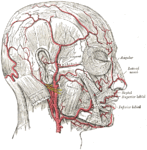
The scalp is the anatomical area bordered by the human face at the front, and by the neck at the sides and back.

Dura mater The outermost layer of meninges

Dura mater is a thick membrane made of dense irregular connective tissue that surrounds the brain and spinal cord. It is the outermost of the three layers of membrane called the meninges that protect the central nervous system. The other two meningeal layers are the arachnoid mater and the pia mater. The dura surrounds the brain and the spinal cord. It envelops the arachnoid mater, which is responsible for keeping in the cerebrospinal fluid. It is derived primarily from the neural crest cell population, with postnatal contributions of the paraxial mesoderm.

Skin grafting

Skin grafting, a type of graft surgery, involves the transplantation of skin. The transplanted tissue is called a skin graft.

Grafting refers to a surgical procedure to move tissue from one site to another on the body, or from another creature, without bringing its own blood supply with it. Instead, a new blood supply grows in after it is placed. A similar technique where tissue is transferred with the blood supply intact is called a flap. In some instances a graft can be an artificially manufactured device. Examples of this are a tube to carry blood flow across a defect or from an artery to a vein for use in hemodialysis.

Otoplasty denotes the surgical and non-surgical procedures for correcting the deformities and defects of the pinna, and for reconstructing a defective, or deformed, or absent external ear, consequent to congenital conditions and trauma. The otoplastic surgeon corrects the defect or deformity by creating an external ear that is of natural proportions, contour, and appearance, usually achieved by the reshaping, the moving, and the augmenting of the cartilaginous support framework of the pinna. Moreover, the occurrence of congenital ear deformities occasionally overlaps with other medical conditions.

Microsurgery

Microsurgery is a general term for surgery requiring an operating microscope. The most obvious developments have been procedures developed to allow anastomosis of successively smaller blood vessels and nerves which have allowed transfer of tissue from one part of the body to another and re-attachment of severed parts. Microsurgical techniques are utilized by several specialties today, such as: general surgery, ophthalmology, orthopedic surgery, gynecological surgery, otolaryngology, neurosurgery, oral and maxillofacial surgery, plastic surgery, podiatric surgery and pediatric surgery.

Rhytidectomy

A facelift, technically known as a rhytidectomy, is a type of cosmetic surgery procedure used to give a more youthful facial appearance. There are multiple surgical techniques and exercise routines. Surgery usually involves the removal of excess facial skin, with or without the tightening of underlying tissues, and the redraping of the skin on the patient's face and neck. Exercise routines tone underlying facial muscles without surgery. Surgical facelifts are effectively combined with eyelid surgery (blepharoplasty) and other facial procedures and are typically performed under general anesthesia or deep twilight sleep.

Buttock augmentation Cosmetic and corrective surgery

Gluteoplasty denotes the plastic surgery and the liposuction procedures for the correction of the congenital, traumatic, and acquired defects and deformities of the buttocks and the anatomy of the gluteal region; and for the aesthetic enhancement of the contour of the buttocks.

Frontal sinus

The frontal sinuses are one of the four pairs of paranasal sinuses that are situated behind the brow ridges. Sinuses are mucosa-lined airspaces within the bones of the face and skull. Each opens into the anterior part of the corresponding middle nasal meatus of the nose through the frontonasal duct which traverses the anterior part of the labyrinth of the ethmoid. These structures then open into the semilunar hiatus in the middle meatus.

The terms free flap, free autologous tissue transfer and microvascular free tissue transfer are synonymous terms used to describe the "transplantation" of tissue from one site of the body to another, in order to reconstruct an existing defect. "Free" implies that the tissue is completely detached from its blood supply at the original location and then transferred to another location and the circulation in the tissue re-established by anastomosis of artery(s) and vein(s). This is in contrast to a "pedicled" flap in which the tissue is left partly attached to the donor site ("pedicle") and simply transposed to a new location; keeping the "pedicle" intact as a conduit to supply the tissue with blood.

Head and neck anatomy

This article describes the anatomy of the head and neck of the human body, including the brain, bones, muscles, blood vessels, nerves, glands, nose, mouth, teeth, tongue, and throat.

Lip reconstruction may be required after trauma or surgical excision. The lips are considered the beginning of the oral cavity and are the most common site of oral cancer. Any reconstruction of the lips must include both functional and cosmetic considerations. The lips are necessary for speech, facial expression, and eating. Because of their prominent location on the face, even small abnormalities can be apparent.

Avulsion injury Tearing off of skin etc.

In medicine, an avulsion is an injury in which a body structure is torn off by either trauma or surgery. The term most commonly refers to a surface trauma where all layers of the skin have been torn away, exposing the underlying structures. This is similar to an abrasion but more severe, as body parts such as an eyelid or an ear can be partially or fully detached from the body.

Nasal reconstruction using a paramedian forehead flap within oral and maxillofacial surgery, is a surgical technique to reconstruct different kinds of nasal defects. In this operation a reconstructive surgeon uses skin from the forehead above the eyebrow and pivots it vertically to replace missing nasal tissue. Throughout history the technique has been modified and adjusted by many different surgeons and it has evolved to become a popular way of repairing nasal defects.

The tint of forehead skin so exactly matches that of the face and nose that it must be first choice. Is not the forehead the crowning feature of the face and important in expression? Why then should we jeopardize its beauty to make a nose? First, because in many instances, the forehead makes far and away the best nose. Second, with some plastic juggling, the forehead defect can be camouflaged effectively.

Flap (surgery) Surgical procedure in which tissue is transferred with intact blood supply

Flap surgery is a technique in plastic and reconstructive surgery where any type of tissue is lifted from a donor site and moved to a recipient site with an intact blood supply. This is distinct from a graft, which does not have an intact blood supply and therefore relies on growth of new blood vessels. This is done to fill a defect such as a wound resulting from injury or surgery when the remaining tissue is unable to support a graft, or to rebuild more complex anatomic structures such as breast or jaw.

The cheek constitutes the facial periphery, plays a key role in the maintenance of oral competence and mastication, is involved in the facial manifestation of human emotion, and supports neighboring primary structures.

Free-flap breast reconstruction is a type of autologous-tissue breast reconstruction applied after mastectomy for breast cancer, without the emplacement of a breast implant prosthesis. As a type of plastic surgery, the free-flap procedure for breast reconstruction employs tissues, harvested from another part of the woman's body, to create a vascularised flap, which is equipped with its own blood vessels. Breast-reconstruction mammoplasty can sometimes be realised with the application of a pedicled flap of tissue that has been harvested from the latissimus dorsi muscle, which is the broadest muscle of the back, to which the pedicle (“foot”) of the tissue flap remains attached until it successfully grafts to the recipient site, the mastectomy wound. Moreover, if the volume of breast-tissue excised was of relatively small mass, breast augmentation procedures, such as autologous-fat grafting, also can be applied to reconstruct the breast lost to mastectomy.

References

  1. 1 2 3 Seitz, Iris A.; Gottlieb, Lawrence J. (2009). "Reconstruction of Scalp and Forehead Defects". Clinics in Plastic Surgery. 36 (3): 355–77. doi:10.1016/j.cps.2009.02.001. PMID   19505608.
  2. Scalp Reconstruction Procedures at eMedicine
  3. 1 2 3 4 Chopra, Karan (2015). "A comprehensive examination of topographic thickness of skin in the human face". Aesthetic Surgery Journal. 35 (8): 1007–13. doi: 10.1093/asj/sjv079 . PMID   26508650.
  4. 1 2 3 Seery Gerard E (2002). "Surgical anatomy of the scalp". Dermatologic Surgery. 28 (7): 581–7. doi:10.1097/00042728-200207000-00010. PMID   12135510.
  5. 1 2 Drake et al, Gray's Anatomy for Students, 2E, Elsevier, 2009[ page needed ]
  6. Frodel, John L.; Ahlstrom, Karen (2004). "Reconstruction of Complex Scalp Defects". Archives of Facial Plastic Surgery. 6 (1): 54–60. doi:10.1001/archfaci.6.1.54. PMID   14732646.
  7. 1 2 Leedy, Jason E.; Janis, Jeffrey E.; Rohrich, Rod J. (2005). "Reconstruction of Acquired Scalp Defects: An Algorithmic Approach". Plastic and Reconstructive Surgery. 116 (4): 54e–72e. doi:10.1097/01.prs.0000179188.25019.6c. PMID   16163072. S2CID   39519720.
  8. 1 2 van Driel, Antoinette A.; Mureau, Marc A. M.; Goldstein, David P.; Gilbert, Ralph W.; Irish, Jonathan C.; Gullane, Patrick J.; Neligan, Peter C.; Hofer, Stefan O. P. (2010). "Aesthetic and Oncologic Outcome after Microsurgical Reconstruction of Complex Scalp and Forehead Defects after Malignant Tumor Resection: An Algorithm for Treatment". Plastic and Reconstructive Surgery. 126 (2): 460–70. doi:10.1097/PRS.0b013e3181de2260. PMID   20679830. S2CID   22302579.
  9. Skin Grafts and Biologic Skin Substitutes at eMedicine
  10. TerKonda, RP; Sykes, JM (1997). "Concepts in scalp and forehead reconstruction". Otolaryngologic Clinics of North America. 30 (4): 519–39. doi:10.1016/S0030-6665(20)30188-2. PMID   9233857.
  11. Lin, Samuel; Hanasono, Matthew; Skoracki, Roman (2008). "Scalp and Calvarial Reconstruction". Seminars in Plastic Surgery. 22 (4): 281–93. doi:10.1055/s-0028-1095887. PMC   2884877 . PMID   20567704.