
Peritonitis is inflammation of the localized or generalized peritoneum, the lining of the inner wall of the abdomen and cover of the abdominal organs. Symptoms may include severe pain, swelling of the abdomen, fever, or weight loss. One part or the entire abdomen may be tender. Complications may include shock and acute respiratory distress syndrome.

Ascites is the abnormal build-up of fluid in the abdomen. Technically, it is more than 25 ml of fluid in the peritoneal cavity, although volumes greater than one liter may occur. Symptoms may include increased abdominal size, increased weight, abdominal discomfort, and shortness of breath. Complications can include spontaneous bacterial peritonitis.
Percussion is a technique of clinical examination.

Paracentesis is a form of body fluid sampling procedure, generally referring to peritoneocentesis in which the peritoneal cavity is punctured by a needle to sample peritoneal fluid.
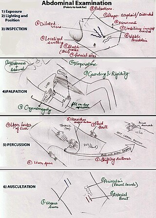
An abdominal examination is a portion of the physical examination which a physician or nurse uses to clinically observe the abdomen of a patient for signs of disease. The physical examination typically occurs after a thorough medical history is taken, that is, after the physician asks the patient the course of their symptoms. The abdominal examination is conventionally split into four different stages: first, inspection of the patient and the visible characteristics of their abdomen. Auscultation (listening) of the abdomen with a stethoscope. Palpation of the patient's abdomen. Finally, percussion (tapping) of the patient's abdomen and abdominal organs. Depending on the need to test for specific diseases such as ascites, special tests may be performed as a part of the physical examination. An abdominal examination may be performed because the physician suspects a disease of the organs inside the abdominal cavity (including the liver, spleen, large or small intestines), or simply as a part of a complete physical examination for other conditions. In a complete physical examination, the abdominal exam classically follows the respiratory examination and cardiovascular examination.
In medicine, Murphy's sign is a maneuver during a physical examination as part of the abdominal examination. It is useful for differentiating pain in the right upper quadrant. Typically, it is positive in cholecystitis, but negative in choledocholithiasis, pyelonephritis, and ascending cholangitis.

A respiratory examination, or lung examination, is performed as part of a physical examination, in response to respiratory symptoms such as shortness of breath, cough, or chest pain, and is often carried out with a cardiac examination.

Costovertebral angle (CVA) tenderness is pain that results from touching the region inside of the costovertebral angle. The CVA is formed by the 12th rib and the spine. Assessing for CVA tenderness is part of the abdominal exam, and CVA tenderness often indicates kidney pathology.

Castell's sign is a medical sign assessed to evaluate splenomegaly and typically part of an abdominal examination. It is an alternative physical examination maneuver to percussion over Traube's space.
In medicine, physiotherapy, chiropractic, and osteopathy the hip examination, or hip exam, is undertaken when a patient has a complaint of hip pain and/or signs and/or symptoms suggestive of hip joint pathology. It is a physical examination maneuver.
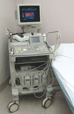
Abdominal ultrasonography is a form of medical ultrasonography to visualise abdominal anatomical structures. It uses transmission and reflection of ultrasound waves to visualise internal organs through the abdominal wall. For this reason, the procedure is also called a transabdominal ultrasound, in contrast to endoscopic ultrasound, the latter combining ultrasound with endoscopy through visualize internal structures from within hollow organs.
In medicine, the fluid wave test or fluid thrill test is a test for ascites. It is performed by having the patient push their hands down on the midline of the abdomen. The examiner then taps one flank, while feeling on the other flank for the tap. The pressure on the midline prevents vibrations through the abdominal wall while the fluid allows the tap to be felt on the other side. The result is considered positive if tap can be felt on the other side. However, even with the midline pressure, transmission through the skin must be excluded. A positive fluid wave test indicates that there is a free fluid (ascites) in the abdomen. When one side of the abdomen is pressed, the other side may also be painful due to the transfer of the fluid in it.

A pelvic examination is the physical examination of the external and internal female pelvic organs. It is frequently used in gynecology for the evaluation of symptoms affecting the female reproductive and urinary tract, such as pain, bleeding, discharge, urinary incontinence, or trauma. It can also be used to assess a woman's anatomy in preparation for procedures. The exam can be done awake in the clinic and emergency department, or under anesthesia in the operating room. The most commonly performed components of the exam are 1) the external exam, to evaluate the external genitalia 2) the internal exam with palpation to examine the uterus, ovaries, and fallopian tubes, and 3) the internal exam using the speculum to visualize the vaginal walls and cervix. During the pelvic exam, sample of cells and fluids may be collected to screen for sexually transmitted infections or cancer.
In gastroenterology, the puddle sign is a physical examination maneuver that can be used to detect the presence of ascites. It is useful for detecting small amounts of ascites—as small as 120 mL; shifting dullness and bulging flanks typically require 500 mL.
A GALS screen is an examination used by doctors and other healthcare professionals to detect locomotor abnormalities and functional disability relating to gait, arms, legs and the spine.
Ballance's sign is used in medical diagnosis. Its indications are dullness to percussion in the left flank LUQ and shifting dullness to percussion in the right flank seen with splenic rupture/hematoma. During trauma assessment of the abdomen, "Ballance's sign" may be observed upon exam.
The liver scratch test is a technique used by medical professionals during a physical exam to locate the inferior border of the liver in order to approximate the size of a patient's liver. The technique was first credited to Burton-Opitz in 1925 where it was used to identify the cardiac silhouette, however there are references of similar techniques used prior to this. The liver scratch test can be used when other exam techniques used to approximate liver size are ineffective or unavailable and is thought to be most useful if the abdomen is distended, too tender for direct palpation, the abdominal muscles are too rigid, or the patient is obese.
The cardiovascular examination is a portion of the physical examination that involves evaluation of the cardiovascular system. The exact contents of the examination will vary depending on the presenting complaint but a complete examination will involve the heart, lungs, belly and the blood vessels.
Lépine's sign is one of the medical signs of gallbladder disease. It is positive when effleurage with crooked third finger at the point of the gallbladder projection to anterior abdominal wall elicits pain. It is not to be confused with the following:

An upper limb neurological examination is part of the neurological examination, and is used to assess the motor and sensory neurons which supply the upper limbs. This assessment helps to detect any impairment of the nervous system, being used both as a screening and an investigative tool. The examination findings when combined with a detailed history of a patient, can help a doctor reach a specific or differential diagnosis. This would enable the doctor to commence treatment if a specific diagnosis has been made, or order further investigations if there are differential diagnoses.
