
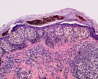
A melanocytic nevus is a type of melanocytic tumor that contains nevus cells.

Nevus is a nonspecific medical term for a visible, circumscribed, chronic lesion of the skin or mucosa. The term originates from nævus, which is Latin for "birthmark", however, a nevus can be either congenital or acquired. Common terms, including mole, birthmark, and beauty mark, are used to describe nevi, but these terms do not distinguish specific types of nevi from one another.
Superficial spreading melanoma (SSM) is usually characterized as the most common form of cutaneous melanoma in Caucasians. The average age at diagnosis is in the fifth decade, and it tends to occur on sun-exposed skin, especially on the backs of males and lower limbs of females.

A dysplastic nevus or atypical mole is a nevus (mole) whose appearance is different from that of common moles. In 1992, the NIH recommended that the term "dysplastic nevus" be avoided in favor of the term "atypical mole". An atypical mole may also be referred to as an atypical melanocytic nevus, atypical nevus, B-K mole, Clark's nevus, dysplastic melanocytic nevus, or nevus with architectural disorder.

Dysplastic nevus syndrome is a cutaneous condition described in certain families, and characterized by unusual nevi and multiple inherited melanomas.

The congenital melanocytic nevus is a type of melanocytic nevus found in infants at birth. This type of birthmark occurs in an estimated 1% of infants worldwide; it is located in the area of the head and neck 15% of the time.
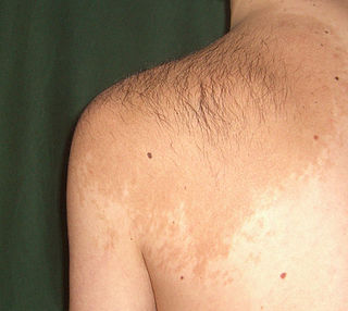
Becker's nevus is a skin disorder predominantly affecting males. The nevus can be present at birth, but more often shows up around puberty. It generally first appears as an irregular pigmentation on the torso or upper arm, and gradually enlarges irregularly, becoming thickened and often hairy (hypertrichosis). The nevus is due to an overgrowth of the epidermis, pigment cells (melanocytes), and hair follicles. This form of nevus was first documented in 1948 by American dermatologist Samuel William Becker (1894–1964).
HMB-45 is a monoclonal antibody that reacts against an antigen present in melanocytic tumors such as melanomas, and stands for Human Melanoma Black. It is used in anatomic pathology as a marker for such tumors. The specific antigen recognized by HMB-45 is now known as Pmel 17.

Blue nevus is a type of melanocytic nevus. The blue colour is caused by the pigment being deeper in the skin than in ordinary nevi. In principle they are harmless but they can sometimes be mimicked by malignant lesions, i.e. some melanomas can look like a blue nevus.
Skin biopsy is a biopsy technique in which a skin lesion is removed to be sent to a pathologist to render a microscopic diagnosis. It is usually done under local anesthetic in a physician's office, and results are often available in 4 to 10 days. It is commonly performed by dermatologists. Skin biopsies are also done by family physicians, internists, surgeons, and other specialties. However, performed incorrectly, and without appropriate clinical information, a pathologist's interpretation of a skin biopsy can be severely limited, and therefore doctors and patients may forgo traditional biopsy techniques and instead choose Mohs surgery. There are four main types of skin biopsies: shave biopsy, punch biopsy, excisional biopsy, and incisional biopsy. The choice of the different skin biopsies is dependent on the suspected diagnosis of the skin lesion. Like most biopsies, patient consent and anesthesia are prerequisites.
Halo nevus is a mole that is surrounded by a depigmented ring or 'halo'.
A Spitz nevus is a benign melanocytic nevus, a type of skin lesion, affecting the epidermis and dermis.
Nevi and melanomas are a group of neoplasia.
Pseudomelanoma is a cutaneous condition in which melantic skin lesions clinically resemble a superficial spreading melanoma at the site of a recent shave removal of a melanocytic nevus.

An acral nevus is a cutaneous condition characterized by a skin lesion that is usually macular or only slightly elevated, and may display uniform brown or dark brown color, but often with linear striations.

A pigmented spindle cell nevus is a skin condition characterized by a dark brown to black macule or papule, usually less than 6 mm.

Nevoid melanoma is a cutaneous condition that may resemble a Spitz nevus or an acquired or congenital melanocytic nevus.
Animal-type melanoma is a cutaneous condition and is characterized by nodules and fascicles of epithelioid melanocytes with pleomorphic nuclei and striking hyperpigmentation, dendritic cells, numerous melanophages and, sometimes, an inflammatory infiltrate of lymphocytes.


