
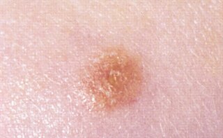
A melanocytic nevus is usually a noncancerous condition of pigment-producing skin cells. It is a type of melanocytic tumor that contains nevus cells. Some sources equate the term mole with "melanocytic nevus", but there are also sources that equate the term mole with any nevus form.

Nevus is a nonspecific medical term for a visible, circumscribed, chronic lesion of the skin or mucosa. The term originates from nævus, which is Latin for "birthmark"; however, a nevus can be either congenital or acquired. Common terms, including mole, birthmark, and beauty mark, are used to describe nevi, but these terms do not distinguish specific types of nevi from one another.

Acral lentiginous melanoma is a type of skin cancer. It typically begins as a uniform brownish mark before becoming darker and wider with a blurred irregular edge, most frequently seen in the foot of a person with darker skin. It may become bumpy and ulcerate. Just under the nail it typically appears as dark longitudinal streaks, and it may spread.
Superficial spreading melanoma (SSM) is a type of skin cancer that typically starts as an irregularly edged dark spot typically on sun-exposed part of the body. The colour may be variable with dark, light and reddish shades; occasionally no color at all. It typically grows in diameter before spreading to deeper tissue, forming a bump or becoming an ulcer. Itching, bleeding and crust formation may occur in some. The backs and shoulders of males and legs of women are particularly prone.

A dysplastic nevus or atypical mole is a nevus (mole) whose appearance is different from that of common moles. In 1992, the NIH recommended that the term "dysplastic nevus" be avoided in favor of the term "atypical mole". An atypical mole may also be referred to as an atypical melanocytic nevus, atypical nevus, B-K mole, Clark's nevus, dysplastic melanocytic nevus, or nevus with architectural disorder.

Dysplastic nevus syndrome, also known as familial atypical multiple mole–melanoma (FAMMM) syndrome, is an inherited cutaneous condition described in certain families, and characterized by unusual nevi and multiple inherited melanomas. First described in 1820, the condition is inherited in an autosomal dominant pattern, and caused by mutations in the CDKN2A gene. In addition to melanoma, individuals with the condition are at increased risk for pancreatic cancer.

The congenital melanocytic nevus is a type of melanocytic nevus found in infants at birth. This type of birthmark occurs in an estimated 1% of infants worldwide; it is located in the area of the head and neck 15% of the time.

A lentigo is a small pigmented spot on the skin with a clearly defined edge, surrounded by normal-appearing skin. It is a harmless (benign) hyperplasia of melanocytes which is linear in its spread. This means the hyperplasia of melanocytes is restricted to the cell layer directly above the basement membrane of the epidermis where melanocytes normally reside. This is in contrast to the "nests" of multi-layer melanocytes found in moles. Because of this characteristic feature, the adjective "lentiginous" is used to describe other skin lesions that similarly proliferate linearly within the basal cell layer.

A blue nevus is a type of coloured mole, typically a single well-defined blue-black bump.
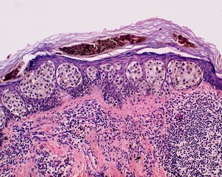
A Spitz nevus is a benign skin lesion. A type of melanocytic nevus, it affects the epidermis and dermis.

Nevus sebaceus or sebaceous nevus is a congenital, hairless plaque that typically occurs on the face or scalp. Such nevi are classified as epidermal nevi and can be present at birth, or early childhood, and affect males and females of all races equally. The condition is named for an overgrowth of sebaceous glands, a relatively uncommon hamartoma, in the area of the nevus. NSJ is first described by Josef Jadassohn in 1895.

Schimmelpenning syndrome is a neurocutaneous condition characterized by one or more sebaceous nevi, usually appearing on the face or scalp, associated with anomalies of the central nervous system, ocular system, skeletal system, cardiovascular system and genitourinary system.
Phakomatosis pigmentokeratotica is a rare neurocutanous condition characterized by the combination of an organoid sebaceous nevus and speckled lentiginous nevus. It is an unusual variant of epidermal naevus syndrome. It was first described by Happle et al. It is often associated with neurological or skeletal anomalies such as hemiatrophy, dysaesthesia and hyperhidrosis in a segmental pattern, mild mental retardation, seizures, deafness, ptosis and strabismus.

A benign melanocytic nevus is a cutaneous condition characterised by well-circumscribed, pigmented, round or ovoid lesions, generally measuring from 2 to 6 mm in diameter. A benign melanocytic nevus may feature hair or pigmentation as well.

Balloon cell nevus is a benign nevus. It appears like a melanocytic nevus.
Oral pigmentation is asymptomatic and does not usually cause any alteration to the texture or thickness of the affected area. The colour can be uniform or speckled and can appear solitary or as multiple lesions. Depending on the site, depth, and quantity of pigment, the appearance can vary considerably.

An acral nevus is a cutaneous condition of the palms, soles, fingers, or toes, characterized by a skin lesion that is usually macular or only slightly elevated, and may display a uniform brown or dark brown color, often with linear striations.
Zosteriform speckled lentiginous nevus is a skin lesion that may be the result of a potentially lethal mutation.
Kamino bodies are eosinophilic globoids. Kamino bodies are commonly observed microscopically with the condition spitz nevi, a benign melanocytic nevus, a type of skin lesion, affecting the epidermis and dermis.

A pigmented spindle cell nevus is a skin condition characterized by a dark brown to black macule or papule, usually less than 6 mm.
