Blue nevus | |
---|---|
Other names | Blue neuronevus, dermal melanocytoma, nevus coeruleus, nevus bleu [1] |
Blue nevus | |
Specialty | Dermatology |
Symptoms | Single well-defined blue-black bump [2] |
Complications | Rarely malignant transformation [3] |
Types | Dendritic, cellular [2] |
Causes | Unclear [3] |
Diagnostic method | Visualisation, dermoscopy [4] |
Differential diagnosis | Dermatofibroma, melanoma [3] [5] |
Treatment | Monitoring, excision [3] |
Prognosis | Good [3] |
Frequency | Female>male [2] |
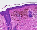
A blue nevus is a type of coloured mole, typically a single well-defined blue-black bump. [1] [2]
The blue colour is caused by the pigment being deep in the skin. [4]
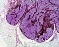
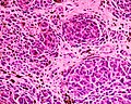
Diagnosis is by visualisation and dermoscopy. [4] A biopsy is sometimes performed, or the whole lesion surgically removed. [3] The outcome is generally good but there is a small chance of cancerous transformation. [3] Differential diagnosis includes dermatofibroma and melanoma. [3]
Blue nevi are more common in females than males. [2] It was first studied in 1906 by Tièche, a student of Josef Jadassohn. [6]
Blue nevi may be divided into the following types: [7] : 701
A melanocytic nevus is usually a noncancerous condition of pigment-producing skin cells. It is a type of melanocytic tumor that contains nevus cells. Some sources equate the term mole with "melanocytic nevus", but there are also sources that equate the term mole with any nevus form.
Nevus is a nonspecific medical term for a visible, circumscribed, chronic lesion of the skin or mucosa. The term originates from nævus, which is Latin for "birthmark"; however, a nevus can be either congenital or acquired. Common terms, including mole, birthmark, and beauty mark, are used to describe nevi, but these terms do not distinguish specific types of nevi from one another.
Acral lentiginous melanoma is an aggressive type of skin cancer. Melanoma is a group of serious skin cancers that arise from pigment cells (melanocytes); acral lentiginous melanoma is a kind of lentiginous skin melanoma. Acral lentiginous melanoma is the most common subtype in people with darker skins and is rare in people with lighter skin types. It is not caused by exposure to sunlight or UV radiation, and wearing sunscreen does not protect against it. Acral lentiginous melanoma is commonly found on the palms, soles, under the nails, and in the oral mucosa. It occurs on non-hair-bearing surfaces of the body, which have not necessarily been exposed to sunlight. It is also found on mucous membranes.
A dysplastic nevus or atypical mole is a nevus (mole) whose appearance is different from that of common moles. In 1992, the NIH recommended that the term "dysplastic nevus" be avoided in favor of the term "atypical mole". An atypical mole may also be referred to as an atypical melanocytic nevus, atypical nevus, B-K mole, Clark's nevus, dysplastic melanocytic nevus, or nevus with architectural disorder.
Dysplastic nevus syndrome, also known as familial atypical multiple mole–melanoma (FAMMM) syndrome, is an inherited cutaneous condition described in certain families, and characterized by unusual nevi and multiple inherited melanomas. First described in 1820, the condition is inherited in an autosomal dominant pattern, and caused by mutations in the CDKN2A gene. In addition to melanoma, individuals with the condition are at increased risk for pancreatic cancer.
Lentigo maligna melanoma is a melanoma that has evolved from a lentigo maligna, as seen as a lentigo maligna with melanoma cells invading below the boundaries of the epidermis. They are usually found on chronically sun damaged skin such as the face and the forearms of the elderly.
Lentigo maligna is where melanocyte cells have become malignant and grow continuously along the stratum basale of the skin, but have not invaded below the epidermis. Lentigo maligna is not the same as lentigo maligna melanoma, as detailed below. It typically progresses very slowly and can remain in a non-invasive form for years.
The congenital melanocytic nevus is a type of melanocytic nevus found in infants at birth. This type of birthmark occurs in an estimated 1% of infants worldwide; it is located in the area of the head and neck 15% of the time.
Melanocytic tumors of uncertain malignant potential (MELTUMP) are melanocytic lesions in the dermis that cannot be classified by morphology as either benign naevi (moles) or malignant melanomas because the mass shows features of both.
Skin biopsy is a biopsy technique in which a skin lesion is removed to be sent to a pathologist to render a microscopic diagnosis. It is usually done under local anesthetic in a physician's office, and results are often available in 4 to 10 days. It is commonly performed by dermatologists. Skin biopsies are also done by family physicians, internists, surgeons, and other specialties. However, performed incorrectly, and without appropriate clinical information, a pathologist's interpretation of a skin biopsy can be severely limited, and therefore doctors and patients may forgo traditional biopsy techniques and instead choose Mohs surgery.
Halo nevus is a mole that is surrounded by a depigmented ring or 'halo'.
A Spitz nevus is a benign skin lesion. A type of melanocytic nevus, it affects the epidermis and dermis.
Nevus cells are a variant of melanocytes. They are larger than typical melanocytes, do not have dendrites, and have more abundant cytoplasm with coarse granules. They are usually located at the dermoepidermal junction or in the dermis of the skin. Dermal nevus cells can be further classified: type A (epithelioid) dermal nevus cells mature into type B (lymphocytoid) dermal nevus cells which mature further into type C (neuroid) dermal nevus cells, through a process involving downwards migration.
A benign melanocytic nevus is a cutaneous condition characterised by well-circumscribed, pigmented, round or ovoid lesions, generally measuring from 2 to 6 mm in diameter. A benign melanocytic nevus may feature hair or pigmentation as well.
Pseudomelanoma is a cutaneous condition in which melanotic skin lesions clinically resemble a superficial spreading melanoma at the site of a recent shave removal of a melanocytic nevus.
Balloon cell nevus is a benign nevus. It appears like a melanocytic nevus.
Oral pigmentation is asymptomatic and does not usually cause any alteration to the texture or thickness of the affected area. The colour can be uniform or speckled and can appear solitary or as multiple lesions. Depending on the site, depth, and quantity of pigment, the appearance can vary considerably.
Melanoma with features of a Spitz nevus is a cutaneous condition characterized histologically with tissue similar to a spitz nevus and with overall symmetry and a dermal nodule of epithelioid melanocytes that do not mature with progressively deeper dermal extension.
A nevoid melanoma is a malignant neoplastic lesion of the skin. It is a type of melanoma, the most dangerous form of skin cancer. Nevoid melanomas are clinically significant because they are difficult to distinguish from a benign nevus of the skin, which requires no treatment and is common on most individuals. Nevoid morphologies represent up to 3% of all cases of melanoma.
Animal-type melanoma is a rare subtype of melanoma that is characterized by heavily pigmented dermal epithelioid and spindled melanocytes. Animal-type melanoma is also known to be called equine-type melanoma, pigment synthesizing melanoma, and pigmented epithelioid melanocytoma (PEM). While melanoma is known as the most aggressive skin cancer, the mortality for PEM is lower than in other melanoma types. Animal-type melanoma earned its name due to the resemblance of melanocytic tumors in grey horses.
{{cite journal}}
: CS1 maint: DOI inactive as of January 2024 (link)