
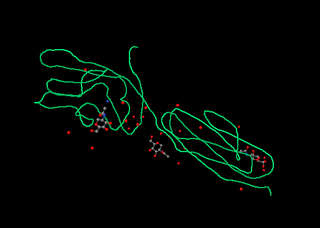
Protein A is a 49 kDa surface protein originally found in the cell wall of the bacteria Staphylococcus aureus. It is encoded by the spa gene and its regulation is controlled by DNA topology, cellular osmolarity, and a two-component system called ArlS-ArlR. It has found use in biochemical research because of its ability to bind immunoglobulins. It is composed of five homologous Ig-binding domains that fold into a three-helix bundle. Each domain is able to bind proteins from many mammalian species, most notably IgGs. It binds the heavy chain within the Fc region of most immunoglobulins and also within the Fab region in the case of the human VH3 family. Through these interactions in serum, where IgG molecules are bound in the wrong orientation, the bacteria disrupts opsonization and phagocytosis.

The restriction endonuclease Fok1, naturally found in Flavobacterium okeanokoites, is a bacterial type IIS restriction endonuclease consisting of an N-terminal DNA-binding domain and a non-specific DNA cleavage domain at the C-terminal. Once the protein is bound to duplex DNA via its DNA-binding domain at the 5'-GGATG-3' recognition site, the DNA cleavage domain is activated and cleaves, without further sequence specificity, the first strand 9 nucleotides downstream and the second strand 13 nucleotides upstream of the nearest nucleotide of the recognition site.

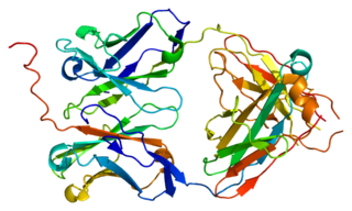
The fragment crystallizable region is the tail region of an antibody that interacts with cell surface receptors called Fc receptors and some proteins of the complement system. This property allows antibodies to activate the immune system. In IgG, IgA and IgD antibody isotypes, the Fc region is composed of two identical protein fragments, derived from the second and third constant domains of the antibody's two heavy chains; IgM and IgE Fc regions contain three heavy chain constant domains in each polypeptide chain. The Fc regions of IgGs bear a highly conserved N-glycosylation site. Glycosylation of the Fc fragment is essential for Fc receptor-mediated activity. The N-glycans attached to this site are predominantly core-fucosylated diantennary structures of the complex type. In addition, small amounts of these N-glycans also bear bisecting GlcNAc and α-2,6 linked sialic acid residues.
CD2 is a cell adhesion molecule found on the surface of T cells and natural killer (NK) cells. It has also been called T-cell surface antigen T11/Leu-5, LFA-2, LFA-3 receptor, erythrocyte receptor and rosette receptor.

The secretory component is a component of immunoglobulin A (IgA). Secretory component is a proteolytic cleavage product of the polymeric immunoglobulin receptor which remains associated with dimeric IgA in sero-mucus secretions. Polymeric IgA binds to the polymeric immunoglobulin receptor on the basolateral surface of epithelial cells and is taken up into the cell via transcytosis. The receptor-IgA complex passes through the cellular compartments before being secreted on the luminal surface of the epithelial cells, still attached to the receptor. Proteolysis of the receptor occurs and the dimeric IgA molecule, along with the secretory component, are free to diffuse throughout the lumen.
Immunoglobulin lambda-like polypeptide 1 is a protein that in humans is encoded by the IGLL1 gene. IGLL1 has also recently been designated CD179B.

Ig mu chain C region is a protein that in humans is encoded by the IGHM gene.

Ig gamma-1 chain C region is a protein that in humans is encoded by the IGHG1 gene.
Basal cell adhesion molecule, also known as Lutheran antigen, is a plasma membrane glycoprotein that in humans is encoded by the BCAM gene. BCAM has also recently been designated CD239.

Immunoglobulin heavy constant alpha 1 is a immunoglobulin gene with symbol IGHA1. It encodes a constant (C) segment of Immunoglobulin A heavy chain. Immunoglobulin A is an antibody that plays a critical role in immune function in the mucous membranes. IgA shows the same typical structure of other antibody classes, with two heavy chains and two light chains, and four distinct domains: one variable region, and three variable regions. As a major class of immunoglobulin in body secretions, IgA plays a role in defending against infection, as well as preventing the access of foreign antigens to the immunologic system.
Ig delta chain C region is a protein that in humans is encoded by the IGHD gene.

Fc receptor-like protein 5 is a protein that in humans is encoded by the FCRL5 gene. FCRL5 has also been designated as CD307.

Leukocyte immunoglobulin-like receptor subfamily B member 3 is a protein that in humans is encoded by the LILRB3 gene.
Ig heavy chain V-III region VH26 is a protein that in humans is encoded by the IGHV@ gene.
Leukocyte immunoglobulin-like receptor subfamily A member 2 is a protein that in humans is encoded by the LILRA2 gene.
Ig gamma-3 chain C region is a protein that in humans is encoded by the IGHG3 gene.

High affinity immunoglobulin epsilon receptor subunit beta is a protein that in humans is encoded by the MS4A2 gene.
In molecular biology SprD is a non-coding RNA expressed on pathogenicity islands in Staphylococcus aureus. It was identified in silico along with a number of other sRNAs (SprA-G) through microarray analysis which were confirmed using a Northern blot. SprD has been found to significantly contribute to causing disease in an animal model.
Zinc finger transcription factors or ZF-TFs, are transcription factors composed of a zinc finger-binding domain and any of a variety of transcription-factor effector-domains that exert their modulatory effect in the vicinity of any sequence to which the protein domain binds.
Although cell wall carbohydrates are ideal immunotherapeutic targets due to their abundance in bacteria and high level of conservation, their poor immunogenicity compared with protein targets complicates their use for the development of protective antibodies. A lysibody is a chimeric antibody in which the Fab region is the binding domain from a bacteriophage lysin, or the binding domain from an autolysin or bacteriocin, all of which bind to bacterial cell wall carbohydrate epitopes. This is linked to the Fc of Immunoglobulin G (IgG). The chimera forms a stable homodimer held together by hinge-region disulfide bonds. Thus, lysibodies are homodimeric hybrid immunoglobulin G molecules that can bind with high affinity and specificity to a carbohydrate substrate in the bacterial cell wall peptidoglycan. Lysibodies behave like authentic IgG by binding at high affinity to their bacterial wall receptor, fix complement and therefore promote phagocytosis by macrophages and neutrophils, protecting mice from infection in model systems. Since cell wall hydrolases, autolysins and bacteriocins are ubiquitous in nature, production of lysibodies specific for difficult to treat pathogenic bacteria is possible.
