The diagnostic tests in cardiology are methods of identifying heart conditions associated with healthy vs. unhealthy, pathologic heart function.

A galvanometer is an electromechanical measuring instrument for electric current. Early galvanometers were uncalibrated, but improved versions, called ammeters, were calibrated and could measure the flow of current more precisely. Galvanometers work by deflecting a pointer in response to an electric current flowing through a coil in a constant magnetic field. The mechanism is also used as an actuator in applications such as hard disks.

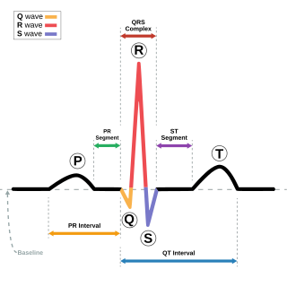
Electrocardiography is the process of producing an electrocardiogram, a recording of the heart's electrical activity through repeated cardiac cycles. It is an electrogram of the heart which is a graph of voltage versus time of the electrical activity of the heart using electrodes placed on the skin. These electrodes detect the small electrical changes that are a consequence of cardiac muscle depolarization followed by repolarization during each cardiac cycle (heartbeat). Changes in the normal ECG pattern occur in numerous cardiac abnormalities, including:

A mirror galvanometer is an ammeter that indicates it has sensed an electric current by deflecting a light beam with a mirror. The beam of light projected on a scale acts as a long massless pointer. In 1826, Johann Christian Poggendorff developed the mirror galvanometer for detecting electric currents. The apparatus is also known as a spot galvanometer after the spot of light produced in some models.

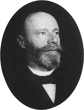
Willem Einthoven was a Dutch medical doctor and physiologist. He invented the first practical electrocardiograph in 1895 and received the Nobel Prize in Physiology or Medicine in 1924 for it.

In medicine, a Holter monitor is a type of ambulatory electrocardiography device, a portable device for cardiac monitoring for at least 24 hours.

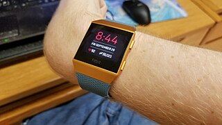
A heart rate monitor (HRM) is a personal monitoring device that allows one to measure/display heart rate in real time or record the heart rate for later study. It is largely used to gather heart rate data while performing various types of physical exercise. Measuring electrical heart information is referred to as electrocardiography.
A flatline is an electrical time sequence measurement that shows no activity and therefore, when represented, shows a flat line instead of a moving one. It almost always refers to either a flatlined electrocardiogram, where the heart shows no electrical activity (asystole), or to a flat electroencephalogram, in which the brain shows no electrical activity. Both of these specific cases are involved in various definitions of death.

Einthoven's triangle is an imaginary formation of three limb leads in a triangle used in the electrocardiography, formed by the two shoulders and the pubis. The shape forms an inverted equilateral triangle with the heart at the center. It is named after Willem Einthoven, who theorized its existence.

Augustus Desiré Waller FRS was a British physiologist and the son of Augustus Volney Waller. He was born in Paris, France.

The Fleming valve, also called the Fleming oscillation valve, was a thermionic valve or vacuum tube invented in 1904 by English physicist John Ambrose Fleming as a detector for early radio receivers used in electromagnetic wireless telegraphy. It was the first practical vacuum tube and the first thermionic diode, a vacuum tube whose purpose is to conduct current in one direction and block current flowing in the opposite direction. The thermionic diode was later widely used as a rectifier — a device that converts alternating current (AC) into direct current (DC) — in the power supplies of a wide range of electronic devices, until beginning to be replaced by the selenium rectifier in the early 1930s and almost completely replaced by the semiconductor diode in the 1960s. The Fleming valve was the forerunner of all vacuum tubes, which dominated electronics for 50 years. The IEEE has described it as "one of the most important developments in the history of electronics", and it is on the List of IEEE Milestones for electrical engineering.

The electrical axis of the heart is the net direction in which the wave of depolarization travels. It is measured using an electrocardiogram (ECG). Normally, this begins at the sinoatrial node ; from here the wave of depolarisation travels down to the apex of the heart. The hexaxial reference system can be used to visualise the directions in which the depolarisation wave may travel.
A Bioamplifier is an electrophysiological device, a variation of the instrumentation amplifier, used to gather and increase the signal integrity of physiologic electrical activity for output to various sources. It may be an independent unit, or integrated into the electrodes.

Cardiac monitoring generally refers to continuous or intermittent monitoring of heart activity to assess a patient's condition relative to their cardiac rhythm. Cardiac monitoring is usually carried out using electrocardiography, which is a noninvasive process that records the heart's electrical activity and displays it in an electrocardiogram. It is different from hemodynamic monitoring, which monitors the pressure and flow of blood within the cardiovascular system. The two may be performed simultaneously on critical heart patients. Cardiac monitoring for ambulatory patients is known as ambulatory electrocardiography and uses a small, wearable device, such as a Holter monitor, wireless ambulatory ECG, or an implantable loop recorder. Data from a cardiac monitor can be transmitted to a distant monitoring station in a process known as telemetry or biotelemetry.

The Human Research Facility Holter Monitor (Holter) is a battery-powered, noninvasive electrocardiogram (ECG) device that accurately measures the heart rate of crew members over an extended period of time. ECG information is stored on a Portable Computer Memory Card International Adapter (PCMCIA) card and downlinked to Earth for analysis after monitoring is complete.
Wireless ambulatory electrocardiography (ECG) is a type of ambulatory electrocardiography with recording devices that use wireless technology, such as Bluetooth and smartphones, for at-home cardiac monitoring (monitoring of heart rhythms). These devices are generally recommended to people who have been previously diagnosed with arrhythmias and want to have them monitored, or for those who have suspected arrhythmias and need to be monitored over an extended period of time in order to be diagnosed.
Bioinstrumentation or biomedical instrumentation is an application of biomedical engineering which focuses on development of devices and mechanics used to measure, evaluate, and treat biological systems. The goal of biomedical instrumentation focuses on the use of multiple sensors to monitor physiological characteristics of a human or animal for diagnostic and disease treatment purposes. Such instrumentation originated as a necessity to constantly monitor vital signs of Astronauts during NASA's Mercury, Gemini, and Apollo missions.
Horatio Burt Williams was an American clinical electrophysiologist.
Bioelectromagnetic medicine deals with the phenomenon of resonance signaling and discusses how specific frequencies modulate cellular function to restore or maintain health. Such electromagnetic (EM) signals are then called medical information, which are used in health informatics.
Alexander Filippovich Samoylov was a Russian physiologist and pioneer of electrophysiology and electrocardiography who applied techniques of using the ECG for diagnostic purposes. He served as a professor at Kazan University from 1903 until his death.


