Subglottic stenosis

Last updated
Subglottic stenosis
Subglottic stenosis (X-ray).jpg
Neck AP x-ray of patient with post-intubation subglottic stenosis, as shown by the narrowing in the tracheal lumen marked by the arrow.
Specialty Pulmonology
Symptoms Difficulty breathing
Usual onsetAny
CausesIntubation, trauma, systemic diseases, cancer
Risk factors Prolonged intubation, nasogastric tube
Diagnostic method CT scan, MRI, OCT

Subglottic stenosis is a congenital or acquired narrowing of the subglottic airway. [1] It can be congenital, acquired, iatrogenic, or very rarely, idiopathic. It is defined as the narrowing of the portion of the airway that lies between the vocal cords and the lower part of the cricoid cartilage. In a normal infant, the subglottic airway is 4.5-5.5 millimeters wide, while in a premature infant, the normal width is 3.5 millimeters. Subglottic stenosis is defined as a diameter of under 4 millimeters in an infant. Acquired cases are more common than congenital cases due to prolonged intubation being introduced in the 1960s. [2] It is most frequently caused by certain medical procedures or external trauma, although infections and systemic diseases can also cause it.

Contents

Signs and symptoms

Symptoms may range from stridor during exercise to complete obstruction of the airway. In idiopathic cases, symptoms may be mistaken for asthma. In congenital cases, symptoms occur soon after birth, and include difficulty breathing, stridor, and air hunger. If the vocal cords are affected, symptoms may include inability to make sound, and an unusual cry. [2]

Causes

Most commonly, acquired cases are caused by trauma or certain medical procedures. The condition is more often caused by external trauma in adults. External injuries occur from vehicular accidents or clothesline injuries. [2] Iatrogenic cases can occur from intubation, tracheostomy, and an endotracheal tube cuff pressure that is too high. [3] 17 hours of intubation in adults and 1 week of intubation in neonates can cause the injury. Infants born prematurely can be intubated for a longer amount of time due to the fact that they have more flexible cartilage and a larynx located high in the airway. 90% of acquired cases in children are due to intubation, due to the ring of cartilage in the upper airway. [2] Less commonly, infections, such as bacterial tracheitis, tuberculosis, histoplasmosis, diphtheria, and laryngeal papillomatosis can cause it. Systemic diseases, such as amyloidosis, granulomatosis with polyangiitis, sarcoidosis, inflammatory bowel disease, scleroderma, relapsing polychondritis, and polyarthritis, can also cause it. Other causes may include aspiration of foreign bodies, burns, and exposure to occupational hazards. [3] Very rarely, the condition may also have no known cause, in which case it is known as idiopathic subglottic stenosis. [2]

Mechanism

High cuff pressure or long-term intubation can cause damage to the tracheal mucosa, causing inflammation, ulceration, and breakdown of cartilage. [3] When the injury heals, scarring occurs, narrowing the airway. [4] Treatment-related risk factors include repeated intubation, the presence of a nasogastric tube, and size of an endotracheal tube. Person-related risk factors include systemic diseases, the likelihood of infection, and inadequate perfusion. Acid reflux may cause narrowing due to scarring resulting from stomach acid damaging the tracheal mucosa. 10-23% of causes result due to narrowing arising from granulomatosis with polyangiitis. In these cases, it is believed that this is more common in people under the age of 20. Around 66% of sarcoidosis cases involve the airway. [3]

Diagnosis

CT scans and MRI (magnetic resonance imaging) can help in diagnosis. X-rays can determine the location and size of the narrowed airway portion. Optical coherence tomography (OCT) can help observe the progression of the injury. Esophageal pH monitoring can help detect any acid reflux, which can worsen the condition. An endoscope can be inserted and used to see the vocal cords, airway, and esophagus. Spirometry is a useful way to measure respiratory function. People affected by subglottic stenosis have a FEV1 of over 10. [2]

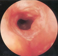
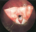
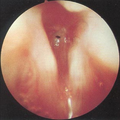
Subglottic stenosis is graded according to the Cotton-Myer classification system from one to four based on the severity of the blockage. Grade 1 is up to 50% obstruction, Grade 2 is 50-70% obstruction, Grade 3 is 70-99% obstruction, and Grade 4 is with no visible lumen. [5]

Treatment

The goal of treatment is to relieve any breathing difficulty and to ensure that the airway remains open in the long term. In mild cases, the disease can be treated with endoscopic balloon dilation. In severe cases, open surgery is done. Owing to the risk of complications, tracheostomy is avoided if possible. [2]

Surgery

In severe cases, the condition frequently recurs after dilation, and the main treatment is open surgery. There are three main surgical procedures that are used when treating subglottic stenosis. These include endoscopy, open neck surgery, and tracheotomy. Endoscopic procedures include dilation via balloon or rigid, incision via laser, and stent placement. [2]

Other

In mild cases, such as cases with only granuloma or thin, web-like tissue, dilation is able to remove the lesion. In severe cases, dilation and laser treatment only works temporarily, and the condition frequently recurs. Repeated procedures, especially stents, come with the risk of increasing the damaged area. Also, laser resection has a risk of damaging the cartilage underneath. For this reason, surgery is typically done. [6] In addition to surgery, mitomycin can be applied topically and glucocorticoids can be injected. [2]

Idiopathic cases are usually treated via endoscopic balloon dilation, and dilation may be improved by injecting corticosteroids. It is also the safest and main treatment in pregnant women. It allows dilation via noninvasive measures such as expanding a balloon catheter via ventilation. [2]

Prognosis

The condition frequently recurs after dilation. The prognosis after surgery is good if it is done carefully to avoid complications. [2]

Epidemiology

The condition has decreased in frequency over time, due to improved management of people on a ventilator. The condition occurs in around 1% of endotracheal tube users. [4]

Related Research Articles

<span class="mw-page-title-main">Trachea</span> Cartilaginous tube that connects the pharynx and larynx to the lungs

The trachea, also known as the windpipe, is a cartilaginous tube that connects the larynx to the bronchi of the lungs, allowing the passage of air, and so is present in almost all animals with lungs. The trachea extends from the larynx and branches into the two primary bronchi. At the top of the trachea the cricoid cartilage attaches it to the larynx. The trachea is formed by a number of horseshoe-shaped rings, joined together vertically by overlying ligaments, and by the trachealis muscle at their ends. The epiglottis closes the opening to the larynx during swallowing.

<span class="mw-page-title-main">Tracheal intubation</span> Placement of a tube into the trachea

Tracheal intubation, usually simply referred to as intubation, is the placement of a flexible plastic tube into the trachea (windpipe) to maintain an open airway or to serve as a conduit through which to administer certain drugs. It is frequently performed in critically injured, ill, or anesthetized patients to facilitate ventilation of the lungs, including mechanical ventilation, and to prevent the possibility of asphyxiation or airway obstruction.

<span class="mw-page-title-main">Tracheotomy</span> Temporary surgical incision to create an airway into the trachea

Tracheotomy, or tracheostomy, is a surgical airway management procedure which consists of making an incision (cut) on the anterior aspect (front) of the neck and opening a direct airway through an incision in the trachea (windpipe). The resulting stoma (hole) can serve independently as an airway or as a site for a tracheal tube or tracheostomy tube to be inserted; this tube allows a person to breathe without the use of the nose or mouth.

<span class="mw-page-title-main">Airway management</span> Medical procedure ensuring an unobstructed airway

Airway management includes a set of maneuvers and medical procedures performed to prevent and relieve airway obstruction. This ensures an open pathway for gas exchange between a patient's lungs and the atmosphere. This is accomplished by either clearing a previously obstructed airway; or by preventing airway obstruction in cases such as anaphylaxis, the obtunded patient, or medical sedation. Airway obstruction can be caused by the tongue, foreign objects, the tissues of the airway itself, and bodily fluids such as blood and gastric contents (aspiration).

<span class="mw-page-title-main">Choanal atresia</span> Congenital disorder where the nasal passage is blocked

Choanal atresia is a congenital disorder where the back of the nasal passage (choana) is blocked, usually by abnormal bony or soft tissue (membranous) due to failed hole development of the nasal fossae during prenatal development. It causes persistent rhinorrhea, and with bilateral choanal atresia and obstructed airway that can cause cyanosis and hypoxia.

Stridor is a high-pitched extra-thoracic breath sound resulting from turbulent air flow in the larynx or lower in the bronchial tree. It is different from a stertor which is a noise originating in the pharynx.

<span class="mw-page-title-main">Laryngomalacia</span> Medical condition

Laryngomalacia is the most common cause of chronic stridor in infancy, in which the soft, immature cartilage of the upper larynx collapses inward during inhalation, causing airway obstruction. It can also be seen in older patients, especially those with neuromuscular conditions resulting in weakness of the muscles of the throat. However, the infantile form is much more common. Laryngomalacia is one of the most common laryngeal congenital disease in infancy and public education about the signs and symptoms of the disease is lacking.

<span class="mw-page-title-main">Aortic valvuloplasty</span>

Aortic valvuloplasty, also known as balloon aortic valvuloplasty (BAV), is a procedure used to improve blood flow through the aortic valve in conditions that cause aortic stenosis, or narrowing of the aortic valve. It can be performed in various patient populations including fetuses, newborns, children, adults, and pregnant women. The procedure involves using a balloon catheter to dilate the narrowed aortic valve by inflating the balloon.

<span class="mw-page-title-main">Gastric outlet obstruction</span> Medical condition

Gastric outlet obstruction (GOO) is a medical condition where there is an obstruction at the level of the pylorus, which is the outlet of the stomach. Individuals with gastric outlet obstruction will often have recurrent vomiting of food that has accumulated in the stomach, but which cannot pass into the small intestine due to the obstruction. The stomach often dilates to accommodate food intake and secretions. Causes of gastric outlet obstruction include both benign causes, as well as malignant causes, such as gastric cancer.

<span class="mw-page-title-main">Laryngotracheal stenosis</span> Medical condition

Laryngotracheal stenosis refers to abnormal narrowing of the central air passageways. This can occur at the level of the larynx, trachea, carina or main bronchi. In a small number of patients narrowing may be present in more than one anatomical location.

<span class="mw-page-title-main">Tracheobronchial injury</span> Damage to the tracheobronchial tree

Tracheobronchial injury is damage to the tracheobronchial tree. It can result from blunt or penetrating trauma to the neck or chest, inhalation of harmful fumes or smoke, or aspiration of liquids or objects.

<span class="mw-page-title-main">Congenital stenosis of vena cava</span> Medical condition

Congenital stenosis of vena cava is a congenital anomaly in which the superior vena cava or inferior vena cava has an aberrant interruption or coarctation.

<span class="mw-page-title-main">Tracheobronchomalacia</span> Medical condition

Tracheobronchomalacia (TBM) is a condition characterized by flaccidity of the tracheal support cartilage which leads to tracheal collapse. This condition can also affect the bronchi. There are two forms of this condition: primary TBM and secondary TBM. Primary TBM is congenital and starts as early as birth. It is mainly linked to genetic causes. Secondary TBM is acquired and starts in adulthood. It is mainly developed after an accident or chronic inflammation.

Megaduodenum is a congenital or acquired dilation and elongation of the duodenum with hypertrophy of all layers that presents as a feeling of gastric fullness, abdominal pain, belching, heartburn, and nausea with vomiting sometimes of food eaten 24 hours prior.

<span class="mw-page-title-main">Aqueductal stenosis</span> Narrowing of the aqueduct of Sylvius

Aqueductal stenosis is a narrowing of the aqueduct of Sylvius which blocks the flow of cerebrospinal fluid (CSF) in the ventricular system. Blockage of the aqueduct can lead to hydrocephalus, specifically as a common cause of congenital and/or obstructive hydrocephalus.

<span class="mw-page-title-main">Advanced airway management</span>

Advanced airway management is the subset of airway management that involves advanced training, skill, and invasiveness. It encompasses various techniques performed to create an open or patent airway – a clear path between a patient's lungs and the outside world.

Laryngeal cysts are cysts involving the larynx or more frequently supraglottic locations, such as epiglottis and vallecula. Usually they do not extend to the thyroid cartilage. They may be present congenitally or may develop eventually due to degenerative cause. They often interfere with phonation.

Laryngotracheal reconstruction is a surgical procedure that involves expanding or removing parts of the airway to widen a narrowing within it, called laryngotracheal stenosis or subglottic stenosis.

Eustachian tube dysfunction (ETD) is a disorder where pressure abnormalities in the middle ear result in symptoms.

<span class="mw-page-title-main">Intubation granuloma</span> Medical condition

Intubation granuloma is a benign growth of granulation tissue in the larynx or trachea, which arises from tissue trauma due to endotracheal intubation. This medical condition is described as a common late complication of tracheal intubation, specifically caused by irritation to the mucosal tissue of the airway during insertion or removal of the patient's intubation tube.

References

  1. "Subglottic Stenosis in Adults: Problem, Etiology, Pathophysiology". 2017-06-21.{{cite journal}}: Cite journal requires |journal= (help)
  2. 1 2 3 4 5 6 7 8 9 10 11 Jagpal, Nitish; Shabbir, Nadeem (2021), "Subglottic Stenosis", StatPearls, Treasure Island (FL): StatPearls Publishing, PMID   33085412 , retrieved 2021-07-27
  3. 1 2 3 4 Aravena, Carlos; Almeida, Francisco A.; Mukhopadhyay, Sanjay; Ghosh, Subha; Lorenz, Robert R.; Murthy, Sudish C.; Mehta, Atul C. (March 2020). "Idiopathic subglottic stenosis: a review". Journal of Thoracic Disease. 12 (3): 1100–1111. doi: 10.21037/jtd.2019.11.43 . ISSN   2072-1439. PMC   7139051 . PMID   32274178.
  4. 1 2 Marston, Alexander P.; White, David R. (2018-12-01). "Subglottic Stenosis". Clinics in Perinatology. 45 (4): 787–804. doi:10.1016/j.clp.2018.07.013. ISSN   0095-5108. PMID   30396418. S2CID   53235038.
  5. Myer Cm, 3rd; O'Connor, D. M.; Cotton, R. T. (April 1994). "Proposed grading system for subglottic stenosis based on endotracheal tube sizes". The Annals of Otology, Rhinology, and Laryngology. 103 (4 Pt 1): 319–23. doi:10.1177/000348949410300410. PMID   8154776. S2CID   12782910.
  6. D’Andrilli, Antonio; Venuta, Federico; Rendina, Erino Angelo (March 2016). "Subglottic tracheal stenosis". Journal of Thoracic Disease. 8 (Suppl 2): S140–S147. doi:10.3978/j.issn.2072-1439.2016.02.03. ISSN   2072-1439. PMC   4775266 . PMID   26981264.