TRIP13

Last updated
TRIP13
Identifiers
Aliases TRIP13 , 16E1BP, thyroid hormone receptor interactor 13, MVA3, OOMD9
External IDs OMIM: 604507 MGI: 1916966 HomoloGene: 3125 GeneCards: TRIP13
Orthologs
SpeciesHumanMouse
Entrez
Ensembl
UniProt
RefSeq (mRNA)

NM_001166260
NM_004237

NM_027182

RefSeq (protein)

NP_001159732
NP_004228

NP_081458

Location (UCSC) Chr 5: 0.89 – 0.92 Mb n/a
PubMed search [2] [3]
Wikidata
View/Edit Human View/Edit Mouse

TRIP13 is a mammalian gene that encodes the thyroid receptor-interacting protein 13. In budding yeast, the analog for TRIP13 is PCH2. TRIP13 is a member of the AAA+ ATPase family, a family known for mechanical forces derived from ATP hydrolase reactions. The TRIP13 gene has been shown to interact with a variety of proteins and implicated in a few diseases, notably interacting with the ligand binding domain of thyroid hormone receptors, and may play a role in early-stage non-small cell lung cancer. [4] However, recent evidence implicates TRIP13 in various cell cycle phases, including meiosis G2/Prophase and during the Spindle Assembly checkpoint (SAC). Evidence shows regulation to occur through the HORMA domains, including Hop1, Rev7, and Mad2. [5] Of note, Mad2's involvement in the SAC is shown to be affected by TRIP13 [6] Due to TRIP13's role in cell cycle arrest and progression, it may present opportunity as a therapeutic candidate for cancers. [7]

Contents

Structure

As an AAA+ ATPase, TRIP13 (and its PCH2 analog) forms homohexamers and interacts with ATP as an energy source. With respect to Hop1, PCH2 binds to and structurally changes Hop1, displacing the Hop1 from DNA. [8] TRIP13/PCH2 interacts with ATP as a hydrolase, hydrolyzing phosphates to derive energy for conformational changes that can induce mechanical force on its substrate, Hop1 in the previous case. [9] TRIP14/PCH2 is believed to have a single AAA+ ATPase domain. [5] TRIP13/PCH2 also functions as a kinetochore protein that interacts with the silencing protein p31-Comet. [10]

Role in meiosis G2/prophase

Meiosis in mammalian cells have a series of checkpoints and steps that need to be properly regulated. TRIP13/PCH2 has been implicated in these processes in budding yeast as well, particularly in the meiosis G2/prophase stage. [11] Double stranded breaks during meiosis is a key part of this phase and is impacted by TRIP13. The homologous recombination that occurs following these breaks requires a protein complex to influence and structure appropriate chromosomal pairing.

In a paper by San-Segundo et al., localization assays and induced mutations in PCH2 in budding yeast was shown to be required for the meiotic checkpoint to prevent chromosome segregation when the recombination or chromosome synapsis are defective. [11] TRIP13, PCH2's analog, was also shown to be required for the formation of the synaptonemal complex – the complex that structures chromosomal pairings. Without TRIP13, meiocytes had pericentric synaptic forks, fewer crossovers, and altered distribution of chiasma (the contact point between homologous chromosomes. [12] For this synaptonemal complex (SC) formation, meiotic HORMADS need to be removed. For example, PCH2 was found to be needed to remove Hop1 from chromosomes during SC formation. [13] Other HORMADs, such as HORMAD1 and HORMAD2, are also depleted from the chromosomal pairs with the help of TRIP13 in mice cells. [14] Research shows a robust and varied role for TRIP13/PCH2 to remove various proteins for SC formation, thus allowing meiosis to continue. Further mechanistic evidence is needed to clarify other proteins affected by TRIP13 in meiosis G2/Prophase, and elucidate the wide ability to affect a multitude of proteins.

Role in spindle assembly checkpoint

Like its role in meiosis, TRIP13/PCH2 is also implicated in mitosis, particularly in the metaphase-to-anaphase transition and the Spindle Assembly Checkpoint (SAC). Its function also has impacts on the Anaphase Promoting Complex (APC). [5] To continue from metaphase to anaphase, the cell must ensure chromosomes are bioriented and properly structured in order for correct and error-free separation of sister chromatids. This process requires many proteins to ensure dynamic timing and consistent response. In order for progression, the APC must be activated, which upon activation degrade securing. The APC is activated by CDC20, a protein that is silenced by the mitotic checkpoint complex (MCC). Of interest in relation to TRIP13 is Mad2, which has two forms (open O-Mad2 and closed C-Mad2) [5] (2). When kinetochores are unattached, O-Mad2 converts to C-Mad2, which is then able to latch to CDC20, and essentially sequester it preventing mitotic progression. [15]

Progression requires the disassembly of the MCC, which is found to be mediated by p31-Comet. [7] This is through to occur in part by structural mimicry, where p31-Comet is structurally similar to C-Mad2. [16] However, this process requires ATP, which is where TRIP13/PCH2 comes into play. Evidence shows that TRIP13/PCH2 uses p31-Comet as an adaptor protein to convert C-Mad2 into O-Mad2. [17] However, the connection between TRIP13/PCH2 and the SAC is more nuanced. Experiments in human HeLa and HCT116 cells show that neither p31-Comet nor TRIP13 was particularly required for unperturbed mitosis, and that depleting P31-Comet only slightly impaired Mad2 inactivation. Additionally, research shows that without TRIP13, Mad2 exists exclusively in the closed form. Interestingly, in TRIP13 deficient cells, the SAC was unable to be inactivated and had a relatively short mitosis. This hints at the possibility that activation of the SAC and the formation of the MCC requires not only C-Mad2 but also the conversion of C-Mad2 to O-Mad2. [6]

Implications in cancer

Given TRIP13/PCH2's role in the correct biorientation of chromosomes during mitosis, it is unsurprising that it is connected to several cancers. In one instance, overexpression of TRIP13 has been shown to affect treatment resistance for Squamous cell carcinoma of the head and neck. [18] Additionally, TRIP13 and Mad2 overexpression are correlated jointly in cancer. In relation to mitotic delays associated with Mad2 overexpression, overexpression of TRIP13 reduced and TRIP13 reduction increased the mitotic delay that Mad2 overexpression brings about. Furthermore, Mad2 over-expression and TRIP13 decrease inhibited proliferation in cells and tumor xenografts – presenting therapeutic value for TRIP13 reduction. [7]

Related Research Articles

Meiosis Type of cell division in sexually-reproducing organisms used to produce gametes

Meiosis is a special type of cell division of germ cells in sexually-reproducing organisms used to produce the gametes, such as sperm or egg cells. It involves two rounds of division that ultimately result in four cells with only one copy of each chromosome (haploid). Additionally, prior to the division, genetic material from the paternal and maternal copies of each chromosome is crossed over, creating new combinations of code on each chromosome. Later on, during fertilisation, the haploid cells produced by meiosis from a male and female will fuse to create a cell with two copies of each chromosome again, the zygote.

Mitosis Part of the cell cycle

In cell biology, mitosis is a part of the cell cycle in which replicated chromosomes are separated into two new nuclei. Cell division gives rise to genetically identical cells in which the total number of chromosomes is maintained. Therefore, mitosis is also known as equational division. In general, mitosis is preceded by the S stage of interphase and is often followed by telophase and cytokinesis; which divides the cytoplasm, organelles and cell membrane of one cell into two new cells containing roughly equal shares of these cellular components. The different stages of Mitosis altogether define the mitotic (M) phase of an animal cell cycle—the division of the mother cell into two daughter cells genetically identical to each other.

Cell division The process resulting in division and partitioning of components of a cell to form more cells; may or may not be accompanied by the physical separation of a cell into distinct, individually membrane-bounded daughter cells.

{{distinguish|all is ronng

Prophase First phase of cell division in both mitosis and meiosis

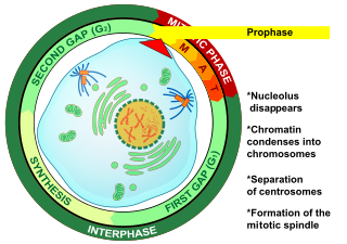
Prophase (from the Greek πρό, "before" and φάσις, "stage") is the first stage of cell division in both mitosis and meiosis. Beginning after interphase, DNA has already been replicated when the cell enters prophase. The main occurrences in prophase are the condensation of the chromatin reticulum and the disappearance of the nucleolus.

Telophase Final stage of a cell division for eukaryotic cells both in mitosis and meiosis

Telophase is the final stage in both meiosis and mitosis in a eukaryotic cell. During telophase, the effects of prophase and prometaphase are reversed. As chromosomes reach the cell poles, a nuclear envelope is re-assembled around each set of chromatids, the nucleoli reappear, and chromosomes begin to decondense back into the expanded chromatin that is present during interphase. The mitotic spindle is disassembled and remaining spindle microtubules are depolymerized. Telophase accounts for approximately 2% of the cell cycle's duration.

Nondisjunction Failure to separate properly during cell division

Nondisjunction is the failure of homologous chromosomes or sister chromatids to separate properly during cell division. There are three forms of nondisjunction: failure of a pair of homologous chromosomes to separate in meiosis I, failure of sister chromatids to separate during meiosis II, and failure of sister chromatids to separate during mitosis. Nondisjunction results in daughter cells with abnormal chromosome numbers (aneuploidy).

Spindle checkpoint

The spindle checkpoint, also known as the metaphase-to-anaphase transition, the spindle assembly checkpoint (SAC), or the mitotic checkpoint, is a cell cycle checkpoint during mitosis or meiosis that prevents the separation of the duplicated chromosomes (anaphase) until each chromosome is properly attached to the spindle. To achieve proper segregation, the two kinetochores on the sister chromatids must be attached to opposite spindle poles. Only this pattern of attachment will ensure that each daughter cell receives one copy of the chromosome. The defining biochemical feature of this checkpoint is the stimulation of the anaphase-promoting complex by M-phase cyclin-CDK complexes, which in turn causes the proteolytic destruction of cyclins and proteins that hold the sister chromatids together.

Kinetochore Protein complex that allows microtubules to attach to chromosomes during cell division

A kinetochore is a disc-shaped protein structure associated with duplicated chromatids in eukaryotic cells where the spindle fibers attach during cell division to pull sister chromatids apart. The kinetochore assembles on the centromere and links the chromosome to microtubule polymers from the mitotic spindle during mitosis and meiosis. The term kinetochore was first used in a footnote in a 1934 Cytology book by Lester W. Sharp and commonly accepted in 1936. Sharp's footnote reads: "The convenient term kinetochore has been suggested to the author by J. A. Moore", likely referring to John Alexander Moore who had joined Columbia University as a freshman in 1932.

Mad2 is an essential spindle checkpoint protein. The spindle checkpoint system is a regulatory system that restrains progression through the metaphase-to-anaphase transition. The Mad2 gene was first identified in the yeast S. cerevisiae in a screen for genes which when mutated would confer sensitivity to microtubule poisons. The human orthologues of Mad2 were first cloned in a search for human cDNAs that would rescue the microtubule poison-sensitivity of a yeast strain in which a kinetochore binding protein was missing. The protein was shown to be present at unattached kinetochores and antibody inhibition studies demonstrated it was essential to execute a block in the metaphase-to-anaphase transition in response to the microtubule poison nocodazole. Subsequent cloning of the Xenopus laevis orthologue, facilitated by the sharing of the human sequence, allowed for the characterization of the mitotic checkpoint in egg extracts.

Aurora A kinase

Aurora kinase A also known as serine/threonine-protein kinase 6 is an enzyme that in humans is encoded by the AURKA gene.

Aurora B kinase Protein

Aurora B kinase is a protein that functions in the attachment of the mitotic spindle to the centromere.

BUB1

Mitotic checkpoint serine/threonine-protein kinase BUB1 also known as BUB1 is an enzyme that in humans is encoded by the BUB1 gene.

BUB3

Mitotic checkpoint protein BUB3 is a protein that in humans is encoded by the BUB3 gene.

AURKC

Serine/threonine-protein kinase 13 is an enzyme that in humans is encoded by the AURKC gene.

HORMAD1

HORMA domain-containing protein 1 (HORMAD1) also known as cancer/testis antigen 46 (CT46) is a protein that in humans is encoded by the HORMAD1 gene.

Meiotic recombination checkpoint

The meiotic recombination checkpoint monitors meiotic recombination during meiosis, and blocks the entry into metaphase I if recombination is not efficiently processed.

In cell biology, Meiomitosis is an aberrant cellular division pathway that combines normal mitosis pathways with ectopically expressed meiotic machinery resulting in genomic instability.

Cdc14 and Cdc14 are a gene and its protein product respectively. Cdc14 is found in most of the eukaryotes. Cdc14 was defined by Hartwell in his famous screen for loci that control the cell cycle of Saccharomyces cerevisiae. Cdc14 was later shown to encode a protein phosphatase. Cdc14 is dual-specificity, which means it has serine/threonine and tyrosine-directed activity. A preference for serines next to proline is reported. Many early studies, especially in the budding yeast Saccharomyces cerevisiae, demonstrated that the protein plays a key role in regulating late mitotic processes. However, more recent work in a range of systems suggests that its cellular function is more complex.

Mad1

Mad1 is a non-essential protein which in yeast has a function in the spindle assembly checkpoint (SAC). This checkpoint monitors chromosome attachment to spindle microtubules and prevents cells from starting anaphase until the spindle is built up. The name Mad refers to the observation that mutant cells are mitotic arrest deficient (MAD) during microtubule depolymerization. Mad1 recruits the anaphase inhibitor Mad2 to unattached kinetochores and is essential for Mad2-Cdc20 complex formation in vivo but not in vitro. In vivo, Mad1 acts as a competitive inhibitor of the Mad2-Cdc20 complex. Mad1 is phosphorylated by Mps1 which then leads together with other activities to the formation of the mitotic checkpoint complex (MCC). Thereby it inhibits the activity of the anaphase-promoting complex/cyclosome (APC/C). Homologs of Mad1 are conserved in eukaryotes from yeast to mammals.

HORMA domain

In molecular biology, the HORMA domain is a protein domain that has been suggested to recognise chromatin states resulting from DNA adducts, double stranded breaks or non-attachment to the spindle and act as an adaptor that recruits other proteins. Hop1 is a meiosis-specific protein, Rev7 is required for DNA damage induced mutagenesis, and MAD2 is a spindle checkpoint protein which prevents progression of the cell cycle upon detection of a defect in mitotic spindle integrity.

References

  1. 1 2 3 GRCh38: Ensembl release 89: ENSG00000071539 - Ensembl, May 2017
  2. "Human PubMed Reference:". National Center for Biotechnology Information, U.S. National Library of Medicine.
  3. "Mouse PubMed Reference:". National Center for Biotechnology Information, U.S. National Library of Medicine.
  4. Makar AB, McMartin KE, Palese M, Tephly TR (June 1975). "Formate assay in body fluids: application in methanol poisoning". Biochemical Medicine. 13 (2): 117–26. doi:10.1016/0006-2944(75)90147-7. PMID   1.
  5. 1 2 3 4 Vader G (September 2015). "Pch2(TRIP13): controlling cell division through regulation of HORMA domains". Chromosoma. 124 (3): 333–9. doi:10.1007/s00412-015-0516-y. PMID   25895724. S2CID   18301840.
  6. 1 2 Ma HT, Poon RY (February 2016). "TRIP13 Regulates Both the Activation and Inactivation of the Spindle-Assembly Checkpoint". Cell Reports. 14 (5): 1086–1099. doi: 10.1016/j.celrep.2016.01.001 . PMID   26832417.
  7. 1 2 3 Marks DH, Thomas R, Chin Y, Shah R, Khoo C, Benezra R (May 2017). "Mad2 Overexpression Uncovers a Critical Role for TRIP13 in Mitotic Exit". Cell Reports. 19 (9): 1832–1845. doi:10.1016/j.celrep.2017.05.021. PMC   5526606 . PMID   28564602.
  8. Chen C, Jomaa A, Ortega J, Alani EE (January 2014). "Pch2 is a hexameric ring ATPase that remodels the chromosome axis protein Hop1". Proceedings of the National Academy of Sciences of the United States of America. 111 (1): E44–53. doi: 10.1073/pnas.1310755111 . PMC   3890899 . PMID   24367111.
  9. Yedidi RS, Wendler P, Enenkel C (2017). "AAA-ATPases in Protein Degradation". Frontiers in Molecular Biosciences. 4: 42. doi: 10.3389/fmolb.2017.00042 . PMC   5476697 . PMID   28676851.
  10. Tipton AR, Wang K, Oladimeji P, Sufi S, Gu Z, Liu ST (June 2012). "Identification of novel mitosis regulators through data mining with human centromere/kinetochore proteins as group queries". BMC Cell Biology. 13 (1): 15. doi:10.1186/1471-2121-13-15. PMC   3419070 . PMID   22712476.
  11. 1 2 San-Segundo PA, Roeder GS (April 1999). "Pch2 links chromatin silencing to meiotic checkpoint control". Cell. 97 (3): 313–24. doi: 10.1016/s0092-8674(00)80741-2 . PMID   10319812. S2CID   16002216.
  12. Roig I, Dowdle JA, Toth A, de Rooij DG, Jasin M, Keeney S (August 2010). "Mouse TRIP13/PCH2 is required for recombination and normal higher-order chromosome structure during meiosis". PLOS Genetics. 6 (8): e1001062. doi:10.1371/journal.pgen.1001062. PMC   2920839 . PMID   20711356.
  13. Rosenberg SC, Corbett KD (November 2015). "The multifaceted roles of the HORMA domain in cellular signaling". The Journal of Cell Biology. 211 (4): 745–55. doi:10.1083/jcb.201509076. PMC   4657174 . PMID   26598612.
  14. Wojtasz L, Daniel K, Roig I, Bolcun-Filas E, Xu H, Boonsanay V, Eckmann CR, Cooke HJ, Jasin M, Keeney S, McKay MJ, Toth A (October 2009). "Mouse HORMAD1 and HORMAD2, two conserved meiotic chromosomal proteins, are depleted from synapsed chromosome axes with the help of TRIP13 AAA-ATPase". PLOS Genetics. 5 (10): e1000702. doi:10.1371/journal.pgen.1000702. PMC   2758600 . PMID   19851446.
  15. Mapelli M, Massimiliano L, Santaguida S, Musacchio A (November 2007). "The Mad2 conformational dimer: structure and implications for the spindle assembly checkpoint" (PDF). Cell. 131 (4): 730–43. doi:10.1016/j.cell.2007.08.049. hdl: 2434/65744 . PMID   18022367. S2CID   17345925.
  16. Yang M, Li B, Tomchick DR, Machius M, Rizo J, Yu H, Luo X (November 2007). "p31comet blocks Mad2 activation through structural mimicry". Cell. 131 (4): 744–55. doi:10.1016/j.cell.2007.08.048. PMC   2144745 . PMID   18022368.
  17. Ye Q, Rosenberg SC, Moeller A, Speir JA, Su TY, Corbett KD (April 2015). "TRIP13 is a protein-remodeling AAA+ ATPase that catalyzes MAD2 conformation switching". eLife. 4. doi:10.7554/eLife.07367. PMC   4439613 . PMID   25918846.
  18. Banerjee R, Russo N, Liu M, Basrur V, Bellile E, Palanisamy N, Scanlon CS, van Tubergen E, Inglehart RC, Metwally T, Mani RS, Yocum A, Nyati MK, Castilho RM, Varambally S, Chinnaiyan AM, D'Silva NJ (July 2014). "TRIP13 promotes error-prone nonhomologous end joining and induces chemoresistance in head and neck cancer". Nature Communications. 5: 4527. doi:10.1038/ncomms5527. PMC   4130352 . PMID   25078033.

Further reading