


Tubulin alpha-1A chain is a protein that in humans is encoded by the TUBA1A gene. [4] [5] [6]
Tubulin alpha-1A chain is a protein that in humans is encoded by the TUBA1A gene. [4] [5] [6]
TUBA1A is a structural gene that encodes for Tubulin, Alpha 1A product. TUBA1A product is an alpha-tubulin that participates in the formation of microtubules - structural proteins that participate in cytoskeletal structure. Specifically, microtubules are composed of a heterodimer of alpha and beta-tubulin molecules. Cowan et al. demonstrated that bα1 is a primary α-tubulin of the human fetal brain, and that it is expressed solely in that structure, by way of Northern blot. [7] Miller et al. further elaborated on the role of α-tubulins and the process of neuronal development and maturation, comparing the expressions of rat α-tubulins Tα1 and T26. These two rat α-tubulins are homologs of bα1 and kα1 showing that a rat homolog of human TUBA1A (Tα1) had elevated expression during the extension of neuronal processes. Culturing of pheochromocytoma cells with Nerve Growth Factor (NGF) induced differentiation and the development of neuronal processes. Northern blot assay showed markedly elevated levels of Tα1 mRNA expression; T26 mRNA expression increased minimally with exposure to NGF. [8] These data suggest that TUBA1A models the brain by participating in the directing of neuronal migration through the ability of microtubules to readily form and break polymers to extend and retract processes to induce nucleokinesis. [9] Poirier et al. used RNA in situ hybridization to show TUBA1A expression in mice embryo; embryo sections from embryonic day 16.5 “showed a strong labeling in the telencephalon, diencephalon, and mesencephalon, the developing cerebellum, the brainstem, the spinal cord, and the dorsal root ganglia”. [10]
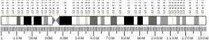
Microtubules of the eukaryotic cytoskeleton perform essential and diverse functions and are composed of a heterodimer of alpha and beta tubulins. The genes encoding these microtubule constituents belong to the tubulin superfamily, which is composed of six distinct families. Genes from the alpha, beta and gamma tubulin families are found in all eukaryotes. The alpha and beta tubulins represent the major components of microtubules, while gamma tubulin plays a critical role in the nucleation of microtubule assembly. There are multiple alpha and beta tubulin genes, which are highly conserved among species. This gene encodes alpha tubulin and is highly similar to mouse and rat Tuba1 gene. Northern blotting studies have shown that the gene expression is predominantly found in morphologically differentiated neurologic cells. This gene is one of three alpha-tubulin genes in a cluster on chromosome 12q. [6]
Mutations to the TUBA1A gene manifest clinically as Type 3 Lissencephaly. In general, lissencephaly is characterized by agyria (lacking of gyri and sulci to the brain – a smooth brain), seizure activity, failure to thrive, as well as intellectual disability and psychomotor retardation, often to a profound degree. [10] The symptoms of Lis3 Lissencephaly are not especially different from generalized lissencephaly (Lis1, related to PAFAH1B1). Diagnosis of lissencephaly generally is made from the symptom profile, while attribution to a specific type is obtained by microarray. Treatment is symptomatic; anti-convulsive drugs for seizure activity, g-button gastrostomy to feed the child, physical therapy for muscle disorders. TUBA1A mutation is common in microlissencephaly
Keays et al. describe a mouse with a mutation of the TUBA1A gene induced by N-ethyl-N-nitrosourea. The relevant point mutation resulted in S140G; [12] the site of the mutation participates in the N-site of the formed α-tubulin, and participates in stabilizing the α-β tubulin polymer by binding GTP at this site. [13] The S140G mutation resulted in the formation of a “compromised GTP binding pocket”. Authors note defects associated with cortical layers II/III and IV, especially in cortical neuronal migration (with respect to wild-type counterparts), showing that the S140G mutation has value as a model for detailing disease associated with the Human TUBA homolog. [12]

Microtubules are polymers of tubulin that form part of the cytoskeleton and provide structure and shape to eukaryotic cells. Microtubules can grow as long as 50 micrometres and are highly dynamic. The outer diameter of a microtubule is between 23 and 27 nm while the inner diameter is between 11 and 15 nm. They are formed by the polymerization of a dimer of two globular proteins, alpha and beta tubulin into protofilaments that can then associate laterally to form a hollow tube, the microtubule. The most common form of a microtubule consists of 13 protofilaments in the tubular arrangement.

Lissencephaly is a set of rare brain disorders where the whole or parts of the surface of the brain appear smooth. It is caused by defective neuronal migration during the 12th to 24th weeks of gestation resulting in a lack of development of brain folds (gyri) and grooves (sulci). It is a form of cephalic disorder. Terms such as agyria and pachygyria are used to describe the appearance of the surface of the brain.
Tubulin in molecular biology can refer either to the tubulin protein superfamily of globular proteins, or one of the member proteins of that superfamily. α- and β-tubulins polymerize into microtubules, a major component of the eukaryotic cytoskeleton. Microtubules function in many essential cellular processes, including mitosis. Tubulin-binding drugs kill cancerous cells by inhibiting microtubule dynamics, which are required for DNA segregation and therefore cell division.
Acetylation is an organic esterification reaction with acetic acid. It introduces an acetyl functional group into a chemical compound. Such compounds are termed acetate esters or acetates. Deacetylation is the opposite reaction, the removal of an acetyl group from a chemical compound.

The tau proteins are a group of six highly soluble protein isoforms produced by alternative splicing from the gene MAPT. They have roles primarily in maintaining the stability of microtubules in axons and are abundant in the neurons of the central nervous system (CNS). They are less common elsewhere but are also expressed at very low levels in CNS astrocytes and oligodendrocytes.

Neuronal migration protein doublecortin, also known as doublin or lissencephalin-X is a protein that in humans is encoded by the DCX gene.
Platelet-activating factor acetylhydrolase IB subunit alpha is an enzyme that in humans is encoded by the PAFAH1B1 gene. The protein is often referred to as Lis1 and plays an important role in regulating the motor protein Dynein.

Class III β-tubulin, otherwise known as βIII-tubulin (β3-tubulin) or β-tubulin III, is a microtubule element of the tubulin family found almost exclusively in neurons, and in testis cells. In humans, it is encoded by the TUBB3 gene.
In enzymology, an alpha-tubulin N-acetyltransferase is an enzyme which is encoded by the ATAT1 gene.

Tubulin alpha-4A chain is a protein that in humans is encoded by the TUBA4A gene.

Tubulin alpha-1B chain is a protein that in humans is encoded by the TUBA1B gene.

Tubulin alpha-3C/D chain is a protein that in humans is encoded by the TUBA3C gene.

Tubulin beta-4A chain is a protein that in humans is encoded by the TUBB4A gene. Two tubulin beta-4 chain proteins are encoded in the human genome by the genes TUBB4A and TUBB4B. Tubulin is the major constituent of microtubules, a key components of the cytoskeleton. It binds two molecules of GTP, one at an exchangeable site on the beta-chain and one at a non-exchangeable site on the alpha-chain. TUBB4A is preferentially and highly expressed in the central nervous system.

Tubulin alpha-8 chain is a protein that in humans is encoded by the TUBA8 gene.
Tubulin alpha-1C chain is a protein that in humans is encoded by the TUBA1C gene.

Tubulin beta chain is a protein that in humans is encoded by the TUBB gene.

Kinesin-like protein KIF1A, also known as axonal transporter of synaptic vesicles or microtubule-based motor KIF1A, is a protein that in humans is encoded by the KIF1A gene.

Microlissencephaly (MLIS) is a rare congenital brain disorder that combines severe microcephaly with lissencephaly. Microlissencephaly is a heterogeneous disorder, i.e. it has many different causes and a variable clinical course. Microlissencephaly is a malformation of cortical development (MCD) that occurs due to failure of neuronal migration between the third and fifth month of gestation as well as stem cell population abnormalities. Numerous genes have been found to be associated with microlissencephaly, however, the pathophysiology is still not completely understood.

Neurotubules are microtubules found in neurons in nervous tissues. Along with neurofilaments and microfilaments, they form the cytoskeleton of neurons. Neurotubules are undivided hollow cylinders that are made up of tubulin protein polymers and arrays parallel to the plasma membrane in neurons. Neurotubules have an outer diameter of about 23 nm and an inner diameter, also known as the central core, of about 12 nm. The wall of the neurotubules is about 5 nm in width. There is a non-opaque clear zone surrounding the neurotubule and it is about 40 nm in diameter. Like microtubules, neurotubules are greatly dynamic and the length of them can be adjusted by polymerization and depolymerization of tubulin.
David Anthony Keays is an Australian neuroscientist who studies magnetoreception and neurodevelopment. He is currently a group leader at the Research Institute of Molecular Pathology (IMP) in Vienna, Austria, Chair of Organismal and Developmental Neurobiology at the Ludwig Maximilians University (LMU) in Munich, and Honorary Associate Professor at the University of Melbourne.
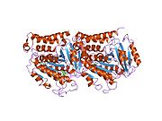
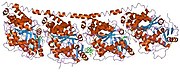
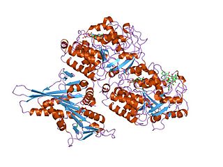
PDB gallery | |
|---|---|
|