TYRP1

Last updated
TYRP1
Identifiers
Aliases TYRP1 , CAS2, CATB, GP75, OCA3, TRP, TRP1, TYRP, b-PROTEIN, tyrosinase related protein 1
External IDs OMIM: 115501 MGI: 98881 HomoloGene: 464 GeneCards: TYRP1
Orthologs
SpeciesHumanMouse
Entrez
Ensembl
UniProt
RefSeq (mRNA)

NM_000550

NM_001282014
NM_001282015
NM_031202

RefSeq (protein)

NP_000541

NP_001268943
NP_001268944
NP_112479

Location (UCSC) Chr 9: 12.69 – 12.71 Mb Chr 4: 80.75 – 80.77 Mb
PubMed search [3] [4]
Wikidata
View/Edit Human View/Edit Mouse

Tyrosinase-related protein 1, also known as TYRP1, is an intermembrane enzyme which in humans is encoded by the TYRP1 gene. [5] [6]

Contents

Function

Tyrp1 is a melanocyte-specific gene product involved in melanin synthesis within melanosomes. [7] Most Tyrp1 possess 5,6-dihydroxyindole-2-carboxylic acid (melanogenic intermediate) oxidase activity. [8] The catalytic function of Tyrp1 in human melanocytes is less clear. Tyrp1 is involved in stabilizing of tyrosinase protein and modulating its catalytic activity. [7] Tyrp1 is also involved in maintenance of melanosome structure and affects melanocyte proliferation and melanocyte cell death. [9] Melanocytes are derived from the neural crest and migrate into the overlying epidermal ectoderm of a developing organism which forms skin and hair. [10] Therefore, Tyrp1 influences the expression of melanin notably in the skin and hair of an organism.

The Tyrp1 gene also has a non-coding function which indirectly promotes melanoma tumor cell proliferation, especially when highly expressed in a cell. [11] Tyrp1 mRNA interacts with miR-16 and affects its ability to repress genes involved in melanoma cell production. [11]

Clinical significance

Mutations in the mouse Tyrp1 gene are associated with brown pelage and in the human TYRP1 gene with oculocutaneous albinism type 3 (OCA3). [9] An allele of TYRP1 common in Solomon Islanders results in blond hair. Although the phenotype is similar to Northern European blond hair, this allele is not found in Europeans. [12] [13] More recent study, Ju et al. 2020 found TYRP1 allele was selected in European population. [14]

Norton et al. 2016 study found TYRP1 is not associated with blond hair color in Melanesians as many populations in Oceania did not carry TYRP1 alleles but still displayed blondism, study indicates that additional unknown alleles contribute to the blondism phenotype in Melanesians. [15]

Alterations of the Tyrp1 gene is responsible for some of the differing phenotypes of skin and coat appearance in various animals. In Dalmatians, black versus "liver" spot color is due to genetic variation of the TYRP1 gene. [16] A particular deletion in the Tyrp1 gene of domestic Chinese-Tibetan swine results in a "brown coloration" of the swine's skin and hair as opposed to the wild-type "black" phenotype. [17] In Oujiang-color carp, mutations of the Tyrp1 gene influenced the expression of "grey" or "brown" phenotypic color of scales. [18]

Elevated levels of Tyrp1 gene expression is also associated with unfavorable patient outcome of those affected by melanoma. [11] The role of Tyrp1 in melanoma progression was determined by comparing "knockout" cell lines which have inactive Tyrp1 to cells with normal and highly expressed Tyrp1. [11] Such studies provide insight to possible clinical usage and treatment of melanoma via regulation of Tyrp1 expression in cells. [11]

Regulation

The expression of TYRP1 is regulated by the microphthalmia-associated transcription factor (MITF). [19] [20]

Interactions

TYRP1 has been shown to interact with GIPC1. [21]

See also

Related Research Articles

<span class="mw-page-title-main">Albinism in humans</span> Condition characterized by partial or complete absence of pigment in the skin, hair and eyes

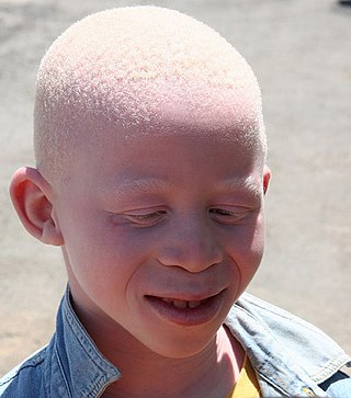
Albinism is a congenital condition characterized in humans by the partial or complete absence of pigment in the skin, hair and eyes. Albinism is associated with a number of vision defects, such as photophobia, nystagmus, and amblyopia. Lack of skin pigmentation makes for more susceptibility to sunburn and skin cancers. In rare cases such as Chédiak–Higashi syndrome, albinism may be associated with deficiencies in the transportation of melanin granules. This also affects essential granules present in immune cells leading to increased susceptibility to infection.

<span class="mw-page-title-main">Human skin color</span> Factors affecting skin color in humans

Human skin color ranges from the darkest brown to the lightest hues. Differences in skin color among individuals is caused by variation in pigmentation, which is the result of genetics, exposure to the sun, natural and sexual selection, or all of these. Differences across populations evolved through natural or sexual selection, because of social norms and differences in environment, as well as regulations of the biochemical effects of ultraviolet radiation penetrating the skin.

<span class="mw-page-title-main">Melanin</span> Group of natural pigments found in most organisms

Melanin is a broad term for a group of natural pigments found in most organisms. Eumelanin is produced through a multistage chemical process known as melanogenesis, where the oxidation of the amino acid tyrosine is followed by polymerization. The melanin pigments are produced in a specialized group of cells known as melanocytes. Functionally, eumelanin serves as protection against UV radiation.

<span class="mw-page-title-main">Melanocyte</span> Melanin-producing cells of the skin

Melanocytes are melanin-producing neural crest-derived cells located in the bottom layer of the skin's epidermis, the middle layer of the eye, the inner ear, vaginal epithelium, meninges, bones, and heart. Melanin is a dark pigment primarily responsible for skin color. Once synthesized, melanin is contained in special organelles called melanosomes which can be transported to nearby keratinocytes to induce pigmentation. Thus darker skin tones have more melanosomes present than lighter skin tones. Functionally, melanin serves as protection against UV radiation. Melanocytes also have a role in the immune system.

<span class="mw-page-title-main">Human hair color</span> Pigmentation of human hair follicles

Human hair color is the pigmentation of human hair follicles due to two types of melanin: eumelanin and pheomelanin. Generally, if more melanin is present, the color of the hair is darker; if less melanin is present, the hair is lighter. The tone of the hair is dependent on the ratio of black or brown eumelanin to yellow or red pheomelanin. Levels of melanin can vary over time causing a person's hair color to change, and it is possible to have hair follicles of more than one color on the same person. Some hair colors are associated with some ethnic groups due to observed higher frequency of particular hair color within their geographical region, e.g. straight dark hair amongst East Asians, a large variety of dark, fair, curly, wavy and bushy hair amongst Europeans, curly, dark, and uniquely helical hair with Africans, whilst gray, white or "silver" hair is often associated with age and wisdom.

<span class="mw-page-title-main">Equine coat color genetics</span> Genetics behind the equine coat color

Equine coat color genetics determine a horse's coat color. Many colors are possible, but all variations are produced by changes in only a few genes. The "base" colors of the horse are determined by the Extension locus, which in recessive form (e) creates a solid chestnut or "red" coat. When dominant (E), a horse is black. The next gene that strongly affects coat color, Agouti, when present on a horse dominant for E, limits the black color to the points, creating a shade known as Bay that is so common and dominant in horses that it is informally grouped as a "base" coat color.

<span class="mw-page-title-main">Tyrosinase</span> Enzyme for controlling the production of melanin

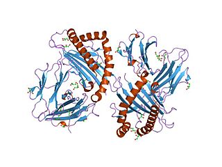
Tyrosinase is an oxidase that is the rate-limiting enzyme for controlling the production of melanin. The enzyme is mainly involved in two distinct reactions of melanin synthesis otherwise known as the Raper Mason pathway. Firstly, the hydroxylation of a monophenol and secondly, the conversion of an o-diphenol to the corresponding o-quinone. o-Quinone undergoes several reactions to eventually form melanin. Tyrosinase is a copper-containing enzyme present in plant and animal tissues that catalyzes the production of melanin and other pigments from tyrosine by oxidation. It is found inside melanosomes which are synthesized in the skin melanocytes. In humans, the tyrosinase enzyme is encoded by the TYR gene.

Oculocutaneous albinism is a form of albinism involving the eyes, the skin, and the hair. Overall, an estimated 1 in 20,000 people worldwide are born with oculocutaneous albinism. OCA is caused by mutations in several genes that control the synthesis of melanin within the melanocytes. Seven types of oculocutaneous albinism have been described, all caused by a disruption of melanin synthesis and all autosomal recessive disorders. Oculocutaneous albinism is also found in non-human animals.

<span class="mw-page-title-main">White horse</span>

A white horse is born predominantly white and stays white throughout its life. A white horse has mostly pink skin under its hair coat, and may have brown, blue, or hazel eyes. "True white" horses, especially those that carry one of the dominant white (W) genes, are rare. Most horses that are commonly referred to as "white" are actually "gray" horses whose hair coats are completely white. Gray horses may be born of any color and their hairs gradually turn white as time goes by and take on a white appearance. Nearly all gray horses have dark skin, except under any white markings present at birth. Skin color is the most common method for an observer to distinguish between mature white and gray horses.

<span class="mw-page-title-main">Microphthalmia-associated transcription factor</span> Mammalian protein found in Homo sapiens

Microphthalmia-associated transcription factor also known as class E basic helix-loop-helix protein 32 or bHLHe32 is a protein that in humans is encoded by the MITF gene.

<span class="mw-page-title-main">Labrador Retriever coat colour genetics</span> Genetics behind Labrador Retriever coat colour

The genetic basis of coat colour in the Labrador Retriever has been found to depend on several distinct genes. The interplay among these genes is used as an example of epistasis.

<span class="mw-page-title-main">PMEL (gene)</span>

Melanocyte protein PMEL also known as premelanosome protein (PMEL) or silver locus protein homolog (SILV) is a protein that in humans is encoded by the PMEL gene. Its gene product may be referred to as PMEL, silver, ME20, gp100 or Pmel17.

<span class="mw-page-title-main">OCA2</span> Protein-coding gene in humans

P protein, also known as melanocyte-specific transporter protein or pink-eyed dilution protein homolog, is a protein that in humans is encoded by the oculocutaneous albinism II (OCA2) gene. The P protein is believed to be an integral membrane protein involved in small molecule transport, specifically of tyrosine - a precursor of melanin. Certain mutations in OCA2 result in type 2 oculocutaneous albinism. OCA2 encodes the human homologue of the mouse p gene.

<span class="mw-page-title-main">MLANA</span> Protein-coding gene in the species Homo sapiens

Protein melan-A also known as melanoma antigen recognized by T cells 1 or MART-1 is a protein that in humans is encoded by the MLANA or "MALENA" gene. A fragment of the protein, usually consisting of the nine amino acids 27 to 35, is bound by MHC class I complexes which present it to T cells of the immune system. These complexes can be found on the surface of melanoma cells. Decameric peptides (26-35) are being investigated as cancer vaccines.

<span class="mw-page-title-main">Membrane-associated transporter protein</span> Protein

Membrane-associated transporter protein (MATP), also known as solute carrier family 45 member 2 (SLC45A2) or melanoma antigen AIM1, is a protein that in humans is encoded by the SLC45A2 gene.

Oculocutaneous albinism type I or type 1A is an autosomal recessive skin disease. This subtype of oculocutaneous albinism is caused when the gene for tyrosinase does not function properly.

<span class="mw-page-title-main">Ocular albinism type 1</span> Most common type of ocular albinism

Ocular albinism type 1(OA1) is the most common type of ocular albinism, with a prevalence rate of 1:50,000. It is an inheritable classical Mendelian type X-linked recessive disorder wherein the retinal pigment epithelium lacks pigment while hair and skin appear normal. Since it is usually an X-linked disorder, it occurs mostly in males, while females are carriers unless they are homozygous. About 60 missense and nonsense mutations, insertions, and deletions have been identified in Oa1. Mutations in OA1 have been linked to defective glycosylation and thus improper intracellular transportation.

<span class="mw-page-title-main">Amelanism</span> Pigmentation abnormality

Amelanism is a pigmentation abnormality characterized by the lack of pigments called melanins, commonly associated with a genetic loss of tyrosinase function. Amelanism can affect fish, amphibians, reptiles, birds, and mammals including humans. The appearance of an amelanistic animal depends on the remaining non-melanin pigments. The opposite of amelanism is melanism, a higher percentage of melanin.

The agouti gene, the Agouti-signaling protein (ASIP) is responsible for variations in color in many species. Agouti works with extension to regulate the color of melanin which is produced in hairs. The agouti protein causes red to yellow pheomelanin to be produced, while the competing molecule α-MSH signals production of brown to black eumelanin. In wildtype mice, alternating cycles of agouti and α-MSH production cause agouti coloration. Each hair has bands of yellow which grew during agouti production, and black which grew during α-MSH production. Wildtype mice also have light-colored bellies. The hairs there are a creamy color the whole length because the agouti protein was produced the whole time the hairs were growing.

References

  1. 1 2 3 GRCh38: Ensembl release 89: ENSG00000107165 - Ensembl, May 2017
  2. 1 2 3 GRCm38: Ensembl release 89: ENSMUSG00000005994 - Ensembl, May 2017
  3. "Human PubMed Reference:". National Center for Biotechnology Information, U.S. National Library of Medicine.
  4. "Mouse PubMed Reference:". National Center for Biotechnology Information, U.S. National Library of Medicine.
  5. EntrezGene 7306
  6. Box NF, Wyeth JR, Mayne CJ, O'Gorman LE, Martin NG, Sturm RA (January 1998). "Complete sequence and polymorphism study of the human TYRP1 gene encoding tyrosinase-related protein 1". Mammalian Genome. 9 (1): 50–53. doi:10.1007/s003359900678. PMID   9434945. S2CID   10020827.
  7. 1 2 Kobayashi T, Imokawa G, Bennett DC, Hearing VJ (November 1998). "Tyrosinase stabilization by Tyrp1 (the brown locus protein)". The Journal of Biological Chemistry. 273 (48): 31801–31805. doi: 10.1074/jbc.273.48.31801 . PMID   9822646.
  8. Jiménez-Cervantes C, Solano F, Kobayashi T, Urabe K, Hearing VJ, Lozano JA, García-Borrón JC (July 1994). "A new enzymatic function in the melanogenic pathway. The 5,6-dihydroxyindole-2-carboxylic acid oxidase activity of tyrosinase-related protein-1 (TRP1)". The Journal of Biological Chemistry. 269 (27): 17993–18000. doi: 10.1016/S0021-9258(17)32408-0 . PMID   8027058.
  9. 1 2 Sarangarajan R, Boissy RE (December 2001). "Tyrp1 and oculocutaneous albinism type 3". Pigment Cell Research. 14 (6): 437–444. doi: 10.1034/j.1600-0749.2001.140603.x . PMID   11775055.
  10. Mayer TC (September 1973). "The migratory pathway of neural crest cells into the skin of mouse embryos". Developmental Biology. 34 (1): 39–46. doi:10.1016/0012-1606(73)90337-0. PMID   4595498.
  11. 1 2 3 4 5 Gilot D, Migault M, Bachelot L, Journé F, Rogiers A, Donnou-Fournet E, et al. (November 2017). "A non-coding function of TYRP1 mRNA promotes melanoma growth" (PDF). Nature Cell Biology. 19 (11): 1348–1357. doi:10.1038/ncb3623. PMID   28991221. S2CID   23539385.
  12. Bhanoo SN (3 May 2012). "Another Genetic Quirk of the Solomon Islands: Blond Hair". The New York Times. Retrieved 3 May 2012.
  13. Kenny EE, Timpson NJ, Sikora M, Yee MC, Moreno-Estrada A, Eng C, et al. (May 2012). "Melanesian blond hair is caused by an amino acid change in TYRP1". Science. 336 (6081): 554. Bibcode:2012Sci...336..554K. doi:10.1126/science.1217849. PMC   3481182 . PMID   22556244.
  14. Ju, Dan; Mathieson, Iain (December 21, 2020). "The evolution of skin pigmentation-associated variation in West Eurasia". Proceedings of the National Academy of Sciences of the United States of America. 118 (1). doi: 10.1073/pnas.2009227118 . PMC   7817156 . PMID   33443182.
  15. Norton HL, Hanna M, Werren E, Friedlaender J (May 2016). "The rs387907171 SNP in TYRP1 is not associated with blond hair color on the Island of Bougainville". American Journal of Human Biology. 28 (3): 431–435. doi:10.1002/ajhb.22795. PMID   26450459. S2CID   13727757.
  16. Cargill EJ, Famula TR, Schnabel RD, Strain GM, Murphy KE (July 2005). "The color of a Dalmatian's spots: linkage evidence to support the TYRP1 gene". BMC Veterinary Research. 1 (1): 1. doi:10.1186/1746-6148-1-1. PMC   1192828 . PMID   16045797.
  17. Ren J, Mao H, Zhang Z, Xiao S, Ding N, Huang L (May 2011). "A 6-bp deletion in the TYRP1 gene causes the brown colouration phenotype in Chinese indigenous pigs". Heredity. 106 (5): 862–868. doi:10.1038/hdy.2010.129. PMC   3186233 . PMID   20978532.
  18. Chen H, Wang J, Du J, Mandal BK, Si Z, Xu X, et al. (August 2021). "Analysis of recently duplicated TYRP1 genes and their effect on the formation of black patches in Oujiang-color common carp (Cyprinus carpio var. color)". Animal Genetics. 52 (4): 451–460. doi:10.1111/age.13071. PMID   33939849. S2CID   233720032.
  19. Fang D, Tsuji Y, Setaluri V (July 2002). "Selective down-regulation of tyrosinase family gene TYRP1 by inhibition of the activity of melanocyte transcription factor, MITF". Nucleic Acids Research. 30 (14): 3096–3106. doi:10.1093/nar/gkf424. PMC   135745 . PMID   12136092.
  20. Hoek KS, Schlegel NC, Eichhoff OM, Widmer DS, Praetorius C, Einarsson SO, et al. (December 2008). "Novel MITF targets identified using a two-step DNA microarray strategy". Pigment Cell & Melanoma Research. 21 (6): 665–676. doi: 10.1111/j.1755-148X.2008.00505.x . PMID   19067971.
  21. Liu TF, Kandala G, Setaluri V (September 2001). "PDZ domain protein GIPC interacts with the cytoplasmic tail of melanosomal membrane protein gp75 (tyrosinase-related protein-1)". The Journal of Biological Chemistry. 276 (38): 35768–35777. doi: 10.1074/jbc.M103585200 . PMID   11441007.

Further reading