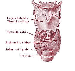
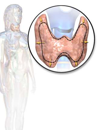
The thyroid, or thyroid gland, is an endocrine gland in vertebrates. In humans, it is in the neck and consists of two connected lobes. The lower two thirds of the lobes are connected by a thin band of tissue called the isthmus (pl.: isthmi). The thyroid gland is a butterfly-shaped gland located in the neck below the Adam's apple. Microscopically, the functional unit of the thyroid gland is the spherical thyroid follicle, lined with follicular cells (thyrocytes), and occasional parafollicular cells that surround a lumen containing colloid. The thyroid gland secretes three hormones: the two thyroid hormones – triiodothyronine (T3) and thyroxine (T4) – and a peptide hormone, calcitonin. The thyroid hormones influence the metabolic rate and protein synthesis and growth and development in children. Calcitonin plays a role in calcium homeostasis. Secretion of the two thyroid hormones is regulated by thyroid-stimulating hormone (TSH), which is secreted from the anterior pituitary gland. TSH is regulated by thyrotropin-releasing hormone (TRH), which is produced by the hypothalamus.

The Bartholin's glands are two pea-sized compound alveolar glands located slightly posterior and to the left and right of the opening of the vagina. They secrete mucus to lubricate the vagina.

The seminal vesicles are a pair of convoluted tubular accessory glands that lie behind the urinary bladder of male mammals. They secrete fluid that largely composes the semen.

A Bartholin's cyst occurs when a Bartholin's gland within the labia becomes blocked. Small cysts may result in minimal or no symptoms. Larger cysts may result in swelling on one side of the vaginal opening, as well as pain during sex or walking. If the cyst becomes infected, an abscess can occur, which is typically red and very painful. If there are no symptoms, no treatment is needed. Bartholin's cysts affect about 2% of women at some point in their life. They most commonly occur during childbearing years.

A thyroglossal cyst or thyroglossal duct cyst is a fibrous cyst that forms from a persistent thyroglossal duct. Thyroglossal cysts can be defined as an irregular neck mass or a lump which develops from cells and tissues left over after the formation of the thyroid gland during developmental stages.

Pseudocysts are like cysts, but lack epithelial or endothelial cells. Initial management consists of general supportive care. Symptoms and complications caused by pseudocysts require surgery. Computed tomography (CT) scans are used for initial imaging of cysts, and endoscopic ultrasounds are used in differentiating between cysts and pseudocysts. Endoscopic drainage is a popular and effective method of treating pseudocysts.
Thyroid disease is a medical condition that affects the function of the thyroid gland. The thyroid gland is located at the front of the neck and produces thyroid hormones that travel through the blood to help regulate many other organs, meaning that it is an endocrine organ. These hormones normally act in the body to regulate energy use, infant development, and childhood development.

A ranula is a mucus extravasation cyst involving a sublingual gland and is a type of mucocele found on the floor of the mouth. Ranulae present as a swelling of connective tissue consisting of collected mucin from a ruptured salivary gland caused by local trauma. If small and asymptomatic further treatment may not be needed, otherwise minor oral surgery may be indicated.

Sialadenitis (sialoadenitis) is inflammation of salivary glands, usually the major ones, the most common being the parotid gland, followed by submandibular and sublingual glands. It should not be confused with sialadenosis (sialosis) which is a non-inflammatory enlargement of the major salivary glands.

A persistent thyroglossal duct is a usually benign medical condition in which the thyroglossal duct, a structure usually only found during embryonic development, fails to atrophy. The duct persists as a midline structure forming an open connection between the back of the tongue and the thyroid gland. This opening can lead to fluid accumulation and infection, which necessitate the removal of the duct.

A branchial cleft cyst or simply branchial cyst is a cyst as a swelling in the upper part of neck anterior to sternocleidomastoid. It can, but does not necessarily, have an opening to the skin surface, called a fistula. The cause is usually a developmental abnormality arising in the early prenatal period, typically failure of obliteration of the second, third, and fourth branchial cleft, i.e. failure of fusion of the second branchial arches and epicardial ridge in lower part of the neck. Branchial cleft cysts account for almost 20% of neck masses in children. Less commonly, the cysts can develop from the first, third, or fourth clefts, and their location and the location of associated fistulas differs accordingly.

Thyroid dysgenesis is a cause of congenital hypothyroidism where the thyroid is missing, ectopic, or severely underdeveloped. It should not be confused with iodine deficiency, or with other forms of congenital hypothyroidism, such as thyroid dyshormonogenesis, where the thyroid is present but not functioning correctly.

Congenital cartilaginous rest of the neck (CCRN) is a minor and very rare congenital cutaneous condition characterized by branchial arch remnants that are considered to be the cervical variant of accessory tragus. It resembles a rudimentary pinna that in most cases is located in the lower anterior part of the neck.
Odontogenic cyst are a group of jaw cysts that are formed from tissues involved in odontogenesis. Odontogenic cysts are closed sacs, and have a distinct membrane derived from rests of odontogenic epithelium. It may contain air, fluids, or semi-solid material. Intra-bony cysts are most common in the jaws, because the mandible and maxilla are the only bones with epithelial components. That odontogenic epithelium is critical in normal tooth development. However, epithelial rests may be the origin for the cyst lining later. Not all oral cysts are odontogenic cysts. For example, mucous cyst of the oral mucosa and nasolabial duct cyst are not of odontogenic origin.
Ectopic thymus is a condition where thymus tissue is found in an abnormal location (ectopia). It usually does not cause symptoms, but may leads to a mass in the neck that may compress the trachea and the esophagus. It is thought to be the result of either a failure of descent or a failure of involution of normal thymus tissue. It may be diagnosed with radiology, such as an ultrasound or magnetic resonance imaging. If it causes illness, surgery can be used to remove it. Recurrence after surgery is very unlikely.

Squamous-cell carcinoma of the thyroid (SCT), or thyroid squamous-cell carcinoma, is rare malignant neoplasm of thyroid gland which shows tumor cells with distinct squamous differentiation. The incidence of SCT is less than 1% out of thyroid malignancies.
The face and neck development of the human embryo refers to the development of the structures from the third to eighth week that give rise to the future head and neck. They consist of three layers, the ectoderm, mesoderm and endoderm, which form the mesenchyme, neural crest and neural placodes. The paraxial mesoderm forms structures named somites and somitomeres that contribute to the development of the floor of the brain and voluntary muscles of the craniofacial region. The lateral plate mesoderm consists of the laryngeal cartilages. The three tissue layers give rise to the pharyngeal apparatus, formed by six pairs of pharyngeal arches, a set of pharyngeal pouches and pharyngeal grooves, which are the most typical feature in development of the head and neck. The formation of each region of the face and neck is due to the migration of the neural crest cells which come from the ectoderm. These cells determine the future structure to develop in each pharyngeal arch. Eventually, they also form the neurectoderm, which forms the forebrain, midbrain and hindbrain, cartilage, bone, dentin, tendon, dermis, pia mater and arachnoid mater, sensory neurons, and glandular stroma.
In CT scan of the thyroid, focal and diffuse thyroid abnormalities are commonly encountered. These findings can often lead to a diagnostic dilemma, as the CT reflects nonspecific appearances. Ultrasound (US) examination has a superior spatial resolution and is considered the modality of choice for thyroid evaluation. Nevertheless, CT detects incidental thyroid nodules (ITNs) and plays an important role in the evaluation of thyroid cancer.
A cervical thymic cyst, also called thymopharyngeal duct cyst, is a fluid-filled mass that occurs when the thymopharyngeal duct, an embryonic structure connecting the nascent thymus with the embryonic pharynx, fails to close and disappear. A thymic cyst is typically a solitary mass on one side of the neck, and is usually found near the carotid sheath. Some cervical thymic cysts may extend into the mediastinum. It is usually asymptomatic. The diagnostic process includes differentiating between other causes of neck masses in infants and children, including branchial cleft cysts and cystic hygromas. The treatment is surgical excision. On histologic examination, the wall of the cyst includes thymic tissue, and may include parathyroid gland tissue because of the parathyroid gland's common embryonic origin with the thymus gland in the third pharyngeal pouch. Fewer than 100 cases of cervical thymic cysts have been reported in the medical literature.