
The mesoderm is the middle layer of the three germ layers that develops during gastrulation in the very early development of the embryo of most animals. The outer layer is the ectoderm, and the inner layer is the endoderm.
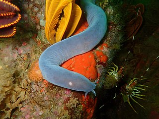
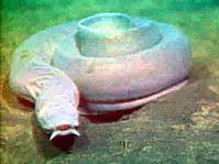
Hagfish, of the class Myxini and order Myxiniformes, are eel-shaped jawless fish. Hagfish are the only known living animals that have a skull but no vertebral column, although they do have rudimentary vertebrae. Hagfish are marine predators and scavengers who can defend themselves against other larger predators by releasing copious amounts of slime from mucous glands in their skin.

The jaws are a pair of opposable articulated structures at the entrance of the mouth, typically used for grasping and manipulating food. The term jaws is also broadly applied to the whole of the structures constituting the vault of the mouth and serving to open and close it and is part of the body plan of humans and most animals.

The occipital bone is a cranial dermal bone and the main bone of the occiput. It is trapezoidal in shape and curved on itself like a shallow dish. The occipital bone overlies the occipital lobes of the cerebrum. At the base of the skull in the occipital bone, there is a large oval opening called the foramen magnum, which allows the passage of the spinal cord.
A craniate is a member of the Craniata, a proposed clade of chordate animals with a skull of hard bone or cartilage. Living representatives are the Myxini (hagfishes), Hyperoartia, and the much more numerous Gnathostomata. Formerly distinct from vertebrates by excluding hagfish, molecular and anatomical research in the 21st century has led to the reinclusion of hagfish as vertebrates, making living craniates synonymous with living vertebrates.

The somites are a set of bilaterally paired blocks of paraxial mesoderm that form in the embryonic stage of somitogenesis, along the head-to-tail axis in segmented animals. In vertebrates, somites subdivide into the dermatomes, myotomes, sclerotomes and syndetomes that give rise to the vertebrae of the vertebral column, rib cage, part of the occipital bone, skeletal muscle, cartilage, tendons, and skin.
A germ layer is a primary layer of cells that forms during embryonic development. The three germ layers in vertebrates are particularly pronounced; however, all eumetazoans produce two or three primary germ layers. Some animals, like cnidarians, produce two germ layers making them diploblastic. Other animals such as bilaterians produce a third layer between these two layers, making them triploblastic. Germ layers eventually give rise to all of an animal's tissues and organs through the process of organogenesis.

A neurula is a vertebrate embryo at the early stage of development in which neurulation occurs. The neurula stage is preceded by the gastrula stage; consequentially, neurulation is preceded by gastrulation. Neurulation marks the beginning of the process of organogenesis.

Pharyngeal slits are filter-feeding organs found among deuterostomes. Pharyngeal slits are repeated openings that appear along the pharynx caudal to the mouth. With this position, they allow for the movement of water in the mouth and out the pharyngeal slits. It is postulated that this is how pharyngeal slits first assisted in filter-feeding, and later, with the addition of gills along their walls, aided in respiration of aquatic chordates. These repeated segments are controlled by similar developmental mechanisms. Some hemichordate species can have as many as 200 gill slits. Pharyngeal clefts resembling gill slits are transiently present during the embryonic stages of tetrapod development. The presence of pharyngeal arches and clefts in the neck of the developing human embryo famously led Ernst Haeckel to postulate that "ontogeny recapitulates phylogeny"; this hypothesis, while false, contains elements of truth, as explored by Stephen Jay Gould in Ontogeny and Phylogeny. However, it is now accepted that it is the vertebrate pharyngeal pouches and not the neck slits that are homologous to the pharyngeal slits of invertebrate chordates. Pharyngeal arches, pouches, and clefts are, at some stage of life, found in all chordates. One theory of their origin is the fusion of nephridia which opened both on the outside and the gut, creating openings between the gut and the environment.

Neural crest cells are a temporary group of cells that arise from the embryonic ectoderm germ layer, and in turn give rise to a diverse cell lineage—including melanocytes, craniofacial cartilage and bone, smooth muscle, peripheral and enteric neurons and glia.

The pharyngeal arches, also known as visceral arches, are structures seen in the embryonic development of vertebrates that are recognisable precursors for many structures. In fish, the arches are known as the branchial arches, or gill arches.
The primitive node is the organizer for gastrulation in most amniote embryos. In birds, it is known as Hensen's node, and in amphibians, it is known as the Spemann-Mangold organizer. It is induced by the Nieuwkoop center in amphibians, or by the posterior marginal zone in amniotes including birds.
Cyclostomi, often referred to as Cyclostomata, is a group of vertebrates that comprises the living jawless fishes: the lampreys and hagfishes. Both groups have jawless mouths with horny epidermal structures that function as teeth called ceratodontes, and branchial arches that are internally positioned instead of external as in the related jawed fishes. The name Cyclostomi means "round mouths". It was named by Joan Crockford-Beattie.

Paraxial mesoderm, also known as presomitic or somitic mesoderm, is the area of mesoderm in the neurulating embryo that flanks and forms simultaneously with the neural tube. The cells of this region give rise to somites, blocks of tissue running along both sides of the neural tube, which form muscle and the tissues of the back, including connective tissue and the dermis.

Human embryonic development or human embryogenesis is the development and formation of the human embryo. It is characterised by the processes of cell division and cellular differentiation of the embryo that occurs during the early stages of development. In biological terms, the development of the human body entails growth from a one-celled zygote to an adult human being. Fertilization occurs when the sperm cell successfully enters and fuses with an egg cell (ovum). The genetic material of the sperm and egg then combine to form the single cell zygote and the germinal stage of development commences. Embryonic development in the human, covers the first eight weeks of development; at the beginning of the ninth week the embryo is termed a fetus. The eight weeks have 23 stages.
The cranial neural crest is one of the four regions of the neural crest.
The chondrocranium is the primitive cartilaginous skeletal structure of the fetal skull that grows to envelop the rapidly growing embryonic brain.

Most bony fishes have two sets of jaws made mainly of bone. The primary oral jaws open and close the mouth, and a second set of pharyngeal jaws are positioned at the back of the throat. The oral jaws are used to capture and manipulate prey by biting and crushing. The pharyngeal jaws, so-called because they are positioned within the pharynx, are used to further process the food and move it from the mouth to the stomach.

The evolution of fish began about 530 million years ago during the Cambrian explosion. It was during this time that the early chordates developed the skull and the vertebral column, leading to the first craniates and vertebrates. The first fish lineages belong to the Agnatha, or jawless fish. Early examples include Haikouichthys. During the late Cambrian, eel-like jawless fish called the conodonts, and small mostly armoured fish known as ostracoderms, first appeared. Most jawless fish are now extinct; but the extant lampreys may approximate ancient pre-jawed fish. Lampreys belong to the Cyclostomata, which includes the extant hagfish, and this group may have split early on from other agnathans.
The face and neck development of the human embryo refers to the development of the structures from the third to eighth week that give rise to the future head and neck. They consist of three layers, the ectoderm, mesoderm and endoderm, which form the mesenchyme, neural crest and neural placodes. The paraxial mesoderm forms structures named somites and somitomeres that contribute to the development of the floor of the brain and voluntary muscles of the craniofacial region. The lateral plate mesoderm consists of the laryngeal cartilages. The three tissue layers give rise to the pharyngeal apparatus, formed by six pairs of pharyngeal arches, a set of pharyngeal pouches and pharyngeal grooves, which are the most typical feature in development of the head and neck. The formation of each region of the face and neck is due to the migration of the neural crest cells which come from the ectoderm. These cells determine the future structure to develop in each pharyngeal arch. Eventually, they also form the neurectoderm, which forms the forebrain, midbrain and hindbrain, cartilage, bone, dentin, tendon, dermis, pia mater and arachnoid mater, sensory neurons, and glandular stroma.

