
The foot is an anatomical structure found in many vertebrates. It is the terminal portion of a limb which bears weight and allows locomotion. In many animals with feet, the foot is a separate organ at the terminal part of the leg made up of one or more segments or bones, generally including claws or nails.

The human leg, in the general word sense, is the entire lower limb of the human body, including the foot, thigh and even the hip or gluteal region. However, the definition in human anatomy refers only to the section of the lower limb extending from the knee to the ankle, also known as the crus or, especially in non-technical use, the shank. Legs are used for standing, and all forms of locomotion including recreational such as dancing, and constitute a significant portion of a person's mass. Female legs generally have greater hip anteversion and tibiofemoral angles, but shorter femur and tibial lengths than those in males.

The femur, or thigh bone, is the proximal bone of the hindlimb in tetrapod vertebrates. The head of the femur articulates with the acetabulum in the pelvic bone forming the hip joint, while the distal part of the femur articulates with the tibia (shinbone) and patella (kneecap), forming the knee joint. By most measures the two femurs are the strongest bones of the body, and in humans, the largest and thickest.

In humans and other primates, the knee joins the thigh with the leg and consists of two joints: one between the femur and tibia, and one between the femur and patella. It is the largest joint in the human body. The knee is a modified hinge joint, which permits flexion and extension as well as slight internal and external rotation. The knee is vulnerable to injury and to the development of osteoarthritis.

The sartorius muscle is the longest muscle in the human body. It is a long, thin, superficial muscle that runs down the length of the thigh in the anterior compartment.

The anterior cruciate ligament (ACL) is one of a pair of cruciate ligaments in the human knee. The two ligaments are also called "cruciform" ligaments, as they are arranged in a crossed formation. In the quadruped stifle joint, based on its anatomical position, it is also referred to as the cranial cruciate ligament. The term cruciate translates to cross. This name is fitting because the ACL crosses the posterior cruciate ligament to form an “X”. It is composed of strong, fibrous material and assists in controlling excessive motion. This is done by limiting mobility of the joint. The anterior cruciate ligament is one of the four main ligaments of the knee, providing 85% of the restraining force to anterior tibial displacement at 30 and 90° of knee flexion. The ACL is the most injured ligament of the four located in the knee.
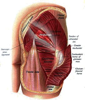
The piriformis muscle is a muscle in the gluteal region of the lower limbs. It is one of the six muscles in the lateral rotator group.

In vertebrate anatomy, hip refers to either an anatomical region or a joint.
The biceps femoris is a muscle of the thigh located to the posterior, or back. As its name implies, it has two parts, one of which forms part of the hamstrings muscle group.

The gracilis muscle is the most superficial muscle on the medial side of the thigh. It is thin and flattened, broad above, narrow and tapering below.

The popliteus muscle in the leg is used for unlocking the knees when walking, by laterally rotating the femur on the tibia during the closed chain portion of the gait cycle. In open chain movements, the popliteus muscle medially rotates the tibia on the femur. It is also used when sitting down and standing up. It is the only muscle in the posterior (back) compartment of the lower leg that acts just on the knee and not on the ankle. The gastrocnemius muscle acts on both joints.

A hip dislocation is when the thighbone (femur) separates from the hip bone (pelvis). Specifically it is when the ball–shaped head of the femur separates from its cup–shaped socket in the hip bone, known as the acetabulum. The joint of the femur and pelvis is very stable, secured by both bony and soft-tissue constraints. With that, dislocation would require significant force which typically results from significant trauma such as from a motor vehicle collision or from a fall from elevation. Hip dislocations can also occur following a hip replacement or from a developmental abnormality known as hip dysplasia.

The iliofemoral ligament is a ligament of the hip joint which extends from the ilium to the femur in front of the joint. It is also referred to as the Y-ligament. the ligament of Bigelow, the ligament of Bertin and any combinations of these names.

The acetabular labrum is a ring of cartilage that surrounds the acetabulum of the hip. The anterior portion is most vulnerable when the labrum tears.

A patellar dislocation is a knee injury in which the patella (kneecap) slips out of its normal position. Often the knee is partly bent, painful and swollen. The patella is also often felt and seen out of place. Complications may include a patella fracture or arthritis.
In medicine, joint locking is a symptom of pathology in a joint. It is a complaint by a person when they are unable to fully flex or fully extend a joint. This term is also used to describe the mechanism of lower limb joints held in full extension without much muscular effort when a person is standing.
Posterolateral corner injuries of the knee are injuries to a complex area formed by the interaction of multiple structures. Injuries to the posterolateral corner can be debilitating to the person and require recognition and treatment to avoid long term consequences. Injuries to the PLC often occur in combination with other ligamentous injuries to the knee; most commonly the anterior cruciate ligament (ACL) and posterior cruciate ligament (PCL). As with any injury, an understanding of the anatomy and functional interactions of the posterolateral corner is important to diagnosing and treating the injury.

The pelvis is either the lower part of the trunk of the human body between the abdomen and the thighs or the skeleton embedded in it.
Medial knee injuries are the most common type of knee injury. The medial ligament complex of the knee is composed of the superficial medial collateral ligament (sMCL), deep medial collateral ligament (dMCL), and the posterior oblique ligament (POL). These ligaments have also been called the medial collateral ligament (MCL), tibial collateral ligament, mid-third capsular ligament, and oblique fibers of the sMCL, respectively. This complex is the major stabilizer of the medial knee. Injuries to the medial side of the knee are most commonly isolated to these ligaments. A thorough understanding of the anatomy and function of the medial knee structures, along with a detailed history and physical exam, are imperative to diagnosing and treating these injuries.

A kick is a skill in association football in which a player strikes the ball with his or her foot. Association football, more commonly referred to as football and also known as soccer, is a sport played world-wide, with up to 265 million people around the world participating on a yearly basis. Kicking is one of the most difficult skills to acquire in football. This skill is also vitally important, as kicking is the way in which passes are made and the primary means by which goals are scored.

