Related Research Articles
In physiology, nociception, also nocioception; from Latin nocere 'to harm/hurt') is the sensory nervous system's process of encoding noxious stimuli. It deals with a series of events and processes required for an organism to receive a painful stimulus, convert it to a molecular signal, and recognize and characterize the signal to trigger an appropriate defensive response.
The sensory nervous system is a part of the nervous system responsible for processing sensory information. A sensory system consists of sensory neurons, neural pathways, and parts of the brain involved in sensory perception and interoception. Commonly recognized sensory systems are those for vision, hearing, touch, taste, smell, balance and visceral sensation. Sense organs are transducers that convert data from the outer physical world to the realm of the mind where people interpret the information, creating their perception of the world around them.

The parietal lobe is one of the four major lobes of the cerebral cortex in the brain of mammals. The parietal lobe is positioned above the temporal lobe and behind the frontal lobe and central sulcus.
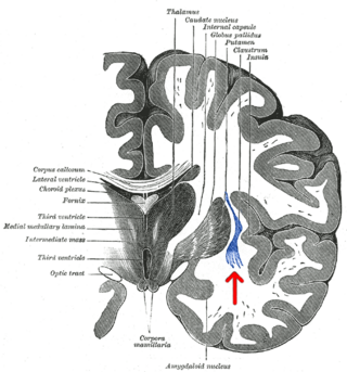
The claustrum is a thin sheet of neurons and supporting glial cells, that connects to the cerebral cortex and subcortical regions including the amygdala, hippocampus and thalamus of the brain. It is located between the insular cortex laterally and the putamen medially, encased by the extreme and external capsules respectively. Blood to the claustrum is supplied by the middle cerebral artery. It is considered to be the most densely connected structure in the brain, and thus hypothesized to allow for the integration of various cortical inputs such as vision, sound and touch, into one experience. Other hypotheses suggest that the claustrum plays a role in salience processing, to direct attention towards the most behaviorally relevant stimuli amongst the background noise. The claustrum is difficult to study given the limited number of individuals with claustral lesions and the poor resolution of neuroimaging.

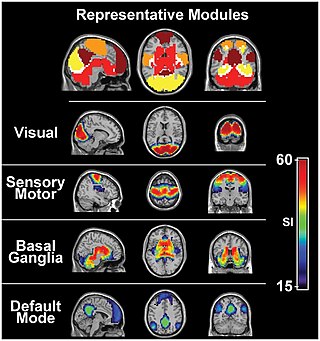
In neuroanatomy, the primary somatosensory cortex is located in the postcentral gyrus of the brain's parietal lobe, and is part of the somatosensory system. It was initially defined from surface stimulation studies of Wilder Penfield, and parallel surface potential studies of Bard, Woolsey, and Marshall. Although initially defined to be roughly the same as Brodmann areas 3, 1 and 2, more recent work by Kaas has suggested that for homogeny with other sensory fields only area 3 should be referred to as "primary somatosensory cortex", as it receives the bulk of the thalamocortical projections from the sensory input fields.
A mirror neuron is a neuron that fires both when an organism acts and when the organism observes the same action performed by another. Thus, the neuron "mirrors" the behavior of the other, as though the observer were itself acting. Mirror neurons are not always physiologically distinct from other types of neurons in the brain; their main differentiating factor is their response patterns. By this definition, such neurons have been directly observed in humans and primate species, and in birds.

The motor cortex is the region of the cerebral cortex involved in the planning, control, and execution of voluntary movements. The motor cortex is an area of the frontal lobe located in the posterior precentral gyrus immediately anterior to the central sulcus.

The insular cortex is a portion of the cerebral cortex folded deep within the lateral sulcus within each hemisphere of the mammalian brain.

The supplementary motor area (SMA) is a part of the motor cortex of primates that contributes to the control of movement. It is located on the midline surface of the hemisphere just in front of the primary motor cortex leg representation. In monkeys the SMA contains a rough map of the body. In humans the body map is not apparent. Neurons in the SMA project directly to the spinal cord and may play a role in the direct control of movement. Possible functions attributed to the SMA include the postural stabilization of the body, the coordination of both sides of the body such as during bimanual action, the control of movements that are internally generated rather than triggered by sensory events, and the control of sequences of movements. All of these proposed functions remain hypotheses. The precise role or roles of the SMA is not yet known.
A topographic map is the ordered projection of a sensory surface, like the retina or the skin, or an effector system, like the musculature, to one or more structures of the central nervous system. Topographic maps can be found in all sensory systems and in many motor systems.
The simulation theory of empathy holds that humans anticipate and make sense of the behavior of others by activating mental processes that, if they culminated in action, would produce similar behavior. This includes intentional behavior as well as the expression of emotions. The theory says that children use their own emotions to predict what others will do; we project our own mental states onto others.
The concept of motor cognition grasps the notion that cognition is embodied in action, and that the motor system participates in what is usually considered as mental processing, including those involved in social interaction. The fundamental unit of the motor cognition paradigm is action, defined as the movements produced to satisfy an intention towards a specific motor goal, or in reaction to a meaningful event in the physical and social environments. Motor cognition takes into account the preparation and production of actions, as well as the processes involved in recognizing, predicting, mimicking, and understanding the behavior of other people. This paradigm has received a great deal of attention and empirical support in recent years from a variety of research domains including embodied cognition, developmental psychology, cognitive neuroscience, and social psychology.

Vittorio Gallese is professor of Psychobiology at the University of Parma, Italy, and was professor in Experimental Aesthetics at the University of London, UK (2016–2018). He is an expert in neurophysiology, cognitive neuroscience, social neuroscience, and philosophy of mind. Gallese is one of the discoverers of mirror neurons. His research attempts to elucidate the functional organization of brain mechanisms underlying social cognition, including action understanding, empathy, language, mindreading and aesthetic experience.

Touch is perceiving the environment using skin. Specialized receptors in the skin send signals to the brain indicating light and soft pressure, hot and cold, body position and pain. It is a subset of the sensory nervous system, which also includes the visual, auditory, olfactory, gustatory and vestibular senses.

Cross modal plasticity is the adaptive reorganization of neurons to integrate the function of two or more sensory systems. Cross modal plasticity is a type of neuroplasticity and often occurs after sensory deprivation due to disease or brain damage. The reorganization of the neural network is greatest following long-term sensory deprivation, such as congenital blindness or pre-lingual deafness. In these instances, cross modal plasticity can strengthen other sensory systems to compensate for the lack of vision or hearing. This strengthening is due to new connections that are formed to brain cortices that no longer receive sensory input.

Christian Keysers is a French and German neuroscientist.
Pain empathy is a specific variety of empathy that involves recognizing and understanding another person's pain.
Neuromorality is an emerging field of neuroscience that studies the connection between morality and neuronal function. Scientists use fMRI and psychological assessment together to investigate the neural basis of moral cognition and behavior. Evidence shows that the central hub of morality is the prefrontal cortex guiding activity to other nodes of the neuromoral network. A spectrum of functional characteristics within this network to give rise to both altruistic and psychopathological behavior. Evidence from the investigation of neuromorality has applications in both clinical neuropsychiatry and forensic neuropsychiatry.
Transcranial random noise stimulation (tRNS) is a non-invasive brain stimulation technique and a form of transcranial electrical stimulation (tES). Terney et al from Göttingen University was the first group to apply tRNS in humans in 2008. They showed that by using an alternate current along with random amplitude and frequency in healthy subjects, the motor cortex excitability increased for up to 60 minutes after 10 minutes of stimulation. The study included all the frequencies up to half of the sampling rate i.e. 640 Hz, however the positive effect was limited only to higher frequencies. Although tRNS has shown positive effects in various studies the optimal parameters, as well as the potential clinical effects of this technique, remain unclear.
Social cognitive neuroscience is the scientific study of the biological processes underpinning social cognition. Specifically, it uses the tools of neuroscience to study "the mental mechanisms that create, frame, regulate, and respond to our experience of the social world". Social cognitive neuroscience uses the epistemological foundations of cognitive neuroscience, and is closely related to social neuroscience. Social cognitive neuroscience employs human neuroimaging, typically using functional magnetic resonance imaging (fMRI). Human brain stimulation techniques such as transcranial magnetic stimulation and transcranial direct-current stimulation are also used. In nonhuman animals, direct electrophysiological recordings and electrical stimulation of single cells and neuronal populations are utilized for investigating lower-level social cognitive processes.
References
- ↑ "Valeria Gazzola - Curriculum Vitae" (PDF). Archived from the original (PDF) on 2022-11-28. Retrieved 2024-01-15.
- 1 2 "Valeria Gazzola elected as member for the Young Academy of Europe". Nederlands Herseninstituut. 2016-06-03. Retrieved 2021-06-06.
- 1 2 "Gazzola". Young Academy of Europe. 2016-12-18. Retrieved 2021-06-06.
- ↑ "University of Groningen, Action in the Brain, Valeria Gazzola" (PDF).
- ↑ Keysers, Christian; Wicker, Bruno; Gazzola, Valeria; Anton, Jean-Luc; Fogassi, Leonardo; Gallese, Vittorio (April 2004). "A Touching Sight". Neuron. 42 (2): 335–346. doi: 10.1016/s0896-6273(04)00156-4 . ISSN 0896-6273. PMID 15091347. S2CID 1414735.
- ↑ "Investigating causality within the Mirror Neuron System using a combination of repetitive transcranial magnetic stimulation and functional magnetic resonance imaging | NWO". www.nwo.nl (in Dutch). March 2010. Retrieved 2021-06-05.
- ↑ Keysers, Christian; Kaas, Jon H.; Gazzola, Valeria (June 2010). "Somatosensation in social perception". Nature Reviews Neuroscience. 11 (6): 417–428. doi:10.1038/nrn2833. ISSN 1471-0048. PMID 20445542. S2CID 12221575.
- ↑ Gazzola, V.; Keysers, C. (2009-06-01). "The Observation and Execution of Actions Share Motor and Somatosensory Voxels in all Tested Subjects: Single-Subject Analyses of Unsmoothed fMRI Data". Cerebral Cortex. 19 (6): 1239–1255. doi:10.1093/cercor/bhn181. ISSN 1047-3211. PMC 2677653 . PMID 19020203.
- 1 2 Meffert, Harma; Gazzola, Valeria; den Boer, Johan A.; Bartels, Arnold A. J.; Keysers, Christian (August 2013). "Reduced spontaneous but relatively normal deliberate vicarious representations in psychopathy". Brain. 136 (8): 2550–2562. doi:10.1093/brain/awt190. ISSN 1460-2156. PMC 3722356 . PMID 23884812.
- 1 2 Valchev, Nikola; Tidoni, Emmanuele; Hamilton, Antonia F. de C.; Gazzola, Valeria; Avenanti, Alessio (May 2017). "Primary somatosensory cortex necessary for the perception of weight from other people's action: A continuous theta-burst TMS experiment". NeuroImage. 152: 195–206. doi:10.1016/j.neuroimage.2017.02.075. PMC 5440175 . PMID 28254507.
- 1 2 V. Gallo; Selene Paracampo; Riccardo Muller-Pinzler; L. Severo; Mario Carlo Blömer; Laila Fernandes-Henriques; Carolina Henschel; Anna Lammes; Balint Kalista Maskaljunas; Tatjana Suttrup; J. Avenanti; Alessio Keysers; Christian Gazzola (2018). "The causal role of the somatosensory cortex in prosocial behaviour". eLife. 7. doi: 10.7554/eLife.32740 . OCLC 1038719949. PMC 5973831 . PMID 29735015.
- ↑ Keysers, Christian; Gazzola, Valeria (April 2014). "Dissociating the ability and propensity for empathy". Trends in Cognitive Sciences. 18 (4): 163–166. doi:10.1016/j.tics.2013.12.011. PMC 4560165 . PMID 24484764.
- ↑ "Do shared circuits really help? Empowering transcranial direct current stimulation to reveal the causal link between emotion sharing and helping | NWO". www.nwo.nl (in Dutch). October 2015. Retrieved 2021-06-05.
- ↑ "Vidi grant for twenty UvA and AMC-UvA researchers". University of Amsterdam. 2015-05-18. Retrieved 2021-06-06.
- ↑ Carrillo, Maria; Han, Yinging; Migliorati, Filippo; Liu, Ming; Gazzola, Valeria; Keysers, Christian (April 2019). "Emotional Mirror Neurons in the Rat's Anterior Cingulate Cortex". Current Biology. 29 (8): 1301–1312.e6. doi:10.1016/j.cub.2019.03.024. ISSN 0960-9822. PMC 6488290 . PMID 30982647.
- ↑ Hernandez-Lallement, Julen; Attah, Augustine Triumph; Soyman, Efe; Pinhal, Cindy M.; Gazzola, Valeria; Keysers, Christian (March 2020). "Harm to Others Acts as a Negative Reinforcer in Rats". Current Biology. 30 (6): 949–961.e7. doi: 10.1016/j.cub.2020.01.017 . hdl: 20.500.11755/ee7ae8ac-7393-4276-84ce-1bad1b8e5e0d . ISSN 0960-9822. PMID 32142701. S2CID 212424287.
- ↑ Abdelgabar, Abdel R; Suttrup, Judith; Broersen, Robin; Bhandari, Ritu; Picard, Samuel; Keysers, Christian; De Zeeuw, Chris I; Gazzola, Valeria (2019-11-21). "Action perception recruits the cerebellum and is impaired in patients with spinocerebellar ataxia". Brain. 142 (12): 3791–3805. doi:10.1093/brain/awz337. ISSN 0006-8950. PMC 7409410 . PMID 31747689.
- ↑ Valchev, Nikola; Gazzola, Valeria; Avenanti, Alessio; Keysers, Christian (2016-03-15). "Primary somatosensory contribution to action observation brain activity—combining fMRI and cTBS". Social Cognitive and Affective Neuroscience. 11 (8): 1205–1217. doi:10.1093/scan/nsw029. ISSN 1749-5024. PMC 4967793 . PMID 26979966.
- ↑ "CUBE – Understanding the Brain with Ultrasound" . Retrieved 2021-06-06.
- 1 2 3 4 5 6 Google Scholar Author page, Accessed Dec. 6, 2021