Feline arterial thromboembolism is a domestic cat disease
Bilateral hindlimb paralysis in a cat with arterial thromboembolism
Feline arterial thromboembolism (FATE syndrome) (German: Feline arterielle Thromboembolie) is a disease of the domestic cat in which blood clots (thrombi) block arteries, causing severe circulatory problems. Relative to the total number of feline patients, the disease is rare, but relatively common in cats with heart disease: about one-sixth of cats with heart disease are affected. Heart disease is the most common underlying cause of arterial thromboembolism. It leads to the formation of blood clots in the heart, which leave it with the bloodstream and obstruct larger blood vessels, in cats mainly the aorta at the outlet of the two external iliac arteries. Arterial thromboembolism occurs suddenly and is very painful. The blockage of the terminal portion of the aorta results in an undersupply of blood to the hind legs. The result is paralysis, cold hind extremities and later severe tissue damage. Rarely, other blood vessels are also affected; the symptoms of failure then depend on the supply area of the affected artery. Since drug thrombolysis in cats does not achieve satisfactory results, the focus today is on the self-dissolution of the clot by the body's own repair processes. Accompanying pain therapy and thrombosis prevention are performed and the underlying disease is treated. The mortality of arterial thromboembolism in cats is very high. Fifty to 60% of affected animals are euthanized without attempted treatment, and only one-quarter to one-third of animals survive such an event. In about half of the recovered cats, thromboembolism recurs despite anticoagulation prophylaxis.
Feline arterial thromboembolism is a rare disease, accounting for approximately 0.1-0.3% of the total number of feline patients.[1][2] The median age at onset of thromboembolism is 12 years (1 to 21 years).[2]
Most common causes and their proportion in cats with arterial thromboembolism (after Smith et al. 2003).
Underlying disease
Frequency
hypertrophic cardiomyopathy
52%
other cardiomyopathy
17%
Hyperthyroidism
9%
Tumor
5%
FATE syndrome develops in approximately 70% of cases as a result of heart disease, most commonly heart disease with cardiac wall thickening (Hypertrophic Cardiomyopathy, HCM). Up to 17% of cats with HCM experience arterial thromboembolism, but cats with other cardiomyopathies are also at increased risk. Cats with abnormally increased hemostasis, which can occur with hyperthyroidism, tumors, extensive inflammation, blood poisoning (sepsis), injury, or disseminated intravascular coagulation, represent another risk group.[3] There is an increased genetic predisposition in male cats, which is related to the higher incidence of heart disease in male cats.[4][2]
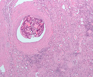
Thrombus in the terminal branch of the aorta in a cat. 1 opened aorta with thrombus, 2 external iliac arteries, 3 common trunk of both internal iliac arteries, 4 circumflex ilium profunda, 5 mesenteric caudal artery, 6 descending colon. circumflexa ilium profunda, 5 A. mesenterica caudalis, 6 Colon descendens
Damage to the endocardium and slowing of blood flow in the enlarged left atrium and atrial auricle are primarily responsible for the formation of blood clots (thrombi). Tissue damage leads to the release of tissue factor and hemostasis.[5] The intact glycocalyx of the endothelial cells of the inner lining of the heart normally reduces contact with blood cells and macromolecules. If endothelial cell injury occurs, reactive oxygen species (ROS), nitric oxide (NO), matrix metalloproteinase, and proinflammatory cytokines are produced in increased amounts and cell adhesion molecules are upregulated. Endothelial cell damage exposes the underlying extracellular matrix, to which platelets attach and form a clot.[6] The clot consists of platelets interconnected by the clotting protein fibrin. As the clot matures, the fibrin content increases and the clot may exhibit stratification.[7] Even in healthy animals, injuries to the endothelium occur spontaneously from time to time, but there is a balance between thrombus formation and breakdown. Substances such as antithrombin III, thrombomodulin, tissue-type plasminogen activator and urokinase dissolve formed blood clots and prostacyclin and nitric oxide inhibit platelet aggregation.[3] Conservative treatment of arterial thromboembolism in cats is also based on this endogenous dissolution of the clot (see below).
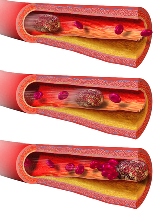
In cats, the blood clots originate mainly in the left atrial auricle.[8] They or parts of them are carried along with the blood flow, enter the aorta via the left ventricle, get stuck at vascular outlets and block them. This condition is called thromboembolism. In cats, this occurs predominantly in the aorta in the area of its terminal branch, i.e., at the outlet of the two external iliac arteries (Aa. iliacae externae). This is also called "saddle thrombus" or "riding thrombus". This results in an ischemia to the rear extremities. In addition, platelets release thromboxane and serotonin, which leads to vasoconstriction and thus to reduced blood flow even to blood vessels that are not directly affected. Serotonin also stimulates nociceptor, which contributes to the high painfulness of the disease.[9] Only in 10% of cases are other blood vessels affected, for example the brachial artery, pulmonary arteries, cerebral circulation, intestinal vessels or coronary arteries.[10][11]
In humans, heart disease (especially atrial fibrillation), increased blood clotting, and atherosclerosis are the most common underlying diseases for the development of arterial thromboembolism. Again, thrombi develop primarily in the left side of the heart. Most commonly, cerebral arteries (cerebral infarction) and arteries of the leg (limb infarction, acute lower limb ischemia) are displaced. Less frequently, thromboembolism of the vessels of the arm, upper mesenteric artery or renal arteries (renal infarction) occurs.[12] In contrast, aortoiliac occlusive disease (aortic bifurcation syndrome), which corresponds to the most common localization in cats, is extremely rare in humans.[13] In domestic dogs, arterial thromboembolism occurs much less frequently than in cats; common underlying diseases in dogs are protein-losing nephropathy, diseases of the immune system, tumors, sepsis, heart disease, protein-losing enteropathy, and hypertension.[14][15] Aortic thrombosis does occasionally occur in dogs, but here the thrombi arise directly at the aortic branch; as a thromboembolic event, as in cats, they are extremely rare.[16] There are also isolated case reports of thromboembolism in the domestic horse,[17] whereas in other species they are of no practical significance. In laboratory animals used in human medical stroke research, thrombi are artificially generated.
Symptoms, clinical diagnosis and laboratory findings
The disease occurs suddenly (peracute) and is accompanied by severe pain. Affected cats meow ("vocalize") and often have hypothermia. The extent of further signs of the disease depends on the location of the clot and whether the vessel is completely or only partially blocked. Occlusion of the iliac arteries results in partial (paresis) or complete paralysis (plegia) of the hind extremities. In most cases, both hind legs are affected.[18] The muscles are hardened and painful after about 10 hours, especially the lower leg muscles.[10] The pulse at the femoral artery (Arteria femoralis) is markedly decreased or absent in 78% of cases. The paws are cold and especially the area of the claws and pads often show bluish discoloration (cyanosis) or are strikingly pale. The reflexes of the hindlimb (patellar reflex, tibialis cranialis reflex, and flexor reflex) are severely reduced or absent. Increased respiratory rate, dyspnea, and syncope are common. Loss of perception may also occur.[19] The main symptoms can be summarized in the "5-P rule" - paresis (Lähmung), pallor (Blässe), pain (Schmerz), pulselessness (Pulsverlust), poikilothermia (Untertemperatur). The tail muscles, anal reflex, and bladder function are mostly unaffected.[7][9]
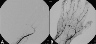
Other occlusions are much less common and the clinical presentation depends on the body part or organ affected. Occlusion of the brachial artery occurs predominantly on the right side and causes sudden paralysis of the forelimb.[20] Thromboembolism of the pulmonary circulation is manifested by increased respiratory frequency and shortness of breath.[21] The clinical picture of occlusion of cerebral circulation (cerebral infarction) depends strongly on the vessel affected and thus on the area of the brain damaged. In most cases, there are unilateral neurological deficits.[22] Occlusion of a coronary artery (myocardial infarction) leads to cardiac arrhythmias, usually with a fatal outcome, and is therefore often no longer presented to a veterinarian at all, so that its frequency is possibly underestimated. Occlusion of renal or intestinal vessels causes severe abdominal pain (acute abdomen) and often also leads quickly to death.[23] There are also case reports of simultaneous occlusion of several vessels with paralysis of all limbs[24] or of cerebellum and kidneys with severe balance disorders.[25]
Large thrombus in the left atrium of a cat, echocardiography
Listening to the heart (auscultation) usually reveals heart murmurs, an irregular heartbeat, palpitations, extrasystoles and a "gallop rhythm" - a sequence of heart sounds reminiscent of a galloping horse. Up to two-thirds of FATE patients are in congestive heart failure,[9] in which the heart no longer pumps enough blood to the body. Atrial fibrillation detectable by ECG is an additional risk factor. Aortic thrombus can often be visualized directly by sonography,[26] and angiography or electromyography may also be performed if necessary. Echocardiography can be used to visualize thrombi and their precursors in the heart and to assess the functional status of the heart. Loss of pulse at the femoral artery can also be detected by doppler sonography, although the pulse is still detectable sonographically if the vessel is incompletely occluded.[27]Infrared thermography can be used to objectify temperature differences between the forelimbs and hindlimbs. The sensitivity of this method is between 80 and 90%, the specificity is 100%.[28] A thromboembolism of the lung often remains undetected; in this case, a chest radiograph can provide initial indications, and a definite diagnosis can be made by means of CT scan[21] or scintigraphy of the lung.[29] If a stroke is suspected, magnetic resonance imaging is indicated.[22]
The activities of the enzymescreatine kinase (CK) and aspartate aminotransferase (AST) are elevated due to the death of muscle cells in the blood.[9] If cardiac disease is present, which is often the case, brain natriuretic peptide is above the reference range.[30] The "kidney values" (creatinine, urea, SDMA) may also be elevated due to the shock-induced reduced renal function (prerenal azotemia). However, all laboratory values are not specific for arterial thromboembolism and play only a minor role in confirming the diagnosis. Determination of blood glucose or lactate concentration in the body compared with that in the paralyzed limb may be helpful.[31] Determination of thyroxine (T4) concentration in the blood is useful for detecting hyperthyroidism; hyperthyroidism was not previously known in 1.7% of cats with thromboembolism.[2]
Diagnosis and differential diagnosis
In most cases, the diagnosis can already be made in the most common location (aortic thrombosis) based on the previous report and clinical signs (peracute posthand paralysis without trauma).[2][32] Existing cardiac disease provides further clues, but cardiac disease is already known in only about 15% of cats with thromboembolism.[33]
The other more common ischemic myopathy, syndrome of Kippfenster, can usually be ruled out by questioning the animal's owner. In addition, tilt window syndrome is not associated with severe pain. Differential diagnosis should continue to exclude primarily trauma to the spinal cord (traffic accident, window fall), which may be due to an event not observed by the owner. A herniated disc or a spinal cord infarction can also lead to sudden signs of paralysis. Tumors in the spinal cord or spinal canal can also cause afterhand paralysis, but these usually develop slowly and signs of loss occur gradually.[34]
The diagnosis of vascular occlusions of the internal organs is more difficult; here, special examinations (CT, MRI) are required to confirm the diagnosis, which are only available in larger facilities.
Therapy
Treatment of arterial thromboembolism in cats consists of pain management, prevention of further clot formation, and treatment of heart failure, if necessary. Intensive medical care is usually required for three days before treatment can be continued at home.[35]
To reduce pain, administration of potent analgesics is indicated, with opioid analgesics such as levomethadone or fentanyl being the most effective.[32] However, both agents are not approved for use in cats in the EU and must therefore be redirected in the sense of a therapeutic emergency. In addition, fentanyl is only effective for about 30 minutes, while levomethadone is effective for about 5 hours, based on data for dogs,[36] which limits further treatment at home. Continuous drip infusion with the combination of fentanyl and lidocaine has been described. Lidocaine, in addition to its analgesic effect, also protects to some extent against damage caused by reopening of the occluded vessel (reperfusion injury). However, the therapeutic range of lidocaine in cats is very narrow; as little as 6 mg/kg can be lethal.[37] The only opioid analgesic approved for cats, buprenorphine, does not have sufficient analgesic effect for initial treatment, at least not when there is total occlusion of both external iliac arteries. It can be used for continuing treatment at home, especially since it can be easily administered through the oral mucosa and has a duration of action of about 8 hours. Non-opioid analgesics do not provide adequate pain relief and may increase circulatory disturbances in animals, causing renal or gastrointestinal damage. Only metamizole is suitable for subsequent further treatment.[38]
Structural formula of clopidogrel
Inhibiting the formation of further blood clots, thrombosis prophylaxis, is the second important pillar of FATE treatment. It should be carried out as early as possible.[32] For this purpose, agents to inhibit hemostasis such as low-molecular-weight heparins are used first, along with agents to prevent platelet aggregation (thrombocyte aggregation) such as aspirin and clopidogrel.[39] For long-term prophylaxis, clopidogrel is preferably administered because it significantly prolongs survival compared with aspirin.[33][40] The use of the active ingredient rivaroxaban as another effective drug is also being discussed.[41] A 2021 study was able to show that the combination of clopidogrel and rivaroxaban is an effective recurrence prophylaxis.[42]
Cats in congestive heart failure are given supplemental oxygen to compensate for the hypoxia. High doses of furosemide are used to reduce preload and afterload and thus unload the heart.[32] In advanced heart disease with ventricular dilatation (DCM) or heart disease with cardiac wall thickening (DCM), pumping efficiency can be improved with pimobendan, possibly also with dobutamine.[43] Pimobendan also increases blood flow in the left atrium and heart ear to a small extent and additionally improves atrial function.[8] On the other hand, if there is no congestive heart failure but reduced blood flow (perfusion), then intravenous therapy are infused.[43] In the case of underlying hyperthyroidism, thyrostatic drugs such as thiamazole or carbimazole are administered.[44]
The benefit of external heat application in cats with hypothermia is controversial. Often the anterior part of the body has a normal temperature and the undertemperature only affects the posterior part and thus also the rectum, where body temperature is normally measured in cats. Measurement in the axillary region or in the ear is unreliable.[45] However, comparison between axillary and rectal temperature at least provides clues to distinguish between local and general undertemperature. In the case of the latter, an application of heat is indicated.[46]
The obvious treatment, reopening of the vessel by drug dissolution (thrombolysis) or invasive removal of the clot (thrombectomy), as long established in human medicine for occlusive diseases such as cerebral infarction or myocardial infarction, provides unsatisfactory results in cats and is therefore no longer recommended.[32] Thrombolysis with streptokinase, urokinase or tissue-type plasminogen activator has not improved treatment success in various studies. This usually results in frequently fatal reperfusion injury, hyperkalemia, metabolic acidosis, kidney failure, and bleeding, so that the survival rate is often lower than with conservative treatment.[47][48] In human medicine, such treatments are only performed in highly specialized facilities (cardiac centers, stroke units) with a high level of personnel and equipment. Surgical removal of the thrombus is also rarely performed in veterinary medicine because of the associated risks, although it can be successful in individual cases.[49] It is associated with the same complications as thrombolysis and is therefore no longer recommended. Therefore, the current focus is on endogenous dissolution of the clot and thus spontaneous revascularization, which occurs quickly enough in just under 40% of cases.[47]
Prognosis and prevention
Spontaneous echocardiographic contrast ('smoke') - a precursor of thrombus formation - in the left atrium of a cat
The treatment outlook (prognosis) for aortic thromboembolism is uncertain to poor. According to a U.S. study, only about one-third of cats survive arterial thromboembolism, with half of those who die being euthanized without attempted treatment.[50] In a U.K. study, about 60% of patients were euthanized. Only 27% survived the first 24 hours. The median survival time was 94 days, and after one year only 2% of the animals were still alive.[2]
Prognosis depends largely on the extent and duration of damage, with bilateral complete occlusions of the iliac arteries having the lowest chance of survival. If only one limb is affected and there is residual motor function, there is a better chance that the cat will recover and continue to live with a good quality of life. If the internal body temperature is above 37.2 °C - the normal temperature in domestic cats is about 39 °C - the prospect of treatment is better than if the temperature is more severely below normal. Excess blood potassium (hyperkalemia) and elevated kidney enzymes (azotemia) are other negative prognostic factors. Even after spontaneous reopening of the blood vessel (revaskularization), relapses often occur due to a new thromboembolism, which even thromboprophylaxis cannot reliably prevent.[2][39][47] In half of the patients, a new thromboembolism occurs despite treatment with clopidogrel.[7] In addition, the extent of the heart disease, in particular the extent of atrial enlargement and the pumping capacity of the left ventricle, determines the further survival of the patient.[51]
In contrast, the prognosis is favorable for occlusion of smaller cerebral arteries. There is often a reduction in the signs of failure within two to three weeks, as other areas of the brain take over the function of the infarcted area.[52] Occlusion of the brachial artery also has a good chance of recovery.[11] The prognosis and mortality of pulmonary thromboembolism is not known, as it is very rare.[53] Anecdotal reports indicate that cats can survive such an event and lung function can return to normal with formation of collaterals.[21] Other occlusions (intestinal, renal, and coronary arteries) are very often fatal.[11][23]
Some small-animalcardiologists recommend coagulation prophylaxis already in the presence of certain cardiac changes, i.e., before the occurrence of thromboembolism. For example, one study showed that a flow velocity in the left heart ear of less than 0.2 m/s was associated with the occurrence of thrombi and spontaneous echocardiographic contrast (smoke). Spontaneous echocardiographic contrast is an aggregation of red blood cells and thus a thrombus precursor that resembles smoke on sonographic imaging.[54] However, prospective studies demonstrating the efficacy of such treatment have yet to be performed.[7]
Related Research Articles
An embolism is the lodging of an embolus, a blockage-causing piece of material, inside a blood vessel. The embolus may be a blood clot (thrombus), a fat globule, a bubble of air or other gas, amniotic fluid, or foreign material.
Thrombosis is the formation of a blood clot inside a blood vessel, obstructing the flow of blood through the circulatory system. When a blood vessel is injured, the body uses platelets (thrombocytes) and fibrin to form a blood clot to prevent blood loss. Even when a blood vessel is not injured, blood clots may form in the body under certain conditions. A clot, or a piece of the clot, that breaks free and begins to travel around the body is known as an embolus.
An embolus is an unattached mass that travels through the bloodstream and is capable of creating blockages. When an embolus occludes a blood vessel, it is called an embolism or embolic event. There are a number of different types of emboli, including blood clots, cholesterol plaque or crystals, fat globules, gas bubbles, and foreign bodies, which can result in different types of embolisms.
A thrombus, colloquially called a blood clot, is the final product of the blood coagulation step in hemostasis. There are two components to a thrombus: aggregated platelets and red blood cells that form a plug, and a mesh of cross-linked fibrin protein. The substance making up a thrombus is sometimes called cruor. A thrombus is a healthy response to injury intended to stop and prevent further bleeding, but can be harmful in thrombosis, when a clot obstructs blood flow through healthy blood vessels in the circulatory system.
Venous thrombosis is the blockage of a vein caused by a thrombus. A common form of venous thrombosis is deep vein thrombosis (DVT), when a blood clot forms in the deep veins. If a thrombus breaks off (embolizes) and flows to the lungs to lodge there, it becomes a pulmonary embolism (PE), a blood clot in the lungs. The conditions of DVT only, DVT with PE, and PE only, are all captured by the term venous thromboembolism (VTE).
Ischemia or ischaemia is a restriction in blood supply to any tissue, muscle group, or organ of the body, causing a shortage of oxygen that is needed for cellular metabolism. Ischemia is generally caused by problems with blood vessels, with resultant damage to or dysfunction of tissue i.e. hypoxia and microvascular dysfunction. It also implies local hypoxia in a part of a body resulting from constriction.
Thrombolysis, also called fibrinolytic therapy, is the breakdown (lysis) of blood clots formed in blood vessels, using medication. It is used in ST elevation myocardial infarction, stroke, and in cases of severe venous thromboembolism.
Vascular surgery is a surgical subspecialty in which vascular diseases involving the arteries, veins, or lymphatic vessels, are managed by medical therapy, minimally-invasive catheter procedures and surgical reconstruction. The specialty evolved from general and cardiovascular surgery where it refined the management of just the vessels, no longer treating the heart or other organs. Modern vascular surgery includes open surgery techniques, endovascular techniques and medical management of vascular diseases - unlike the parent specialities. The vascular surgeon is trained in the diagnosis and management of diseases affecting all parts of the vascular system excluding the coronaries and intracranial vasculature. Vascular surgeons also are called to assist other physicians to carry out surgery near vessels, or to salvage vascular injuries that include hemorrhage control, dissection, occlusion or simply for safe exposure of vascular structures.
Infarction is tissue death (necrosis) due to inadequate blood supply to the affected area. It may be caused by artery blockages, rupture, mechanical compression, or vasoconstriction. The resulting lesion is referred to as an infarct (from the Latin infarctus, "stuffed into").
Hypertrophic cardiomyopathy is a condition in which muscle tissues of the heart become thickened without an obvious cause. The parts of the heart most commonly affected are the interventricular septum and the ventricles. This results in the heart being less able to pump blood effectively and also may cause electrical conduction problems. Specifically, within the bundle branches that conduct impulses through the interventricular septum and into the Purkinje fibers, as these are responsible for the depolarization of contractile cells of both ventricles.
An embolus, is described as a free-floating mass, located inside blood vessels that can travel from one site in the blood stream to another. An embolus can be made up of solid, liquid, or gas. Once these masses get "stuck" in a different blood vessel, it is then known as an "embolism." An embolism can cause ischemia—damage to an organ from lack of oxygen. A paradoxical embolism is a specific type of embolism in which the embolus travels from the right side of the heart to the left side of the heart and lodges itself in a blood vessel known as an artery. Thus, it is termed "paradoxical" because the embolus lands in an artery, rather than a vein.
A watershed stroke is defined as a brain ischemia that is localized to the vulnerable border zones between the tissues supplied by the anterior, posterior and middle cerebral arteries. The actual blood stream blockage/restriction site can be located far away from the infarcts. Watershed locations are those border-zone regions in the brain supplied by the major cerebral arteries where blood supply is decreased. Watershed strokes are a concern because they comprise approximately 10% of all ischemic stroke cases. The watershed zones themselves are particularly susceptible to infarction from global ischemia as the distal nature of the vasculature predisposes these areas to be most sensitive to profound hypoperfusion.
Ocular ischemic syndrome is the constellation of ocular signs and symptoms secondary to severe, chronic arterial hypoperfusion to the eye. Amaurosis fugax is a form of acute vision loss caused by reduced blood flow to the eye; it may be a warning sign of an impending stroke, as both stroke and retinal artery occlusion can be caused by thromboembolism due to atherosclerosis elsewhere in the body. Consequently, those with transient blurring of vision are advised to urgently seek medical attention for a thorough evaluation of the carotid artery. Anterior segment ischemic syndrome is a similar ischemic condition of anterior segment usually seen in post-surgical cases. Retinal artery occlusion leads to rapid death of retinal cells, thereby resulting in severe loss of vision.
Embolectomy is the emergency interventional or surgical removal of emboli which are blocking blood circulation. It usually involves removal of thrombi, and is then referred to as thromboembolectomy or thrombectomy. Embolectomy is an emergency procedure often as the last resort because permanent occlusion of a significant blood flow to an organ leads to necrosis. Other involved therapeutic options are anticoagulation and thrombolysis.
Central aortic blood pressure is the blood pressure at the root of aorta. Studies have shown the importance of central aortic pressure, especially as compared to peripheral blood pressure, and its implications in assessing the efficacy of antihypertensive treatment with respect to cardiovascular risk factors, kidney disease, and mortality. There is an emerging movement for clinicians to begin using central aortic blood pressure, instead of peripheral blood pressure, as a guide for clinical decisions.
Arterial embolism is a sudden interruption of blood flow to an organ or body part due to an embolus adhering to the wall of an artery blocking the flow of blood, the major type of embolus being a blood clot (thromboembolism). Sometimes, pulmonary embolism is classified as arterial embolism as well, in the sense that the clot follows the pulmonary artery carrying deoxygenated blood away from the heart. However, pulmonary embolism is generally classified as a form of venous embolism, because the embolus forms in veins. Arterial embolism is the major cause of infarction.
A limb infarction is an area of tissue death of an arm or leg. It may cause skeletal muscle infarction, avascular necrosis of bones, or necrosis of a part of or an entire limb.
Blood vessel disorder generally refers to the narrowing, hardening or enlargement of arteries and veins. It is often due to the build-up of fatty deposits in the lumen of blood vessels or infection of the vessel wall. This can occur in various locations such as coronary blood vessels, peripheral arteries and veins. The narrowed arteries would block the blood supply to different organs and tissues. In severe conditions, it may develop into more critical health problems like myocardial infarction, stroke or heart failure, which are some of the major reasons of death.
Arterial occlusion is a condition involving partial or complete blockage of blood flow through an artery. Arteries are blood vessels that carry oxygenated blood to body tissues. An occlusion of arteries disrupts oxygen and blood supply to tissues, leading to ischemia. Depending on the extent of ischemia, symptoms of arterial occlusion range from simple soreness and pain that can be relieved with rest, to a lack of sensation or paralysis that could require amputation.
Feline hyperthyroidism is a disorder of the endocrine system in domestic cats. It is characterized by hyperthyroidism and is the most common hormonal disorder (endocrinopathy) in cats over ten years of age. In contrast, hyperthyroidism is much less common in other pets. The disease often manifests itself as weight loss despite increased food intake, is usually detected by blood tests, and is easily treatable.
References
↑ Hogan, Daniel F.; Brainard, Benjamin M. (2015). "Cardiogenic embolism in the cat". Journal of Veterinary Cardiology. 17: S202–S214. doi:10.1016/j.jvc.2015.10.006. PMID26776579.
1 2 Sänger, Florian; Dörfelt, Rene (2020). "Feline arterielle Thromboembolie – Aktueller Stand der Diagnostik und Therapie". Kleintierpraxis. 65 (4). doi:10.2377/0023-2076-65-220.
↑ Smith, Stephanie A. (2003). "Arterial thromboembolism in cats: acute crisis in 127 cases (1992–2001) and long-term management with low-dose aspirin in 24 cases". J Vet Intern Med. 17 (1): 73–83. doi:10.1111/j.1939-1676.2003.tb01326.x. PMID12564730.
↑ Sänger, Florian; Dörfelt, Rene (2020). "Feline arterielle Thromboembolie – Aktueller Stand der Diagnostik und Therapie". Kleintierpraxis. 65 (4): 222. doi:10.2377/0023-2076-65-220.
↑ Winter, R. L. (2012). "Aortic thrombosis in dogs: presentation, therapy, and outcome in 26 cases". Journal of Veterinary Cardiology. 14 (2): 333–342. doi:10.1016/j.jvc.2012.02.008. PMID22591640.
↑ Ross, M. W. (1997). "First-pass radionuclide angiography in the diagnosis of aortoiliac thromboembolism in a horse". Veterinary Radiology & Ultrasound. 38 (3): 226–230. doi:10.1111/j.1740-8261.1997.tb00845.x. PMID9238795.
↑ Jaggy, Andre (2005). Atlas und Lehrbuch der Kleintierneurologie. Schlütersche. p.272. ISBN3-87706-739-5.
↑ Wendy A. Ware (2008), Richard W. Nelson und C. Guillermo Couto (ed.), "Systemic arterial thromboembolism in cats", Small Animal Internal Medicine (4ed.), Elsevier Health Sciences, pp.194–200, ISBN978-0-323-06512-2
1 2 Garosi, L. S. (2010). "Cerebrovascular disease in dogs and cats". The Veterinary Clinics of North America. Small Animal Practice. 40 (1): 65–79. doi:10.1016/j.cvsm.2009.09.001. PMID19942057.
1 2 Galloway, Pru (2001). "Feline Aortic Thromboembolism". Proc of the Companion Animal Society of the NZVA. 214.
↑ Bowles, D. B. (2010). "Cardiogenic arterial thromboembolism causing non-ambulatory tetraparesis in a cat". Journal of Feline Medicine and Surgery. 12 (2): 144–150. doi:10.1016/j.jfms.2009.06.004. PMID19692276. S2CID37119533.
↑ Alan Kovacevic (2017), Nadja Siegrist (ed.), "Kardiologische Notfälle", Notfallmedizin für Hund und Katze, Stuttgart: Enke, pp.231–255, ISBN978-3-13-205281-9
↑ Smith, S. A.; Tobias, A. H. (2004). "Smith Feline arterial thromboembolism: an update". The Veterinary Clinics of North America. Small Animal Practice. 34 (5): 1245–1271. doi:10.1016/j.cvsm.2004.05.006. PMID15325481.
↑ Pouzot-Nevoret, C. (2018). "Infrared thermography: a rapid and accurate technique to detect feline aortic thromboembolism". Journal of Feline Medicine and Surgery. 20 (8): 780–785. doi:10.1177/1098612X17732485. PMID28948905. S2CID9199383.
↑ Pouchelon, J. L. (1997). "Diagnosis of pulmonary thromboembolism in a cat using echocardiography and pulmonary scintigraphy". The Journal of Small Animal Practice. 38 (7): 306–310. doi:10.1111/j.1748-5827.1997.tb03472.x. PMID9239634.
1 2 Hogan, Daniel F. (2015). "Secondary prevention of cardiogenic arterial thromboembolism in the cat: The double-blind, randomized, positive-controlled feline arterial thromboembolism; clopidogrel vs. aspirin trial (FAT CAT)". J Vet Cardiol. 17: 306–317. doi:10.1016/j.jvc.2015.10.004. PMID26776588.
↑ Jan-Gerd Kresken; Ralph T. Wendt; Peter Modler, eds. (2019), "Feline Arterielle Thromboembolie", Praxis der Kardiologie Hund und Katze, Stuttgart: Thieme, pp.389–392, ISBN978-3-13-242994-9
↑ Sänger, Florian; Dörfelt, Rene (2020). "Feline arterielle Thromboembolie – Aktueller Stand der Diagnostik und Therapie". Kleintierpraxis. 65 (4): 232. doi:10.2377/0023-2076-65-220.
↑ Wolfgang Löscher (2006), Wolfgang Löscher, Fritz Rupert Ungemach und Reinhard Kroker (ed.), "Vollsynthetische Morphinabkömmlinge", Pharmakotherapie bei Haus- und Nutztieren (7ed.), Berlin: Paul Parey, pp.95–96, ISBN978-3-8304-4160-1
1 2 Sänger, Florian; Dörfelt, Rene (2020). "Feline arterielle Thromboembolie – Aktueller Stand der Diagnostik und Therapie". Kleintierpraxis. 65 (4): 229. doi:10.2377/0023-2076-65-220.
↑ Peterson, M. E.; Aucoin, D. P. (1993). "Comparison of disposition of carbimazole and methimazole in clinically normal cats". Res. Vet. Sci. 54 (3): 351–355. doi:10.1016/0034-5288(93)90134-2. PMID8337482.
↑ Sänger, Florian; Dörfelt, Rene (2020). "Feline arterielle Thromboembolie – Aktueller Stand der Diagnostik und Therapie". Kleintierpraxis. 65 (4): 230. doi:10.2377/0023-2076-65-220.
1 2 3 Reimer, S. B. (2006). "Use of rheolytic thrombectomy in the treatment of feline distal aortic thromboembolism". Journal of Veterinary Internal Medicine. 20 (2): 290–296. doi:10.1111/j.1939-1676.2006.tb02859.x. PMID16594585.
↑ Guillaumin, J. (2019). "Thrombolysis with tissue plasminogen activator (TPA) in feline acute aortic thromboembolism: a retrospective study of 16 cases". Journal of Feline Medicine and Surgery. 21 (4): 340–346. doi:10.1177/1098612X18778157. PMID29807505. S2CID44135779.
↑ Hogan, D. F. (2017). "Feline Cardiogenic Arterial Thromboembolism: Prevention and Therapy". The Veterinary Clinics of North America. Small Animal Practice. 47 (5): 1065–1082. doi:10.1016/j.cvsm.2017.05.001. PMID28662872.
↑ Boudreau, C. E. (2018). "An Update on Cerebrovascular Disease in Dogs and Cats". The Veterinary Clinics of North America. Small Animal Practice. 48 (1): 45–62. doi:10.1016/j.cvsm.2017.08.009. PMID29056397.
↑ Schober, Karsten E.; Maerz, Imke (2006). "Assessment of left atrial appendage flow velocity and its relation to spontaneous echocardiographic contrast in 89 cats with myocardial disease". J Vet Intern Med. 20 (1): 120–130. doi:10.1892/0891-6640(2006)20[120:aolaaf]2.0.co;2. PMID16496931.
Bibliography
Dominik Faissler (2005), Andre Jaggy (ed.), "Ischämische Myopathie", Atlas und Lehrbuch der Kleintierneurologie, Hannover: Schlütersche, pp.272–273, ISBN3-87706-739-5
Florian Sänger und Rene Dörfelt: Feline arterielle Thromboembolie – Aktueller Stand der Diagnostik und Therapie. In: Kleintierpraxis Band 65, Nummer 4, April 2020, S. 220–235. doi:10.2377/0023-2076-65-220
Lisa Joy Miriam Keller (2019), Markus Killich (ed.), "Hypertrophe Kardiomyopathie der Katze", Kleintierkardiologie., Stuttgart: Georg Thieme, pp.369–370, ISBN978-3-13-219991-0
Alan Kovacevic (2017), Nadja Siegrist (ed.), "Kardiologische Notfälle", Notfallmedizin für Hund und Katze, Stuttgart: Enke, pp.231–255, ISBN978-3-13-205281-9
This page is based on this Wikipedia article Text is available under the CC BY-SA 4.0 license; additional terms may apply. Images, videos and audio are available under their respective licenses.