
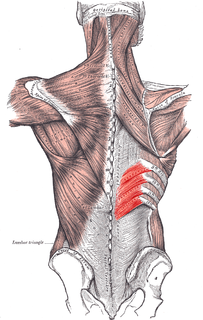
Grynfeltt-Lesshaft hernia is a herniation of abdominal contents through the back, specifically through the superior lumbar triangle, which is defined by the quadratus lumborum muscle, twelfth rib, and internal oblique muscle.
Grynfeltt-Lesshaft hernia is a herniation of abdominal contents through the back, specifically through the superior lumbar triangle, which is defined by the quadratus lumborum muscle, twelfth rib, and internal oblique muscle.
Grynfeltt described a hernia through the superior lumbar triangle in 1866 (Grynfeltt, 1866). In 1870, Lesshaft independently reported a similar case (Lesshaft, 1870).

A hernia is the abnormal exit of tissue or an organ, such as the bowel, through the wall of the cavity in which it normally resides. Hernias come in a number of types. Most commonly they involve the abdomen, specifically the groin. Groin hernias are most commonly of the inguinal type but may also be femoral. Other hernias include hiatus, incisional, and umbilical hernias. Symptoms are present in about 66% of people with groin hernias. This may include pain or discomfort, especially with coughing, exercise or going to the bathroom. Often, it gets worse throughout the day and improves when lying down. A bulging area may appear that becomes larger when bearing down. Groin hernias occur more often on the right than left side. The main concern is strangulation, where the blood supply to part of the bowel is blocked. This usually produces severe pain and tenderness in the area. Hiatus, or hiatal, hernias often result in heartburn but may also cause chest pain or pain with eating.

The latissimus dorsi is a large, flat muscle on the back that stretches to the sides, behind the arm, and is partly covered by the trapezius on the back near the midline. The word latissimus dorsi comes from Latin and means "broadest [muscle] of the back", from "latissimus" ' and "dorsum". The pair of muscles are commonly known as "lats", especially among bodybuilders. The latissimus dorsi is the largest muscle in the upper body.

The thoracic diaphragm, or simply the diaphragm, is a sheet of internal skeletal muscle in humans and other mammals that extends across the bottom of the thoracic cavity. The diaphragm separates the thoracic cavity, containing the heart and lungs, from the abdominal cavity and performs an important function in respiration: as the diaphragm contracts, the volume of the thoracic cavity increases, creating a negative pressure there, which draws air into the lungs.

The inguinal ligament, also known as Poupart's ligament or groin ligament, is a band running from the pubic tubercle to the anterior superior iliac spine. It forms the base of the inguinal canal through which an indirect inguinal hernia may develop.
The serratus posterior inferior muscle, also known as the posterior serratus muscle, is a muscle of the human body.
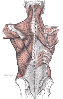
Petit's hernia is a hernia that protrudes through the lumbar triangle. This triangle lies in the posterolateral abdominal wall and is bounded anteriorly by the free margin of external oblique muscle, posteriorly by the latissimus dorsi and inferiorly by the iliac crest. The neck is large, and therefore this hernia has a lower risk of strangulating than some other hernias.
Athletic pubalgia, also called sports hernia, core injury, hockey hernia, hockey groin, Gilmore's groin, or groin disruption is a medical condition of the pubic joint affecting athletes.
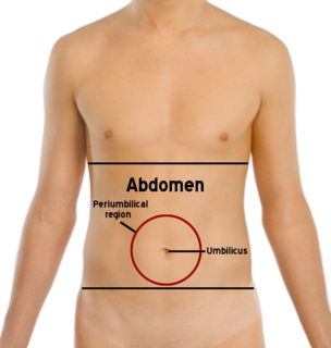
The abdomen is the part of the body between the thorax (chest) and pelvis, in humans and in other vertebrates. The abdomen is the front part of the abdominal segment of the trunk. The area occupied by the abdomen is called the abdominal cavity. In arthropods it is the posterior tagma of the body; it follows the thorax or cephalothorax.

In human anatomy, the inguinal triangle is a region of the abdominal wall. It is also known by the eponym Hesselbach's triangle, after Franz Kaspar Hesselbach.

In human anatomy, inferior epigastric artery refers to the artery that arises from the external iliac artery. It anastomoses with the superior epigastric artery. Along its course, it is accompanied by a similarly named vein, the inferior epigastric vein. These epigastric vessels form the lateral border of Hesselbach's triangle, which outlines the area through which direct inguinal hernias protrude.

The lumbar plexus is a web of nerves in the lumbar region of the body which forms part of the larger lumbosacral plexus. It is formed by the divisions of the first four lumbar nerves (L1-L4) and from contributions of the subcostal nerve (T12), which is the last thoracic nerve. Additionally, the ventral rami of the fourth lumbar nerve pass communicating branches, the lumbosacral trunk, to the sacral plexus. The nerves of the lumbar plexus pass in front of the hip joint and mainly support the anterior part of the thigh.
The iliohypogastric nerve is a nerve that originates from the lumbar plexus that supplies sensation to skin over the lateral gluteal and hypogastric regions and motor to the internal oblique muscles and transverse abdominal muscles.

The erector spinae or spinal erectors is a set of muscles that straighten and rotate the back.
The obturator artery is a branch of the internal iliac artery that passes antero-inferiorly on the lateral wall of the pelvis, to the upper part of the obturator foramen, and, escaping from the pelvic cavity through the obturator canal, it divides into both an anterior and a posterior branch.
The lumbar triangle can refer to either the inferior lumbar (Petit) triangle, which lies superficially, or the superior lumbar (Grynfeltt) triangle, which is deep and superior to the inferior triangle. Of the two, the superior triangle is the more consistently found in cadavers, and is more commonly the site of herniation; however, the inferior lumbar triangle is often simply called the lumbar triangle, perhaps owing to its more superficial location and ease in demonstration.

The arcuate line of rectus sheath, the linea semicircularis, the arcuate line, or the semicircular line of Douglas, is a horizontal line that demarcates the lower limit of the posterior layer of the rectus sheath. It is commonly known simply as the arcuate line. It is also where the inferior epigastric vessels perforate the rectus abdominis.

The pectineal ligament, sometimes known as the inguinal ligament of Cooper, is an extension of the lacunar ligament. It runs on the pectineal line of the pubic bone. The pectineal ligament is the posterior border of the femoral ring.

The Lumbocostal triangle or Bochdalek's foramen is a space between the costal and lumbar parts of the diaphragm. The base of this triangular space is formed by muscle attachments originating from the XII rib and muscle fibers attaching to the lateral arcuate ligament. The apex of the triangle is oriented towards the tendinous centre of the diaphragm. Parietal pleura and renal capsule are in contact in this space, so possible infection can be transmitted through this space.
The TESSYS method is a minimally-invasive, endoscopic spinal procedure for the treatment of a herniated disc. It was a further development of the YESS method by the Dutch Dr Thomas Hoogland in the Alpha Klinik in Munich in 1989 and was first called THESSYS. The procedure involves performing a small foramenotomy and removal of soft tissue compressing the nerve root.

In the vertebrate spinal column, each vertebra is an irregular bone with a complex structure composed of bone and some hyaline cartilage, the proportions of which vary according to the segment of the backbone and the species of vertebrate.
| | This surgery article is a stub. You can help Wikipedia by expanding it. |