List of vaginal tumors

Last updated

Vaginal tumors are neoplasms (tumors) found in the vagina. They can be benign or malignant. [1] [lower-alpha 1] A neoplasm is an abnormal growth of tissue that usually forms a tissue mass. [2] [3] [4] Vaginal neoplasms may be solid, cystic or of mixed type. [5]

Contents

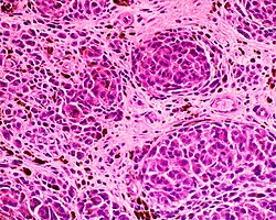
Vaginal cancers arise from vaginal tissue, with vaginal sarcomas develop from bone, cartilage, fat, muscle, blood vessels or other connective or supportive tissue. [6] [7] Tumors in the vagina may also be metastases (malignant tissue that has spread to the vagina from other parts of the body). [8] [7] Cancer that has spread from the colon, bladder, and stomach is far more common than cancer that originates in the vagina itself. [9] Some benign tumors may later progress to become malignant tumors, such as vaginal cancers. [10] [11] Some neoplastic growths of the vagina are sufficiently rare as to be only described in case studies. [3]

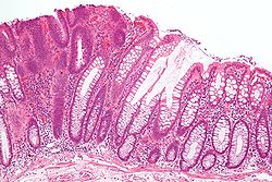
Signs and symptoms may include a feeling of pressure, painful intercourse or bleeding. [12] Most vaginal tumors are located during a pelvic exam. Ultrasonography, CT and MRI imaging is used to establish the location and presence or absence of fluid in a tumor. [13] [14] Biopsy provides a more definitive diagnosis. [15]

Vaginal tumors

Micrograph showing the yolk sac component of a mixed germ cell tumour. H&E stain. Mixed germ cell tumour - high mag.jpg
Micrograph showing the yolk sac component of a mixed germ cell tumour. H&E stain.
Micrograph of an H&E stained section of a peripheral PNET. PNET Histopathology HE 200x.jpg
Micrograph of an H&E stained section of a peripheral PNET.
Blue nevus SkinTumors-456.jpg
Blue nevus
Micrograph of a small-cell carcinoma showing cells with nuclear moulding, minimal amount of cytoplasm and stippled chromatin. Small cell lung cancer - cytology.jpg
Micrograph of a small-cell carcinoma showing cells with nuclear moulding, minimal amount of cytoplasm and stippled chromatin.
Micrograph of a mucinous adenocarcinoma Tubular adenoma 2 intermed mag.jpg
Micrograph of a mucinous adenocarcinoma
Micrograph of fibroepithelial polyp SkinTumors-P9250819.jpg
Micrograph of fibroepithelial polyp
Vaginal tumorsBenignSynonyms and notesReferences
Yolk sac tumor no Endodermal sinus tumor [16] [17] [18] [19] [20] [21] [22]
Peripheral primitive neuroectodermal tumor no Ewing's sarcoma [16] [17] [18] [23]
Vaginal melanoma no Melanocytic tumor [16] [17] [18] [24] [25]
Blue nevus yes [26] Melanocytic tumor, blue mole, nevus bleu,

melancytic nevus

[16] [17] [18] [25]
Carcinosarcoma noMalignant Mullerian Mixed tumors;

metaplastic carcinoma

[18]
Sarcoma botryoides nobotryoid sarcoma, botryoid rhabdomyosarcoma;

subtype of embryonal rhabdomyosarcoma

[9] [16] [17] [18] [22] [27] [28]
Leimyosarcoma nolocalized tumor of leukemic cells [18] [29]
Endometrioid stromal sarcomano endometrial stromal sarcoma [18] [30]
Undifferentiated vaginal sarcoma [18]
Leiomyoma yesfibromyoma [6] [13] [16] [17] [18]
Genital rhabdomyoma [16] [17] [18] [31] [32] [33]
Deep angiomyoxoma [16] [17] [18]
Spindle cell nodule Vaginal Solitary Fibrous Tumor [9] [16] [17] [18] [34]
Undifferentiated carcinoma [16] [17] [18]
Small cell carcinoma no [16] [17] [18]
Carcinoid no [18]
Adenoid basal carcinoma [18]
Adenosquamous carcinoma no [18]
Adenoma yes [35]
Mucinous adenocarcinoma [18]
Squamous papilloma yesvaginal micropapillomatosis [16] [17] [18]
Endometrioid adenocarcinoma no [18]
Mesonephric adenocarcinoma [18]
Clear cell adenocarcinoma no [16] [17] [18]
Fibroepithelial polyp yes [16] [17] [18]
Squamous intraepithelial neoplasia [16] [17] [18]
Genital wart yes Condylomata acuminata [6] [16] [17] [18]
Squamous cell carcinoma noKeratinizing, Nonkeratininzing, Basalaoid,

Verrucous, Warty

[16] [17] [18]
Mesenchymal tumors [18]
Alveolar soft part sarcoma [18]
Mixed epithelial and mesenchymal Tumors [18]
Malignant mixed Tumors resembling synovial sarcoma [18]
Benign mixed tumors [18]
Adenomatoid Tumor yes [18]
Malignant lymphoma no [18]
Granulocytic sarcoma [18]
Fibroepithelial polyp yes [6] [17]
Verrucous carcinoma no [9]
Squamotransitional cell carcinoma [9]

Other animals

Vaginal tumors also can be found in domesticated animals:

See also

Notes

  1. There are four main groups of vaginal neoplasms: benign neoplasms, in situ neoplasms, malignant neoplasms, and neoplasms of uncertain or unknown behavior. Malignant neoplasms are also simply known as cancers. Tumor (American English) or tumour (British English), Latin for swelling, one of the cardinal signs of inflammation, originally meant any form of swelling, neoplastic or not. Current English, however, both medical and non-medical, uses tumor as a synonym for a neoplasm (a solid or fluid-filled cystic lesion that may or may not be formed by an abnormal growth of neoplastic cells) that appears enlarged in size.Some neoplasms do not form a tumor; these include leukemia and most forms of carcinoma in situ. Tumor is also not synonymous with cancer. While cancer is by definition malignant, a tumor can be benign, precancerous, or malignant

Related Research Articles

<span class="mw-page-title-main">Teratoma</span> Type of germ cell tumor

A teratoma is a tumor made up of several different types of tissue, such as hair, muscle, teeth, or bone. Teratomata typically form in the tailbone, ovary, or testicle.

<span class="mw-page-title-main">Rhabdomyosarcoma</span> Medical condition

Rhabdomyosarcoma (RMS) is a highly aggressive form of cancer that develops from mesenchymal cells that have failed to fully differentiate into myocytes of skeletal muscle. Cells of the tumor are identified as rhabdomyoblasts.

<span class="mw-page-title-main">Surface epithelial-stromal tumor</span> Medical condition

Surface epithelial-stromal tumors are a class of ovarian neoplasms that may be benign or malignant. Neoplasms in this group are thought to be derived from the ovarian surface epithelium or from ectopic endometrial or fallopian tube (tubal) tissue. Tumors of this type are also called ovarian adenocarcinoma. This group of tumors accounts for 90% to 95% of all cases of ovarian cancer; however is mainly only found in postmenopausal women with the exception of the United States where 7% of cases occur in women under the age of 40. Serum CA-125 is often elevated but is only 50% accurate so it is not a useful tumor marker to assess the progress of treatment. 75% of women with epithelial ovarian cancer are found within the advanced-stages; however younger patients are more likely to have better prognoses than older patients.

<span class="mw-page-title-main">Neoplasm</span> Tumor or other abnormal growth of tissue

A neoplasm is a type of abnormal and excessive growth of tissue. The process that occurs to form or produce a neoplasm is called neoplasia. The growth of a neoplasm is uncoordinated with that of the normal surrounding tissue, and persists in growing abnormally, even if the original trigger is removed. This abnormal growth usually forms a mass, which may be called a tumour or tumor.

<span class="mw-page-title-main">Grading (tumors)</span> Measure of the cell appearance in tumors and other neoplasms

In pathology, grading is a measure of the cell appearance in tumors and other neoplasms. Some pathology grading systems apply only to malignant neoplasms (cancer); others apply also to benign neoplasms. The neoplastic grading is a measure of cell anaplasia in the sampled tumor and is based on the resemblance of the tumor to the tissue of origin. Grading in cancer is distinguished from staging, which is a measure of the extent to which the cancer has spread.

<span class="mw-page-title-main">Eye neoplasm</span> Medical condition

An Eye neoplasm is a tumor of the eye. A rare type of tumor, eye neoplasms can affect all parts of the eye, and can either be benign or malignant (cancerous), in which case it is known as eye cancer. Eye cancers can be primary or metastatic cancer. The two most common cancers that spread to the eye from another organ are breast cancer and lung cancer. Other less common sites of origin include the prostate, kidney, thyroid, skin, colon and blood or bone marrow.

Breast diseases make up a number of conditions. The most common symptoms are a breast mass, breast pain, and nipple discharge.

Vaginal cancer is an extraordinarily rare form of cancer that develops in the tissue of the vagina. Primary vaginal cancer originates from the vaginal tissue – most frequently squamous cell carcinoma, but primary vaginal adenocarcinoma, sarcoma, and melanoma have also been reported – while secondary vaginal cancer involves the metastasis of a cancer that originated in a different part of the body. Secondary vaginal cancer is more common. Signs of vaginal cancer may include abnormal vaginal bleeding, dysuria, tenesmus, or pelvic pain, though as many as 20% of women diagnosed with vaginal cancer are asymptomatic at the time of diagnosis. Vaginal cancer occurs more frequently in women over age 50, and the mean age of diagnosis of vaginal cancer is 60 years. It often can be cured if found and treated in early stages. Surgery alone or surgery combined with pelvic radiation is typically used to treat vaginal cancer.

Sarcoma botryoides or botryoid sarcoma is a subtype of embryonal rhabdomyosarcoma, that can be observed in the walls of hollow, mucosa lined structures such as the nasopharynx, common bile duct, urinary bladder of infants and young children or the vagina in females, typically younger than age 8. The name comes from the gross appearance of "grape bunches".

Malignant ectomesenchymoma(MEM) is a rare, fast-growing tumor of the nervous system or soft tissue that occurs in children and young adults. MEM is part of a group of small round blue cell tumors which includes neuroblastoma, rhabdomyosarcoma, non-Hodgkin's lymphoma, and the Ewing's family of tumors.

<span class="mw-page-title-main">Mesoblastic nephroma</span> Medical condition

Congenital mesoblastic nephroma, while rare, is the most common kidney neoplasm diagnosed in the first three months of life and accounts for 3-5% of all childhood renal neoplasms. This neoplasm is generally non-aggressive and amenable to surgical removal. However, a readily identifiable subset of these kidney tumors has a more malignant potential and is capable of causing life-threatening metastases. Congenital mesoblastic nephroma was first named as such in 1967 but was recognized decades before this as fetal renal hamartoma or leiomyomatous renal hamartoma.

Ectomesenchymoma is a rare, fast-growing tumor of the nervous system or soft tissue that occurs mainly in children, although cases have been reported in patients up to age 60. Ectomesenchymomas may form in the head and neck, abdomen, perineum, scrotum, or limbs. Also called malignant ectomesenchymoma.

The Bethesda system (TBS), officially called The Bethesda System for Reporting Cervical Cytology, is a system for reporting cervical or vaginal cytologic diagnoses, used for reporting Pap smear results. It was introduced in 1988 and revised in 1991, 2001, and 2014. The name comes from the location of the conference, sponsored by the National Institutes of Health, that established the system.

<span class="mw-page-title-main">Endodermal sinus tumor</span> Medical condition

Endodermal sinus tumor (EST) is a member of the germ cell tumor group of cancers. It is the most common testicular tumor in children under three, and is also known as infantile embryonal carcinoma. This age group has a very good prognosis. In contrast to the pure form typical of infants, adult endodermal sinus tumors are often found in combination with other kinds of germ cell tumor, particularly teratoma and embryonal carcinoma. While pure teratoma is usually benign, endodermal sinus tumor is malignant.

A rhabdomyoblast is a cell type which is found in some rhabdomyosarcomas. When found histologically, a rhabdomyoblast aids the diagnosis of embryonal, alveolar, spindle cell/sclerosing, and pleomorphic rhabdomyosarcomas; however, in a tumor, expression of the rhabdomyoblast phenotype is not the only factor in diagnosing a rhabdomyosarcoma. Mesenchymal malignancies can exhibit this phenotype as well. Immunohistochemistry techniques allow for the sensitive detection of desmin, vimentin, muscle specific actin, and MyoD1. Similarly the rhabdomyoblast phenotype can be detected morphologically. Rhabdomyoblasts are early stage mesenchymal cells, having the potential to differentiate into a wide range of skeletal cells. Each stage of differentiation exhibits unique and distinguishable histological characteristics. In its initial from, stellate cells with amphiphilic cytoplasm and ovular central nuclei are observed. Commonly referred to as rhabdoid features, the maturing rhabdomyoblast will likely exhibit low levels of eosinophilic cytoplasm in proximal distances to the nucleus. As maturation and differentiation progress, the cell's cytoplasmic levels of white blood cells increase; additionally, elongated shapes, commonly depicted as “tadpole”, “strap” and "spider cells", are observed. In the concluding phase of differentiation, the white blood cell rich cytoplasm appears bright and exhibits cross-striation. The highly regulated organization of actin and myosin microfilaments in contractile proteins results in this appearance.

<span class="mw-page-title-main">Vaginal cysts</span> Benign growths of the vaginal epithelium

Vaginal cysts are uncommon benign cysts that develop in the vaginal wall. The type of epithelial tissue lining a cyst is used to classify these growths. They can be congenital. They can present in childhood and adulthood. The most common type is the squamous inclusion cyst. It develops within vaginal tissue present at the site of an episiotomy or other vaginal surgical sites. In most instances they do not cause symptoms and present with few or no complications. A vaginal cyst can develop on the surface of the vaginal epithelium or in deeper layers. Often, they are found by the woman herself and as an incidental finding during a routine pelvic examination. Vaginal cysts can mimic other structures that protrude from the vagina such as a rectocele and cystocele. Some cysts can be distinguished visually but most will need a biopsy to determine the type. Vaginal cysts can vary in size and can grow as large as 7 cm. Other cysts can be present on the vaginal wall though mostly these can be differentiated. Vaginal cysts can often be palpated (felt) by a clinician. Vaginal cysts are one type of vaginal mass, others include cancers and tumors. The prevalence of vaginal cysts is uncertain since many go unreported but it is estimated that 1 out of 200 women have a vaginal cyst. Vaginal cysts may initially be discovered during pregnancy and childbirth. These are then treated to provide an unobstructed delivery of the infant. Growths that originate from the urethra and other tissue can present as cysts of the vagina.

Vulvar tumors are those neoplasms of the vulva. Vulvar and vaginal neoplasms make up a small percentage (3%) of female genital cancers. They can be benign or malignant. Vulvar neoplasms are divided into cystic or solid lesions and other mixed types. Vulvar cancers are those malignant neoplasms that originate from vulvar epithelium, while vulvar sarcomas develop from non-epithelial cells such as bone, cartilage, fat, muscle, blood vessels, or other connective or supportive tissue. Epithelial and mesenchymal tissue are the origin of vulvar tumors.

<span class="mw-page-title-main">Proliferative fasciitis and proliferative myositis</span> Medical condition

Proliferative fasciitis and proliferative myositis (PF/PM) are rare benign soft tissue lesions that increase in size over several weeks and often regress over the ensuing 1–3 months. The lesions in PF/PM are typically obvious tumors or swellings. Historically, many studies had grouped the two descriptive forms of PF/PM as similar disorders with the exception that proliferative fasciitis occurs in subcutaneous tissues while proliferative myositis occurs in muscle tissues. In 2020, the World Health Organization agreed with this view and defined these lesions as virtually identical disorders termed proliferative fasciitis/proliferative myositis or proliferative fasciitis and proliferative myositis. The Organization also classified them as one of the various forms of the fibroblastic and myofibroblastic tumors.

Fibroblastic and myofibroblastic tumors (FMTs) develop from the mesenchymal stem cells which differentiate into fibroblasts and/or the myocytes/myoblasts that differentiate into muscle cells. FMTs are a heterogeneous group of soft tissue neoplasms. The World Health Organization (2020) defined tumors as being FMTs based on their morphology and, more importantly, newly discovered abnormalities in the expression levels of key gene products made by these tumors' neoplastic cells. Histopathologically, FMTs consist of neoplastic connective tissue cells which have differented into cells that have microscopic appearances resembling fibroblasts and/or myofibroblasts. The fibroblastic cells are characterized as spindle-shaped cells with inconspicuous nucleoli that express vimentin, an intracellular protein typically found in mesenchymal cells, and CD34, a cell surface membrane glycoprotein. Myofibroblastic cells are plumper with more abundant cytoplasm and more prominent nucleoli; they express smooth muscle marker proteins such as smooth muscle actins, desmin, and caldesmon. The World Health Organization further classified FMTs into four tumor forms based on their varying levels of aggressiveness: benign, intermediate, intermediate, and malignant.

The FET protein family consists of three similarly structured and functioning proteins. They and the genes in the FET gene family which encode them are: 1) the EWSR1 protein encoded by the EWSR1 gene located at band 12.2 of the long arm of chromosome 22; 2) the FUS protein encoded by the FUS gene located at band 16 on the short arm of chromosome 16; and 3) the TAF15 protein encoded by the TAF15 gene located at band 12 on the long arm of chromosome 7 The FET in this protein family's name derives from the first letters of FUS, EWSR1, and TAF15.

References

  1. "NCI Dictionary of Cancer Terms - Benign". National Cancer Institute. 2011-02-02. Retrieved 2018-03-13.
  2. "NCI Dictionary of Cancer Terms - Tumor". National Cancer Institute. 2011-02-02. Retrieved 2018-03-13.
  3. 1 2 Stedman's medical dictionary (28th ed.). Philadelphia: Lippincott Williams & Wilkins. 2006. p. Neoplasm. ISBN   978-0781733908.
  4. "NCI Dictionary of Cancer Terms - Neoplasm". National Cancer Institute. 2011-02-02. Retrieved 2018-03-13.
  5. "What Are Tumors?". pathology.jhu.edu. Retrieved 2018-03-13.
  6. 1 2 3 4 "Benign Neoplasms of the Vagina | GLOWM". www.glowm.com. Retrieved 2018-03-01.
  7. 1 2 "NCI Dictionary of Cancer Terms - Sarcoma". National Cancer Institute. 2011-02-02. Retrieved 2018-03-13.
  8. "NCI Dictionary of Cancer Terms - Metastasis". National Cancer Institute. 2011-02-02. Retrieved 2018-03-13.
  9. 1 2 3 4 5 Dey, Pranab (2017-02-06). Essentials of Gynecologic Pathology. JP Medical Ltd. pp. 41–48. ISBN   9789386261205.
  10. Taylor, Elizabeth J. (2000). Dorland's Illustrated medical dictionary (29th ed.). Philadelphia: Saunders. p.  1184. ISBN   978-0721662541.
  11. Cooper GM (1992). Elements of human cancer. Boston: Jones and Bartlett Publishers. p. 16. ISBN   978-0-86720-191-8.
  12. "Vaginal Bleeding - Gynecology and Obstetrics - Merck Manuals Professional Edition". Merck Manuals Professional Edition. Retrieved 2018-03-02.
  13. 1 2 Shobeiri, S. Abbas; Rostaminia, Ghazaleh; White, Dena; Quiroz, Lieschen H.; Nihira, Mikio A. (2013-08-01). "Evaluation of Vaginal Cysts and Masses by 3-Dimensional Endovaginal and Endoanal Sonography". Journal of Ultrasound in Medicine. 32 (8): 1499–1507. doi:10.7863/ultra.32.8.1499. ISSN   1550-9613. PMID   23887963. S2CID   34402240.
  14. Hamm, Bernd; Forstner, Rosemarie (2007-01-19). MRI and CT of the Female Pelvis. Springer Science & Business Media. ISBN   9783540682127.
  15. Nelson, Philippa (2018-01-23). "Endometriosis presenting as a vaginal mass". BMJ Case Reports. 2018: bcr–2017–222431. doi:10.1136/bcr-2017-222431. ISSN   1757-790X. PMC   5786902 . PMID   29367370.
  16. 1 2 3 4 5 6 7 8 9 10 11 12 13 14 15 16 17 Humphrey, Peter A.; Dehner, Louis P.; Pfeifer, John D. (22 February 2018). The Washington Manual of Surgical Pathology. Lippincott Williams & Wilkins. ISBN   9780781765275 . Retrieved 22 February 2018 via Google Books.
  17. 1 2 3 4 5 6 7 8 9 10 11 12 13 14 15 16 17 18 "Tumours of the Vagina; Chapter Six" (PDF). International Agency for Research on Cancer, World Health Organization. pp. 291–311. Archived from the original (PDF) on 2015-09-08.
  18. 1 2 3 4 5 6 7 8 9 10 11 12 13 14 15 16 17 18 19 20 21 22 23 24 25 26 27 28 29 30 31 32 33 34 35 "Vulva and Vagina tumors: an overview". atlasgeneticsoncology.org. Retrieved 2018-02-22.
  19. "NCI Dictionary of Cancer Terms - Mature Teratoma". National Cancer Institute. 2011-02-02.
  20. Tang QL, Jiang XF, Yuan XP, Liu Y, Zhang L, Tang XF, Zhou JJ, Li HG, Fang JP, Xue L (2014). "Prognosis of eight Chinese cases of primary vaginal yolk sac tumor with a review of the literature". Asian Pacific Journal of Cancer Prevention. 15 (21): 9395–404. doi: 10.7314/APJCP.2014.15.21.9395 . PMID   25422231.
  21. Bhatt MD, Braga LH, Stein N, Terry J, Portwine C (July 2015). "Vaginal Yolk Sac Tumor in an Infant: A Case Report and Literature Review of the Last 30 Years". Journal of Pediatric Hematology/Oncology. 37 (5): e336–40. doi:10.1097/MPH.0000000000000325. PMID   25851552. S2CID   7605939.
  22. 1 2 Coran, Arnold G.; Caldamone, Anthony; Adzick, N. Scott; Krummel, Thomas M.; Laberge, Jean-Martin; Shamberger, Robert (2012-01-25). Pediatric Surgery E-Book. Elsevier Health Sciences. ISBN   978-0323091619.
  23. Baldini EH, Demetri GD, Fletcher CD, Foran J, Marcus KC, Singer S (July 1999). "Adults with Ewing's sarcoma/primitive neuroectodermal tumor: adverse effect of older age and primary extraosseous disease on outcome". Annals of Surgery. 230 (1): 79–86. doi:10.1097/00000658-199907000-00012. PMC   1420848 . PMID   10400040.
  24. Kalampokas E, Kalampokas T, Damaskos C (January 2017). "Primary Vaginal Melanoma, A Rare and Aggressive Entity. A Case Report and Review of the Literature". In Vivo. 31 (1): 133–139. doi:10.21873/invivo.11036. PMC   5354139 . PMID   28064232.
  25. 1 2 "Blue naevus | DermNet New Zealand". www.dermnetnz.org.
  26. but mimics other malignant tumors
  27. PDQ Pediatric Treatment Editorial Board (2018). "Childhood Rhabdomyosarcoma Treatment (PDQ®): Health Professional Version". PDQ Cancer Information Summaries - Childhood Rhabdomyosarcoma Treatment. Bethesda (MD): National Cancer Institute (US). PMID   26389243.
  28. "Ewing Sarcoma Treatment". National Cancer Institute. 1980-01-01. Retrieved 2018-02-28.
  29. Keller NA, Godoy H (2015). "Leiomyosarcoma of the Vagina: An Exceedingly Rare Diagnosis". Case Reports in Obstetrics and Gynecology. 2015: 363895. doi: 10.1155/2015/363895 . PMC   4689886 . PMID   26783476.
  30. "What Is Uterine Sarcoma?". www.cancer.org. Retrieved 2018-02-28.
  31. Schoolmeester JK, Xing D, Keeney GL, Sukov WR (July 2017). "Genital Rhabdomyoma of the Lower Female Genital Tract: A Study of 12 Cases With Molecular Cytogenetic Findings". International Journal of Gynecological Pathology. 37 (4): 349–355. doi:10.1097/PGP.0000000000000428. PMID   28700439. S2CID   43341442.
  32. Folpe, Andrew L.; Inwards, Carrie Y. (2010-01-01). Bone and Soft Tissue Pathology. Elsevier Health Sciences. ISBN   978-0443066887.
  33. Andrassy RJ, Wiener ES, Raney RB, Hays DM, Arndt CA, Lobe TE, Lawrence W, Anderson JR, Qualman SJ, Crist WM (May 1999). "Progress in the surgical management of vaginal rhabdomyosarcoma: a 25-year review from the Intergroup Rhabdomyosarcoma Study Group". Journal of Pediatric Surgery. 34 (5): 731–4, discussion 734–5. doi:10.1016/S0022-3468(99)90365-2. PMID   10359173.
  34. Rekhi B, Bapat P, Shetty O (2019). "A Rare Case of a Vaginal Solitary Fibrous Tumor, Presenting as a Cystic Mass, Showing NAB2ex4-STAT6ex2 Fusion and STAT6 Immunostaining". International Journal of Gynecological Pathology. 38 (1): 21–26. doi:10.1097/PGP.0000000000000479. PMID   29257041. S2CID   19130435.
  35. Mierau GW, Lovell MA, Wyatt-Ashmead J, Goin L (May 2005). "Benign müllerian papilloma of childhood". Ultrastructural Pathology. 29 (3–4): 209–16. doi:10.1080/01913120590951211. PMID   16036876. S2CID   22146300.
  36. Baba, Alecsandru Ioan; Câtoi, Cornel (22 February 2018). "FEMALE GENITAL TRACT TUMORS". The Publishing House of the Romanian Academy. Retrieved 22 February 2018 via www.ncbi.nlm.nih.gov.