Oligosaccharyltransferase

Last updated
STT3
OST 6EZN.png
Structure of yeast OST [1]
Identifiers
SymbolSTT3
Pfam PF02516
Pfam clan CL0111
InterPro IPR003674
OPM superfamily 242
OPM protein 3rce
CAZy GT66
Membranome 275
Available protein structures:
Pfam   structures / ECOD  
PDB RCSB PDB; PDBe; PDBj
PDBsum structure summary

Oligosaccharyltransferase or OST (EC 2.4.1.119) is a membrane protein complex that transfers a 14-sugar oligosaccharide from dolichol to nascent protein. It is a type of glycosyltransferase. The sugar Glc3Man9GlcNAc2 (where Glc=Glucose, Man=Mannose, and GlcNAc=N-acetylglucosamine) is attached to an asparagine (Asn) residue in the sequence Asn-X-Ser or Asn-X-Thr where X is any amino acid except proline. This sequence is called a glycosylation sequon. The reaction catalyzed by OST is the central step in the N-linked glycosylation pathway.

Contents

Location

ER Translocon complex. Many protein complexes are involved in protein synthesis. The actual production takes place in the ribosomes (grey and light blue). Through the ER translocon (green: Sec61, blue: TRAP complex, and red: oligosaccharyl transferase complex) the newly synthesized protein is transported across the membrane (gray) into the interior of the ER. Sec61 is the protein-conducting channel and the OST adds sugar moieties to the nascent protein. OST PM-1.jpg
ER Translocon complex. Many protein complexes are involved in protein synthesis. The actual production takes place in the ribosomes (grey and light blue). Through the ER translocon (green: Sec61, blue: TRAP complex, and red: oligosaccharyl transferase complex) the newly synthesized protein is transported across the membrane (gray) into the interior of the ER. Sec61 is the protein-conducting channel and the OST adds sugar moieties to the nascent protein.

OST is a component of the translocon in the endoplasmic reticulum (ER) membrane. A lipid-linked core-oligosaccharide is assembled at the membrane of the endoplasmic reticulum and transferred to selected asparagine residues of nascent polypeptide chains by the oligosaccharyl transferase complex. [3] The active site of OST is located about 4 nm from the lumenal face of the ER membrane. [4]

It usually acts during translation as the nascent protein is entering the ER, but this cotranslational glycosylation is nevertheless called a posttranslational modification. A few examples have been found of OST activity after translation is complete. [5] [6] Current opinion is that post-translational activity may occur if the protein is poorly folded or folds slowly. [6]

Structure and function

Yeast OST is composed of eight different membrane-spanning proteins in three subcomplexes (one of them is OST4). [7] [8] These octomers do not form higher order oligomers, and three of the eight proteins are glycosylated themselves. [7] OST in mammals is known to have a similar composition. [9] [10]

OST is thought to require many subunits because it must: [11]

  1. Position itself near the translocon pore.
  2. Recognize and bind oligosaccharyldolichol.
  3. Scan the nascent protein in order to recognize and bind sequons.
  4. Move these two large substrates into their proper locations and conformations.
  5. Activate the Asn amide nitrogen atom for the actual transfer of oligosaccharide.
  6. Release its substrates.

The catalytically active subunit of the OST is called STT3. Two paralogs exist in eukaryotes, termed STT3A and STT3B. STT3A is primarily responsible for cotranslational glycosylation of the nascent polypeptide as it enters the lumen of the endoplasmic reticulum whereas STT3B can also mediate posttranslational glycosylation. [12] The structure of PglB, the prokaryotic homolog of STT3 has been solved. [13] The high sequence similarity between the prokaryotic and the eukaryotic STT3 suggests that their structures are similar.

Clinical significance

CDG syndromes are genetic disorders of the glycosylation pathway. They are labelled "Type I" if the defective gene is for an enzyme involved in the assembly or transfer of the Glc3Man9GlcNAc2-dolichol precursor. They are labelled "Type II" if the defective step occurs after the action of OST in the N-linked glycosylation pathway or involves O-linked glycosylation. [14]

See also

Related Research Articles

<span class="mw-page-title-main">Endoplasmic reticulum</span> Cell organelle that synthesizes, folds and processes proteins

The endoplasmic reticulum (ER) is, in essence, the transportation system of the eukaryotic cell, and has many other important functions such as protein folding. It is a type of organelle made up of two subunits – rough endoplasmic reticulum (RER), and smooth endoplasmic reticulum (SER). The endoplasmic reticulum is found in most eukaryotic cells and forms an interconnected network of flattened, membrane-enclosed sacs known as cisternae, and tubular structures in the SER. The membranes of the ER are continuous with the outer nuclear membrane. The endoplasmic reticulum is not found in red blood cells, or spermatozoa.

A congenital disorder of glycosylation is one of several rare inborn errors of metabolism in which glycosylation of a variety of tissue proteins and/or lipids is deficient or defective. Congenital disorders of glycosylation are sometimes known as CDG syndromes. They often cause serious, sometimes fatal, malfunction of several different organ systems in affected infants. The most common sub-type is PMM2-CDG where the genetic defect leads to the loss of phosphomannomutase 2 (PMM2), the enzyme responsible for the conversion of mannose-6-phosphate into mannose-1-phosphate.

A signal peptide is a short peptide present at the N-terminus of most newly synthesized proteins that are destined toward the secretory pathway. These proteins include those that reside either inside certain organelles, secreted from the cell, or inserted into most cellular membranes. Although most type I membrane-bound proteins have signal peptides, most type II and multi-spanning membrane-bound proteins are targeted to the secretory pathway by their first transmembrane domain, which biochemically resembles a signal sequence except that it is not cleaved. They are a kind of target peptide.

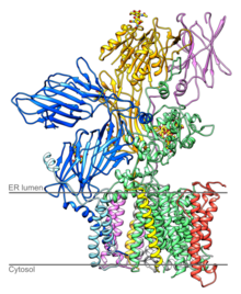
The translocon is a complex of proteins associated with the translocation of polypeptides across membranes. In eukaryotes the term translocon most commonly refers to the complex that transports nascent polypeptides with a targeting signal sequence into the interior space of the endoplasmic reticulum (ER) from the cytosol. This translocation process requires the protein to cross a hydrophobic lipid bilayer. The same complex is also used to integrate nascent proteins into the membrane itself. In prokaryotes, a similar protein complex transports polypeptides across the (inner) plasma membrane or integrates membrane proteins. In either case, the protein complex are formed from Sec proteins, with the heterotrimeric Sec61 being the channel. In prokaryotes, the homologous channel complex is known as SecYEG.

An oligosaccharide is a saccharide polymer containing a small number of monosaccharides. Oligosaccharides can have many functions including cell recognition and cell adhesion.

Dolichol refers to any of a group of long-chain mostly unsaturated organic compounds that are made up of varying numbers of isoprene units terminating in an α-saturated isoprenoid group, containing an alcohol functional group.

<span class="mw-page-title-main">Ribophorin</span>

Ribophorins are dome shaped transmembrane glycoproteins which are located in the membrane of the rough endoplasmic reticulum, but are absent in the membrane of the smooth endoplasmic reticulum. There are two types of ribophorines: ribophorin I and II. These act in the protein complex oligosaccharyltransferase (OST) as two different subunits of the named complex. Ribophorin I and II are only present in eukaryote cells.

<span class="mw-page-title-main">Dolichol kinase</span> Protein-coding gene in the species Homo sapiens

In enzymology, a dolichol kinase is an enzyme that catalyzes the chemical reaction

<span class="mw-page-title-main">DPAGT1</span> Protein-coding gene in the species Homo sapiens

UDP-N-acetylglucosamine—dolichyl-phosphate N-acetylglucosaminephosphotransferase is an enzyme that in humans is encoded by the DPAGT1 gene.

<span class="mw-page-title-main">RPN1</span> Protein-coding gene in the species Homo sapiens

Dolichyl-diphosphooligosaccharide—protein glycosyltransferase subunit 1 is an enzyme that in humans is encoded by the RPN1 gene.

<span class="mw-page-title-main">RPN2</span> Protein-coding gene in the species Homo sapiens

Dolichyl-diphosphooligosaccharide—protein glycosyltransferase subunit 2, also called ribophorin ǁ is an enzyme that in humans is encoded by the RPN2 gene.

<span class="mw-page-title-main">SIL1</span> Protein-coding gene in the species Homo sapiens

Nucleotide exchange factor SIL1 is a protein that in humans is encoded by the SIL1 gene.

<span class="mw-page-title-main">DPM1</span> Protein-coding gene in the species Homo sapiens

Dolichol-phosphate mannosyltransferase is an enzyme that in humans is encoded by the DPM1 gene.

In cell biology, membrane bound polyribosomes are attached to a cell's endoplasmic reticulum. When certain proteins are synthesized by a ribosome they can become "membrane-bound". The newly produced polypeptide chains are inserted directly into the endoplasmic reticulum by the ribosome and are then transported to their destinations. Bound ribosomes usually produce proteins that are used within the cell membrane or are expelled from the cell via exocytosis.

<i>N</i>-linked glycosylation Attachment of an oligosaccharide to a nitrogen atom

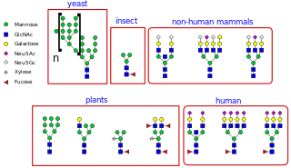
N-linked glycosylation, is the attachment of an oligosaccharide, a carbohydrate consisting of several sugar molecules, sometimes also referred to as glycan, to a nitrogen atom, in a process called N-glycosylation, studied in biochemistry. The resulting protein is called an N-linked glycan, or simply an N-glycan.

<span class="mw-page-title-main">DPM3</span> Protein-coding gene in the species Homo sapiens

dolichyl-phosphate mannosyltransferase polypeptide 3, also known as DPM3, is a human gene.

<span class="mw-page-title-main">Oligosaccaryltransferase</span>

In molecular biology, OST4 is a subunit of the oligosaccharyltransferase complex. OST4 is a very short, approximately 30 amino acids, protein found from fungi to vertebrates. It appears to be an integral membrane protein that mediates the en bloc transfer of a pre-assembled high-mannose oligosaccharide onto asparagine residues of nascent polypeptides as they enter the lumen of the rough endoplasmic reticulum.

Mannosyl-oligosaccharide glucosidase (MOGS) (EC 3.2.1.106, processing α-glucosidase I,Glc3Man9NAc2 oligosaccharide glucosidase, trimming glucosidase I, GCS1) is an enzyme with systematic name mannosyl-oligosaccharide glucohydrolase. MOGS is a transmembrane protein found in the membrane of the endoplasmic reticulum of eukaryotic cells. Biologically, it functions within the N-glycosylation pathway.

<span class="mw-page-title-main">Stt3a, catalytic subunit of the oligosaccharyltransferase complex</span> Protein-coding gene in the species Homo sapiens

STT3A, catalytic subunit of the oligosaccharyltransferase complex is a protein that in humans is encoded by the STT3A gene.

William Joseph Lennarz was a biochemist at Stony Brook University. He was born in May 1934 in New York City. Before Lennarz began his tenure at Stony Brook, he studied chemistry and organic chemistry. After working as a postdoctoral researcher at Harvard, he developed an interest in biochemistry. He has focused the majority of his research on biochemical processes in cells.

References

  1. Wild R, Kowal J, Eyring J, Ngwa EM, Aebi M, Locher KP (2018). "Structure of the yeast oligosaccharyltransferase complex gives insight into eukaryotic N-glycosylation". Science. 359 (6375): 545–550. Bibcode:2018Sci...359..545W. doi: 10.1126/science.aar5140 . hdl: 20.500.11850/228327 . PMID   29301962.
  2. Pfeffer S, Dudek J, Gogala M, Schorr S, Linxweiler J, Lang S, Becker T, Beckmann R, Zimmermann R, Förster F (2014). "Structure of the mammalian oligosaccharyl-transferase complex in the native ER protein translocon". Nat. Commun. 5 (5): 3072. Bibcode:2014NatCo...5.3072P. doi: 10.1038/ncomms4072 . PMID   24407213.
  3. Zufferey R, Knauer R, Burda P, Stagljar I, te Heesen S, Lehle L, Aebi M (October 1995). "STT3, a highly conserved protein required for yeast oligosaccharyl transferase activity in vivo". EMBO J. 14 (20): 4949–60. doi:10.1002/j.1460-2075.1995.tb00178.x. PMC   394598 . PMID   7588624.
  4. Nilsson IM, von Heijne G (March 1993). "Determination of the distance between the oligosaccharyltransferase active site and the endoplasmic reticulum membrane". J. Biol. Chem. 268 (8): 5798–801. doi: 10.1016/S0021-9258(18)53389-5 . PMID   8449946.
  5. Pless DD, Lennarz WJ; Lennarz (January 1977). "Enzymatic conversion of proteins to glycoproteins". Proc. Natl. Acad. Sci. U.S.A. 74 (1): 134–8. Bibcode:1977PNAS...74..134P. doi: 10.1073/pnas.74.1.134 . PMC   393212 . PMID   264667.
  6. 1 2 Duvet S, Op De Beeck A, Cocquerel L, Wychowski C, Cacan R, Dubuisson J (February 2002). "Glycosylation of the hepatitis C virus envelope protein E1 occurs posttranslationally in a mannosylphosphoryldolichol-deficient CHO mutant cell line". Glycobiology. 12 (2): 95–101. doi: 10.1093/glycob/12.2.95 . PMID   11886842.
  7. 1 2 Knauer R, Lehle L (January 1999). "The oligosaccharyltransferase complex from yeast". Biochim. Biophys. Acta. 1426 (2): 259–73. doi:10.1016/S0304-4165(98)00128-7. PMID   9878773.
  8. Dempski RE, Imperiali B (December 2002). "Oligosaccharyl transferase: gatekeeper to the secretory pathway". Curr Opin Chem Biol. 6 (6): 844–50. doi:10.1016/S1367-5931(02)00390-3. PMID   12470740.
  9. Nilsson I, Kelleher DJ, Miao Y, Shao Y, Kreibich G, Gilmore R, von Heijne G, Johnson AE (May 2003). "Photocross-linking of nascent chains to the STT3 subunit of the oligosaccharyltransferase complex". J. Cell Biol. 161 (4): 715–25. doi:10.1083/jcb.200301043. PMC   2199356 . PMID   12756234.
  10. Karaoglu D, Kelleher DJ, Gilmore R (October 2001). "Allosteric regulation provides a molecular mechanism for preferential utilization of the fully assembled dolichol-linked oligosaccharide by the yeast oligosaccharyltransferase". Biochemistry. 40 (40): 12193–206. doi:10.1021/bi0111911. PMID   11580295.
  11. Imperiali B (November 1997). "Protein Glycosylation: The Clash of the Titans". Accounts of Chemical Research. 30 (11): 452–459. doi:10.1021/ar950226k.
  12. Ruiz-Canada C, Kelleher DJ, Gilmore R (January 2009). "Cotranslational and posttranslational N-glycosylation of polypeptides by distinct mammalian OST isoforms". Cell. 136 (2): 272–83. doi:10.1016/j.cell.2008.11.047. PMC   2859625 . PMID   19167329.
  13. Lizak C, Gerber S, Numao S, Aebi M, Locher KP (June 2011). "X-ray structure of a bacterial oligosaccharyltransferase". Nature. 474 (7351): 350–5. doi:10.1038/nature10151. PMID   21677752. S2CID   205225231.
  14. Marquardt T, Denecke J (June 2003). "Congenital disorders of glycosylation: review of their molecular bases, clinical presentations and specific therapies". Eur. J. Pediatr. 162 (6): 359–79. doi:10.1007/s00431-002-1136-0. PMID   12756558. S2CID   3196550.