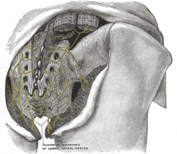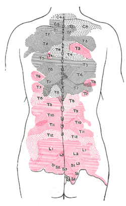
The sacrum, in human anatomy, is a large, triangular bone at the base of the spine that forms by the fusing of sacral vertebrae S1–S5 between 18 and 30 years of age.

The sciatic nerve is a large nerve in humans and other animals. It begins in the lower back and runs through the buttock and down the lower limb. It is the longest and widest single nerve in the human body, going from the top of the leg to the foot on the posterior aspect. The sciatic nerve provides the connection to the nervous system for nearly the whole of the skin of the leg, the muscles of the back of the thigh, and those of the leg and foot. It is derived from spinal nerves L4 to S3. It contains fibers from both the anterior and posterior divisions of the lumbosacral plexus.

A spinal nerve is a mixed nerve, which carries motor, sensory, and autonomic signals between the spinal cord and the body. In the human body there are 31 pairs of spinal nerves, one on each side of the vertebral column. These are grouped into the corresponding cervical, thoracic, lumbar, sacral and coccygeal regions of the spine. There are eight pairs of cervical nerves, twelve pairs of thoracic nerves, five pairs of lumbar nerves, five pairs of sacral nerves, and one pair of coccygeal nerves. The spinal nerves are part of the peripheral nervous system.
In anatomy, a foramen is any opening. Foramina inside the body of humans and other animals typically allow muscles, nerves, arteries, veins, or other structures to connect one part of the body with another.

The filum terminale is a delicate strand of fibrous tissue, about 20 cm in length, proceeding downward from the apex of the conus medullaris. It is one of the modifications of pia mater. It gives longitudinal support to the spinal cord and consists of two parts:
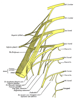
In human anatomy, the sacral plexus is a nerve plexus which provides motor and sensory nerves for the posterior thigh, most of the lower leg and foot, and part of the pelvis. It is part of the lumbosacral plexus and emerges from the lumbar vertebrae and sacral vertebrae (L4-S4). A sacral plexopathy is a disorder affecting the nerves of the sacral plexus, usually caused by trauma, nerve compression, vascular disease, or infection. Symptoms may include pain, loss of motor control, and sensory deficits.

The posterior cutaneous nerve of the thigh provides innervation to the skin of the posterior surface of the thigh and leg, as well as to the skin of the perineum.

The inferior gluteal nerve is the main motor neuron that innervates the gluteus maximus muscle. It is responsible for the movement of the gluteus maximus in activities requiring the hip to extend the thigh, such as climbing stairs. Injury to this nerve is rare but often occurs as a complication of posterior approach to the hip during hip replacement. When damaged, one would develop gluteus maximus lurch, which is a gait abnormality which causes the individual to 'lurch' backwards to compensate lack in hip extension.

The obturator nerve in human anatomy arises from the ventral divisions of the second, third, and fourth lumbar nerves in the lumbar plexus; the branch from the third is the largest, while that from the second is often very small.

The perforating cutaneous nerve is a cutaneous nerve that supplies skin over the gluteus maximus muscle.

The lateral sacral arteries arise from the posterior division of the internal iliac artery; there are usually two, a superior and an inferior.

The inferior gluteal artery, the smaller of the two terminal branches of the anterior trunk of the internal iliac artery, is distributed chiefly to the buttock and back of the thigh.
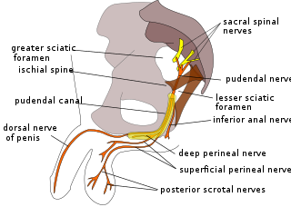
The perineal nerve is a nerve arising from the pudendal nerve that supplies the perineum.

The pelvic cavity is a body cavity that is bounded by the bones of the pelvis. Its oblique roof is the pelvic inlet. Its lower boundary is the pelvic floor.

The anterior division of the twelfth thoracic nerve is larger than the others; it runs along the lower border of the twelfth rib, often gives a communicating branch to the first lumbar nerve, and passes under the lateral lumbocostal arch.

The Inferior rectal nerves usually branch from the pudendal nerve but occasionally arises directly from the sacral plexus; they cross the ischiorectal fossa along with the inferior rectal artery and veins, toward the anal canal and the lower end of the rectum, and is distributed to the Sphincter ani externus and to the integument (skin) around the anus.

The nerve to piriformis is the peripheral nerve that innervates the piriformis muscle.

The nerve to obturator internus is a nerve that innervates the obturator internus and gemellus superior muscles.
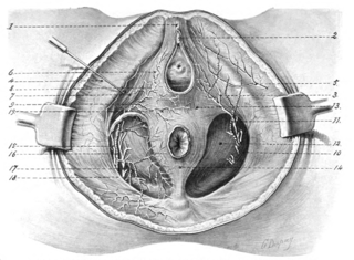
The dorsal nerve of the clitoris is a nerve in females that branches off the pudendal nerve to innervate the clitoris.

The medial clunial nerves innervate the skin of the buttocks closest to the midline of the body. Those nerves arise from the posterior rami of sacral spinal nerves.
The public domain consists of all the creative works to which no exclusive intellectual property rights apply. Those rights may have expired, been forfeited, expressly waived, or may be inapplicable.

Gray's Anatomy is an English language textbook of human anatomy originally written by Henry Gray and illustrated by Henry Vandyke Carter. Earlier editions were called Anatomy: Descriptive and Surgical, Anatomy of the Human Body and Gray's Anatomy: Descriptive and Applied, but the book's name is commonly shortened to, and later editions are titled, Gray's Anatomy. The book is widely regarded as an extremely influential work on the subject, and has continued to be revised and republished from its initial publication in 1858 to the present day. The latest edition of the book, the 41st, was published in September 2015.
