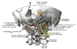
The greater occipital nerve is a nerve of the head. It is a spinal nerve, specifically the medial branch of the dorsal primary ramus of cervical spinal nerve 2. It arises from between the first and second cervical vertebrae, ascends, and then passes through the semispinalis muscle. It ascends further to supply the skin along the posterior part of the scalp to the vertex. It supplies sensation to the scalp at the top of the head, over the ear and over the parotid glands.

The lesser occipital nerve is a cutaneous spinal nerve of the cervical plexus. It arises from second cervical (spinal) nerve (C2). It innervates the skin of the back of the upper neck and of the scalp posterior to the ear.

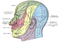
The scalp is the anatomical area bordered by the face at the front, and by the neck at the sides and back.
The posterior tibial artery of the lower limb is an artery that carries blood to the posterior compartment of the leg and plantar surface of the foot. It branches from the popliteal artery via the tibial-fibular trunk.
The anterior tibial artery is an artery of the leg. It carries blood to the anterior compartment of the leg and dorsal surface of the foot, from the popliteal artery.

In human anatomy, the superficial temporal artery is a major artery of the head. It arises from the external carotid artery when it splits into the superficial temporal artery and maxillary artery.

The occipital artery is a branch of the external carotid artery that provides arterial supply to the back of the scalp, sternocleidomastoid muscles, and deep muscles of the back and neck.

The anterior ethmoidal artery is a branch of the ophthalmic artery in the orbit. It exits the orbit through the anterior ethmoidal foramen alongside the anterior ethmoidal nerve. It contributes blood supply to the ethmoid sinuses, frontal sinuses, the dura mater, lateral nasal wall, and nasal septum. It issues a meningeal branch, and nasal branches.

The frontal vein begins on the forehead in a venous plexus which communicates with the frontal branches of the superficial temporal vein. The veins converge to form a single trunk, which runs downward near the middle line of the forehead parallel with the vein of the opposite side. The two veins are joined, at the root of the nose, by a transverse branch, called the nasal arch, which receives some small veins from the dorsum of the nose. At the root of the nose the veins diverge, and, each at the medial angle of the orbit, joins the supraorbital vein, to form the angular vein. Occasionally the frontal veins join to form a single trunk, which bifurcates at the root of the nose into the two angular veins.

The medial plantar artery, much smaller than the lateral plantar artery, passes forward along the medial side of the foot.
The anterior medial malleolar artery is an artery in the ankle. It arises about 5 cm. above the ankle-joint from the anterior tibial artery.

The buccal branches of the facial nerve, are of larger size than the rest of the branches, pass horizontally forward to be distributed below the orbit and around the mouth.
The posterior auricular nerve is a nerve of the head. It is a branch of the facial nerve. It communicates with branches from the vagus nerve, the great auricular nerve, and the lesser occipital nerve. Its auricular branch supplies the posterior auricular muscle, the intrinsic muscles of the auricle, and gives sensation to the auricle. Its occipital branch supplies the occipitalis muscle.
The cervical branch of the facial nerve is a nerve in the neck. It is a branch of the facial nerve (VII). It supplies the platysma muscle, among other functions.
The digastric branch of facial nerve provides motor innervation to the posterior belly of the digastric muscle. It branches from the facial nerve near to the stylomastoid foramen as the CN VII exits the facial canal. It commonly arises in common with the stylohyoid branch of facial nerve.
The medial tarsal arteries are two or three small branches which ramify on the medial border of the foot and join the medial malleolar network.
The plantar arch is a circulatory anastomosis formed from:
The parotid plexus or plexus parotideus is the branch point of the facial nerve (extratemporal) after it leaves the stylomastoid foramen. This division takes place within the parotid gland.
The proper plantar digital arteries are arteries of the foot.
The common plantar digital arteries are arteries of the foot.
This page is based on this
Wikipedia article Text is available under the
CC BY-SA 4.0 license; additional terms may apply.
Images, videos and audio are available under their respective licenses.