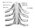
The middle divisions of the posterior branches run close to the articular processes of the vertebrae and end in the multifidus muscle. The outer branches supply the erector spinae muscles.
The anterior divisions of the lumbar nerves (Latin: rami anteriores) increase in size from above downward.
The anterior divisions communicate with the sympathetic trunk. Near the origin of the divisions, they are joined by gray rami communicantes from the lumbar ganglia of the sympathetic trunk. These rami consist of long, slender branches which accompany the lumbar arteries around the sides of the vertebral bodies, beneath the Psoas major. Their arrangement is somewhat irregular: one ganglion may give rami to two lumbar nerves, or one lumbar nerve may receive rami (branches) from two ganglia. The first and second, and sometimes the third and fourth lumbar nerves are each connected with the lumbar part of the sympathetic trunk by a white ramus communicans.
As the nerves travel forward, they create nervous plexuses. The first three lumbar nerves, and the greater part of the fourth together form the lumbar plexus. The smaller part of the fourth joins with the fifth to form the lumbosacral trunk, which assists in the formation of the sacral plexus.
The fourth nerve is named the furcal nerve, from the fact that it is subdivided between the two plexuses.
L1 supplies many muscles, either directly or through nerves originating from L1. They may be innervated with L1 as single origin, or be innervated partly by L1 and partly by other spinal nerves. The muscles are:
The second lumbar spinal nerve (L2)[2] originates from the spinal column from below the lumbar vertebra 2 (L2).
L2 supplies many muscles, either directly or through nerves originating from L2. They may be innervated with L2 as single origin, or be innervated partly by L2 and partly by other spinal nerves. The muscles are:
The third lumbar spinal nerve (L3)[3] originates from the spinal column from below the lumbar vertebra 3 (L3).
L3 supplies many muscles, either directly or through nerves originating from L3. They may be innervated with L3 as single origin, or be innervated partly by L3 and partly by other spinal nerves. The muscles are:
The fourth lumbar spinal nerve (L4)[4] originates from the spinal column from below the lumbar vertebra 4 (L4).
L4 supplies many muscles, either directly or through nerves originating from L4. They are not innervated with L4 as single origin, but partly by L4 and partly by other spinal nerves. The muscles are:
The fifth lumbar spinal nerve 5 (L5)[5] originates from the spinal column from below the lumbar vertebra 5 (L5).
L5 supplies many muscles, either directly or through nerves originating from L5. They are not innervated with L5 as single origin, but partly by L5 and partly by other spinal nerves. The muscles are:
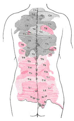
Areas of distribution of the cutaneous branches of the posterior divisions of the spinal nerves. The areas of the medial branches are in black, those of the lateral in red.
This page is based on this Wikipedia article Text is available under the CC BY-SA 4.0 license; additional terms may apply. Images, videos and audio are available under their respective licenses.