Gray's Anatomy is a reference book of human anatomy written by Henry Gray, illustrated by Henry Vandyke Carter and first published in London in 1858. It has had multiple revised editions and the current edition, the 42nd, remains a standard reference, often considered "the doctors' bible".

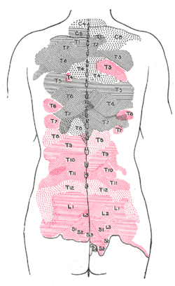
A spinal nerve is a mixed nerve, which carries motor, sensory, and autonomic signals between the spinal cord and the body. In the human body there are 31 pairs of spinal nerves, one on each side of the vertebral column. These are grouped into the corresponding cervical, thoracic, lumbar, sacral and coccygeal regions of the spine. There are eight pairs of cervical nerves, twelve pairs of thoracic nerves, five pairs of lumbar nerves, five pairs of sacral nerves, and one pair of coccygeal nerves. The spinal nerves are part of the peripheral nervous system.

The occipital artery is a branch of the external carotid artery that provides arterial supply to the back of the scalp, sternocleidomastoid muscles, and deep muscles of the back and neck.

The urethral artery arises from the internal pudendal artery a branch of the internal iliac artery. The internal pudendal artery has numerous branches including the artery of the bulb of the penis immediately before the urethral and the dorsal artery of the penis more distally.

The right colic artery is an artery of the abdomen, a branch of the superior mesenteric artery supplying the ascending colon. It divides into two terminal branches - an ascending branch and a descending branch - which form anastomoses with the middle colic artery, and ileocolic artery (respectively).

The middle colic artery is an artery of the abdomen; a branch of the superior mesenteric artery distributed to parts of the ascending and transverse colon. It usually divides into two terminal branches - a left one and a right one - which go on to form anastomoses with the left colic artery, and right colic artery (respectively), thus participating in the formation of the marginal artery of the colon.
The left colic artery is a branch of the inferior mesenteric artery distributed to the descending colon, and left part of the transverse colon. It ends by dividing into an ascending branch and a descending branch; the terminal branches of the two branches go on to form anastomoses with the middle colic artery, and a sigmoid artery (respectively).

The medial circumflex femoral artery is an artery in the upper thigh that arises from the profunda femoris artery. It supplies arterial blood to several muscles in the region, as well as the femoral head and neck.

The lacrimal artery is an artery of the orbit. It is a branch of the ophthalmic artery. It accompanies the lacrimal nerve along the upper border of the lateral rectus muscle, travelling forward to reach the lacrimal gland. It supplies the lacrimal gland, two rectus muscles of the eye, the eyelids, and the conjunctiva.

The deep auricular artery is a branch of the maxillary artery. The deep auricular artery pierces the external acoustic meatus. It provides arterial supply to the skin of the external acoustic meatus, and contributes arterial supply to the tympanic membrane, and the temporomandibular joint.
The posterior ethmoidal artery is an artery of the head which arises from the ophthalmic artery to supply the posterior ethmoidal air cells, and the meninges. It is smaller than the anterior ethmoidal artery.

The appendicular artery, also known as the appendiceal artery, commonly arises from the terminal branch of the ileocolic artery, or less commonly from the posterior cecal artery or an ileal artery. It descends behind the termination of the ileum and enters the mesoappendix of the vermiform appendix. It runs near the free margin of the mesoappendix and ends in branches which supply the appendix.
The supraorbital artery is a branch of the ophthalmic artery. It passes anteriorly within the orbit to exit the orbit through the supraorbital foramen or notch alongside the supraorbital nerve, splitting into two terminal branches which go on to form anastomoses with arteries of the head.
The medial palpebral arteries are arteries of the head that contribute arterial blood supply to the eyelids. They are derived from the ophthalmic artery; a single medial palpebral artery issues from the ophthalmic artery before splitting into a superior and an inferior medial palpebral artery, each supplying one eyelid.

The palmar branch of the ulnar nerve arises about five cm proximal to the wrist from where the ulnar nerve splits into palmar and dorsal branches. It supplies sensory innervation to a small area in the palmar surface of the wrist.
The palmar branch of the median nerve is a branch of the median nerve which arises at the distal part of the forearm.

The digastric branch of facial nerve provides motor innervation to the posterior belly of the digastric muscle. It branches from the facial nerve near to the stylomastoid foramen as the CN VII exits the facial canal. It commonly arises in common with the stylohyoid branch of facial nerve.
The stylohyoid branch of facial nerve provides motor innervation to the stylohyoid muscle. It frequently arises from the facial nerve in common with the digastric branch of facial nerve.
The salivatory nuclei are two parasympathetic general visceral efferent cranial nerve nuclei - the superior salivatory nucleus and the inferior salivatory nucleus - that innervate the salivary glands. Both are located in the pontine tegmentum of the brainstem.
Crossing the under surface of the sphenoid the sphenopalatine artery ends on the nasal septum as the posterior septal branches; these anastomose with the ethmoidal arteries and the septal branch of the superior labial; one branch descends in a groove on the vomer to the incisive canal and anastomoses with the descending palatine artery.