
In anatomy, the epidural space is the potential space between the dura mater and vertebrae (spine).

The posterior cranial fossa is the part of the cranial cavity located between the foramen magnum, and tentorium cerebelli. It is formed by the sphenoid bones, temporal bones, and occipital bone. It lodges the cerebellum, and parts of the brainstem.
The emissary veins connect the extracranial venous system with the intracranial venous sinuses. They connect the veins outside the cranium to the venous sinuses inside the cranium. They drain from the scalp, through the skull, into the larger meningeal veins and dural venous sinuses. They may also connect to diploic veins within the skull.

The cavernous sinus within the human head is one of the dural venous sinuses creating a cavity called the lateral sellar compartment bordered by the temporal bone of the skull and the sphenoid bone, lateral to the sella turcica.
The confluence of sinuses, torcular Herophili, or torcula is the connecting point of the superior sagittal sinus, straight sinus, and occipital sinus. It is below the internal occipital protuberance of the skull. It drains venous blood from the brain into the transverse sinuses. It may be affected by arteriovenous fistulas, a thrombus, major trauma, or surgical damage, and may be imaged with many radiology techniques.

The occipital sinus is the smallest of the dural venous sinuses. It is usually unpaired, and is sometimes altogether absent. It is situated in the attached margin of the falx cerebelli. It commences near the foramen magnum, and ends by draining into the confluence of sinuses.
The inferior petrosal sinuses are two small sinuses situated on the inferior border of the petrous part of the temporal bone, one on each side. Each inferior petrosal sinus drains the cavernous sinus into the internal jugular vein.

The inferior ophthalmic vein is a vein of the orbit that - together with the superior ophthalmic vein - represents the principal drainage system of the orbit. It begins from a venous network in the front of the orbit, then passes backwards through the lower orbit. It drains several structures of the orbit. It may end by splitting into two branches, one draining into the pterygoid venous plexus and the other ultimately into the cavernous sinus.

The pterygoid plexus is a fine venous plexus upon and within the lateral pterygoid muscle. It drains by a short maxillary vein.

The carotid canal is a passage in the petrous part of the temporal bone of the skull through which the internal carotid artery and its internal carotid (nervous) plexus pass from the neck into the cranial cavity.

The occipital vein is a vein of the scalp. It originates from a plexus around the external occipital protuberance and superior nuchal line to the back part of the vertex of the skull. It usually drains into the internal jugular vein, but may also drain into the posterior auricular vein. It drains part of the scalp.

The mastoid foramen is a hole in the posterior border of the temporal bone. It transmits an emissary vein between the sigmoid sinus and the suboccipital venous plexus, and a small branch of the occipital artery, the posterior meningeal artery to the dura mater.
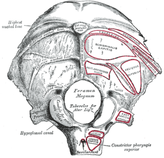
The condylar canal is a canal in the condyloid fossa of the lateral parts of occipital bone behind the occipital condyle. Resection of the rectus capitis posterior major and minor muscles reveals the bony recess leading to the condylar canal, which is situated posterior and lateral to the occipital condyle. It is immediately superior to the extradural vertebral artery, which makes a loop above the posterior C1 ring to enter the foramen magnum. The anteriomedial wall of the condylar canal thickens to join the foramen magnum rim and connect to the occipital condyle.

The internal vertebral venous plexuses lie within the vertebral canal in the epidural space, embedded within epidural fat. They receive tributaries from bones, red bone marrow, and spinal cord. They are arranged into four interconnected, vertically oriented vessels - two situated anteriorly, and two posteriorly:

The spinal veins are situated in the pia mater and form a minute, tortuous, venous plexus.

In vertebrates, a venous plexus is a normal congregation anywhere in the body of multiple veins.
Anterior spinal veins are veins that receive blood from the anterior spinal cord.
The cerebrospinal venous system (CSVS) consists of the interconnected venous systems of the brain and the spine.
The venous plexus of hypoglossal canal is a small venous plexus surrounding the hypoglossal nerve as it passes through the hypoglossal canal. The plexus connects with the occipital sinus (intercranially), inferior petrosal sinus (intercranially), internal jugular vein (extracranially), condylar vein, and paravertebral venous plexus.
The marginal sinus is a dural venous sinus surrounding the margin of the foramen magnum inside the skull, accommodated by the groove for marginal sinus. It usually drains into either the sigmoid sinus, or the jugular bulb. It communicates with the basilar venous plexus anteriorly, and the occipital sinus posteriorly ; it may form extracranial communications with the internal vertebral venous plexuses, or deep cervical veins.
