
The tongue is a muscular organ in the mouth of a typical tetrapod. It manipulates food for chewing and swallowing as part of the digestive process, and is the primary organ of taste. The tongue's upper surface (dorsum) is covered by taste buds housed in numerous lingual papillae. It is sensitive and kept moist by saliva and is richly supplied with nerves and blood vessels. The tongue also serves as a natural means of cleaning the teeth. A major function of the tongue is the enabling of speech in humans and vocalization in other animals.
Articles related to anatomy include:

The hypoglossal nerve, also known as the twelfth cranial nerve, cranial nerve XII, or simply CN XII, is a cranial nerve that innervates all the extrinsic and intrinsic muscles of the tongue except for the palatoglossus, which is innervated by the vagus nerve. CN XII is a nerve with a sole motor function. The nerve arises from the hypoglossal nucleus in the medulla as a number of small rootlets, pass through the hypoglossal canal and down through the neck, and eventually passes up again over the tongue muscles it supplies into the tongue.

Taste buds are clusters of taste receptor cells, which are also known as gustatory cells. The taste receptors are located around the small structures known as papillae found on the upper surface of the tongue, soft palate, upper esophagus, the cheek, and epiglottis. These structures are involved in detecting the five elements of taste perception: saltiness, sourness, bitterness, sweetness and savoriness (umami). A popular myth assigns these different tastes to different regions of the tongue; in fact, these tastes can be detected by any area of the tongue. Via small openings in the tongue epithelium, called taste pores, parts of the food dissolved in saliva come into contact with the taste receptors. These are located on top of the taste receptor cells that constitute the taste buds. The taste receptor cells send information detected by clusters of various receptors and ion channels to the gustatory areas of the brain via the seventh, ninth and tenth cranial nerves.
Black hairy tongue syndrome (BHT) is a condition of the tongue in which the small bumps on the tongue elongate with black or brown discoloration, giving a black and hairy appearance. The appearance may be alarming, but it is a harmless condition. Predisposing factors include smoking, xerostomia, soft diet, poor oral hygiene and certain medications. Management is facilitated by improving oral hygiene, especially scraping or brushing the tongue.

Glossitis can mean soreness of the tongue, or more usually inflammation with depapillation of the dorsal surface of the tongue, leaving a smooth and erythematous (reddened) surface,. In a wider sense, glossitis can mean inflammation of the tongue generally. Glossitis is often caused by nutritional deficiencies and may be painless or cause discomfort. Glossitis usually responds well to treatment if the cause is identified and corrected. Tongue soreness caused by glossitis is differentiated from burning mouth syndrome, where there is no identifiable change in the appearance of the tongue, and there are no identifiable causes.

The frenulumof the tongue, tongue web, lingual frenulum, frenulum linguae, or fraenulum is a small fold of mucous membrane extending from the floor of the mouth to the midline of the underside of the human tongue.

The hyoglossus is a thin and quadrilateral extrinsic muscle of the tongue. It originates from the hyoid bone; it inserts onto the side of the tongue. It is innervated by the hypoglossal nerve. It acts to depress and retract the tongue.

The lingual artery arises from the external carotid artery between the superior thyroid artery and facial artery. It can be located easily in the tongue.

The inferior alveolar artery is an artery of the head. It is a branch of the maxillary artery. It descends through the infratemporal fossa as part of a neurovascular bundle with the inferior alveolar nerve and vein to the mandibular foramen where it enters and passes anteriorly inside the mandible, supplying the body of mandible and the dental pulp of the lower molar and premolar teeth. Its terminal incisor branch supplies the rest of the lower teeth. Its mental branch exits the mandibula anteriorly through the mental foramen to supply adjacent lip and skin.
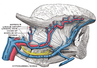
The lingual veins are multiple veins of the tongue with two distinct courses: one group drains into the lingual artery; another group drains either into the lingual artery, (common) facial vein, or internal jugular vein.

This article describes the anatomy of the head and neck of the human body, including the brain, bones, muscles, blood vessels, nerves, glands, nose, mouth, teeth, tongue, and throat.

The following outline is provided as an overview of and topical guide to human anatomy:
The deep lingual vein is one of the lingual veins. It commences near the apex of the tongue. It passes posterior-ward close to the inferior surface of the tongue. It terminates near the anterior border of the hyoglossus muscle by uniting with the sublingual vein to form the vena comitans of the hypoglossal nerve ; this vein then passes posterior-ward alongside the nerve to empty into either a lingual vein, the (common) facial vein, or the internal jugular vein.

Tongue diseases can be congenital or acquired, and are multiple in number. Considered according to a surgical sieve, some example conditions which can involve the tongue are discussed below. Glossitis is a general term for tongue inflammation, which can have various etiologies, e.g. infection.

Median rhomboid glossitis is a condition characterized by an area of redness and loss of lingual papillae on the central dorsum of the tongue, sometimes including lesions of the tongue and palate. It is seen in patients using inhaled steroids and smokers, and is usually a kind of chronic atrophic oral candidiasis, but hematinic deficiency and diabetes should be excluded.

The sublingual space is a fascial space of the head and neck. It is a potential space located below the mouth and above the mylohyoid muscle, and is part of the suprahyoid group of fascial spaces.

Lingual papillae are small structures on the upper surface of the tongue that give it its characteristic rough texture. The four types of papillae on the human tongue have different structures and are accordingly classified as circumvallate, fungiform, filiform, and foliate. All except the filiform papillae are associated with taste buds.
