| Choroid veins | |
|---|---|
 | |
| Details | |
| Identifiers | |
| Latin | venae choroideae |
| Anatomical terminology | |
The choroid veins are the superior choroid vein, and the inferior choroid vein of the lateral ventricle. Both veins drain different parts of the choroid plexus.
| Choroid veins | |
|---|---|
| | |
| Details | |
| Identifiers | |
| Latin | venae choroideae |
| Anatomical terminology | |
The choroid veins are the superior choroid vein, and the inferior choroid vein of the lateral ventricle. Both veins drain different parts of the choroid plexus.
The superior choroid vein runs along the length of the choroid plexus in the lateral ventricle. It drains the choroid plexus, and also the hippocampus, fornix, and corpus callosum. It unites with the superior thalamostriate vein to form the internal cerebral vein. [1] [2]
The inferior choroid vein drains the inferior choroid plexus into the basal vein. [1]

Cerebrospinal fluid (CSF) is a clear, colorless body fluid found within the tissue that surrounds the brain and spinal cord of all vertebrates.
The ventricular system is a set of four interconnected cavities known as cerebral ventricles in the brain. Within each ventricle is a region of choroid plexus which produces the circulating cerebrospinal fluid (CSF). The ventricular system is continuous with the central canal of the spinal cord from the fourth ventricle, allowing for the flow of CSF to circulate.

The third ventricle is one of the four connected ventricles of the ventricular system within the mammalian brain. It is a slit-like cavity formed in the diencephalon between the two thalami, in the midline between the right and left lateral ventricles, and is filled with cerebrospinal fluid (CSF).
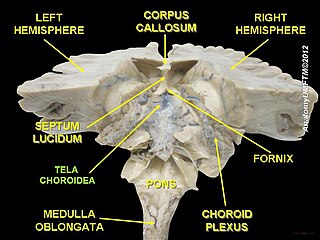
The choroid plexus, or plica choroidea, is a plexus of cells that arises from the tela choroidea in each of the ventricles of the brain. Regions of the choroid plexus produce and secrete most of the cerebrospinal fluid (CSF) of the central nervous system. The choroid plexus consists of modified ependymal cells surrounding a core of capillaries and loose connective tissue. Multiple cilia on the ependymal cells move to circulate the cerebrospinal fluid.

The omohyoid muscle is a muscle that depresses the hyoid. It is located in the front of the neck, and consists of two bellies separated by an intermediate tendon. The omohyoid muscle is proximally attached to the scapula and distally attached to the hyoid bone, stabilising it. Its superior belly serves as the most lateral member of the infrahyoid muscles, located lateral to both the sternothyroid muscles and the thyrohyoid muscles.

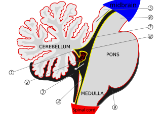
The fourth ventricle is one of the four connected fluid-filled cavities within the human brain. These cavities, known collectively as the ventricular system, consist of the left and right lateral ventricles, the third ventricle, and the fourth ventricle. The fourth ventricle extends from the cerebral aqueduct to the obex, and is filled with cerebrospinal fluid (CSF).
The median aperture is an opening of the fourth ventricle at the caudal portion of the roof of the fourth ventricle. It allows flow of cerebrospinal fluid (CSF) from the fourth ventricle into the cisterna magna. The other two openings of the fourth ventricle are the lateral apertures - one on either side. Nonetheless, the median aperture accounts for most of the outflow of CSF out of the fourth ventricle. The median aperture varies in size.

The lateral ventricles are the two largest ventricles of the brain and contain cerebrospinal fluid (CSF). Each cerebral hemisphere contains a lateral ventricle, known as the left or right lateral ventricle, respectively.

In the brain, the interventricular foramina are channels that connect the paired lateral ventricles with the third ventricle at the midline of the brain. As channels, they allow cerebrospinal fluid (CSF) produced in the lateral ventricles to reach the third ventricle and then the rest of the brain's ventricular system. The walls of the interventricular foramina also contain choroid plexus, a specialized CSF-producing structure, that is continuous with that of the lateral and third ventricles above and below it.

The cisterna magna is the largest of the subarachnoid cisterns. It occupies the space created by the angle between the caudal/inferior surface of the cerebellum, and the dorsal/posterior surface of the medulla oblongata. The fourth ventricle communicates with the cistern via the unpaired midline median aperture. It is continuous inferiorly with the subarachnoid space of the spinal canal.

The anterior choroidal artery is a bilaterally paired artery of the brain. It is typically a branch of the internal carotid artery which supplies the choroid plexus of lateral ventricle and third ventricle as well as numerous structures of the brain.

The cavernous sinus within the human head is one of the dural venous sinuses creating a cavity called the lateral sellar compartment bordered by the temporal bone of the skull and the sphenoid bone, lateral to the sella turcica.

The carotid sheath is a condensation of the deep cervical fascia enveloping multiple vital neurovascular structures of the neck, including the common and internal carotid arteries, the internal jugular vein, the vagus nerve, and ansa cervicalis. The carotid sheath helps protects the structures contained therein.

The posterior inferior cerebellar artery (PICA) is the largest branch of the vertebral artery. It is one of the three main arteries that supply blood to the cerebellum, a part of the brain. Blockage of the posterior inferior cerebellar artery can result in a type of stroke called lateral medullary syndrome.

The inferior ophthalmic vein is a vein of the orbit that - together with the superior ophthalmic vein - represents the principal drainage system of the orbit. It begins from a venous network in the front of the orbit, then passes backwards through the lower orbit. It drains several structures of the orbit. It may end by splitting into two branches, one draining into the pterygoid venous plexus and the other ultimately into the cavernous sinus.

The rectal venous plexus is the venous plexus surrounding the rectum. It consists of an internal and an external rectal plexus. It is drained by the superior, middle, and inferior rectal veins. It forms a portosystemic (portocaval) anastomosis. This allows rectally administered medications to bypassing first pass metabolism.

The tela choroidea is a region of meningeal pia mater that adheres to the underlying ependyma, and gives rise to the choroid plexus in each of the brain’s four ventricles. Tela is Latin for woven and is used to describe a web-like membrane or layer. The tela choroidea is a very thin part of the loose connective tissue of pia mater overlying and closely adhering to the ependyma. It has a rich blood supply. The ependyma and vascular pia mater – the tela choroidea, form regions of minute projections known as a choroid plexus that projects into each ventricle. The choroid plexus produces most of the cerebrospinal fluid of the central nervous system that circulates through the ventricles of the brain, the central canal of the spinal cord, and the subarachnoid space. The tela choroidea in the ventricles forms from different parts of the roof plate in the development of the embryo.

The superior medullary velum is a thin, transparent lamina of white matter which - together with the inferior medullary velum - forms the roof of the fourth ventricle. It extends between the two superior cerebellar peduncles. The lingula of cerebellum covers - and adheres to - its dorsal surface.

Lamina affixa is a layer of epithelium growing on the surface of the thalamus and forming the floor of the central part of lateral ventricle, on whose medial margin is attached the choroid plexus of the lateral ventricle; it covers the superior thalamostriate vein and the superior choroid vein. The torn edge of this plexus is called the tela choroidea.

The Infratemporal space is a fascial space of the head and neck. It is a potential space in the side of the head, and is paired on either side. It is located posterior to the maxilla, between the lateral pterygoid plate of the sphenoid bone medially and by the base of skull superiorly. The term is derived from infra- meaning below and temporal which refers to the temporalis muscle.
| | This cardiovascular system article is a stub. You can help Wikipedia by expanding it. |