Articles related to anatomy include:

The internal carotid artery is an artery in the neck which supplies the anterior circulation of the brain.

The great cerebral vein is one of the large blood vessels in the skull draining the cerebrum of the brain. It is also known as the vein of Galen, named for its discoverer, the Greek physician Galen.

A carotid-cavernous fistula results from an abnormal communication between the arterial and venous systems within the cavernous sinus in the skull. It is a type of arteriovenous fistula. As arterial blood under high pressure enters the cavernous sinus, the normal venous return to the cavernous sinus is impeded and this causes engorgement of the draining veins, manifesting most dramatically as a sudden engorgement and redness of the eye of the same side.

The cavernous sinus within the human head is one of the dural venous sinuses creating a cavity called the lateral sellar compartment bordered by the temporal bone of the skull and the sphenoid bone, lateral to the sella turcica.
The confluence of sinuses, torcular Herophili, or torcula is the connecting point of the superior sagittal sinus, straight sinus, and occipital sinus. It is below the internal occipital protuberance of the skull. It drains venous blood from the brain into the transverse sinuses. It may be affected by arteriovenous fistulas, a thrombus, major trauma, or surgical damage, and may be imaged with many radiology techniques.
The straight sinus, also known as tentorial sinus or the sinus rectus, is an area within the skull beneath the brain. It receives blood from the inferior sagittal sinus and the great cerebral vein, and drains into the confluence of sinuses.
The superior sagittal sinus, within the human head, is an unpaired area along the attached margin of the falx cerebri. It allows blood to drain from the lateral aspects of anterior cerebral hemispheres to the confluence of sinuses. Cerebrospinal fluid drains through arachnoid granulations into the superior sagittal sinus and is returned to venous circulation.

A dural arteriovenous fistula (DAVF) or malformation is an abnormal direct connection (fistula) between a meningeal artery and a meningeal vein or dural venous sinus.
The sigmoid sinuses, also known as the pars sigmoid, are paired dural venous sinuses within the skull that receive blood from posterior transverse sinuses.
The transverse sinuses, within the human head, are two areas beneath the brain which allow blood to drain from the back of the head. They run laterally in a groove along the interior surface of the occipital bone. They drain from the confluence of sinuses to the sigmoid sinuses, which ultimately connect to the internal jugular vein. See diagram : labeled under the brain as "SIN. TRANS.".

The angular vein is a vein of the face. It is the upper part of the facial vein, above its junction with the superior labial vein. It is formed by the junction of the supratrochlear vein and supraorbital vein, and joins with the superior labial vein. It drains the medial canthus, and parts of the nose and the upper lip. It can be a route of spread of infection from the danger triangle of the face to the cavernous sinus.

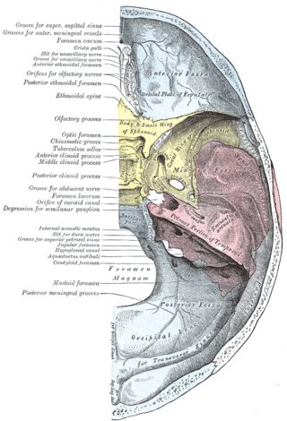
The middle cranial fossa is formed by the sphenoid bones, and the temporal bones. It lodges the temporal lobes, and the pituitary gland. It is deeper than the anterior cranial fossa, is narrow medially and widens laterally to the sides of the skull. It is separated from the posterior cranial fossa by the clivus and the petrous crest.

The cerebellar veins are veins which drain the cerebellum. They consist of the superior cerebellar veins and the inferior cerebellar veins. The superior cerebellar veins drain to the straight sinus and the internal cerebral veins. The inferior cerebellar veins drain to the transverse sinus, the superior petrosal sinus, and the occipital sinus.

The nasofrontal vein is a vein in the orbit around the eye. It drains into the superior ophthalmic vein. It can be used for endovascular access to the cavernous sinus.
The inferior petrosal sulcus is the groove containing the inferior petrosal sinus.

The following outline is provided as an overview of and topical guide to human anatomy:

Vein of Galen aneurysmal malformations(VGAMs) and Vein of Galen aneurysmal dilations (VGADs) are the most frequent arteriovenous malformations in infants and fetuses. A VGAM consists of a tangled mass of dilated vessels supplied by an enlarged artery. The malformation increases greatly in size with age, although the mechanism of the increase is unknown. Dilation of the great cerebral vein of Galen is a secondary result of the force of arterial blood either directly from an artery via an arteriovenous fistula or by way of a tributary vein that receives the blood directly from an artery. There is usually a venous anomaly downstream from the draining vein that, together with the high blood flow into the great cerebral vein of Galen causes its dilation. The right sided cardiac chambers and pulmonary arteries also develop mild to severe dilation.
Interventional neuroradiology (INR) also known as neurointerventional surgery (NIS), endovascular therapy (EVT), endovascular neurosurgery, and interventional neurology is a medical subspecialty of neurosurgery, neuroradiology, intervention radiology and neurology specializing in minimally invasive image-based technologies and procedures used in diagnosis and treatment of diseases of the head, neck, and spine.