The brachiocephalic artery,brachiocephalic trunk, or innominate artery is an artery of the mediastinum that supplies blood to the right arm, head, and neck.

The left and right brachiocephalic veins are major veins in the upper chest, formed by the union of the ipsilateral internal jugular vein and subclavian vein behind the sternoclavicular joint. The left brachiocephalic vein is more than twice the length of the right brachiocephalic vein.

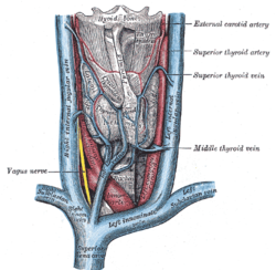
The external carotid artery is a major artery of the head and neck. It arises from the common carotid artery when it splits into the external and internal carotid artery. The external carotid artery supplies blood to the face, brain and neck.

The internal jugular vein is a paired jugular vein that collects blood from the brain and the superficial parts of the face and neck. This vein runs in the carotid sheath with the common carotid artery and vagus nerve.
In anatomy, the left and right common carotid arteries (carotids) are arteries that supply the head and neck with oxygenated blood; they divide in the neck to form the external and internal carotid arteries.

The thyrohyoid muscle is a small skeletal muscle of the neck. Above, it attaches onto the greater cornu of the hyoid bone; below, it attaches onto the oblique line of the thyroid cartilage. It is innervated by fibres derived from the cervical spinal nerve 1 that run with the hypoglossal nerve to reach this muscle. The thyrohyoid muscle depresses the hyoid bone and elevates the larynx during swallowing. By controlling the position and shape of the larynx, it aids in making sound.

The superior ophthalmic vein is a vein of the orbit that drains venous blood from structures of the upper orbit. It is formed by the union of the angular vein, and supraorbital vein. It passes backwards within the orbit alongside the ophthalmic artery, then exits the orbit through the superior orbital fissure to drain into the cavernous sinus.
The inferior thyroid veins appear two, frequently three or four, in number, and arise in the venous plexus on the thyroid gland, communicating with the middle and superior thyroid veins. While the superior and middle thyroid veins serve as direct tributaries to the internal jugular vein, the inferior thyroid veins drain directly to the brachiocephalic veins.
The middle thyroid vein collects the blood from the lower portion of the thyroid gland. It receives tributaries that drain the larynx, and trachea. It passes anterior to the common carotid artery to reach and drain into the internal jugular vein.
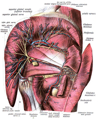
The superior gluteal veins are venæ comitantes of the superior gluteal artery. They receive tributaries from the buttock corresponding with the branches of the artery. They enter the pelvis through the greater sciatic foramen, superior to the piriformis. They drain into internal iliac vein.

The inferior thyroid artery is an artery in the neck. It arises from the thyrocervical trunk and passes upward, in front of the vertebral artery and longus colli muscle. It then turns medially behind the carotid sheath and its contents, and also behind the sympathetic trunk, the middle cervical ganglion resting upon the vessel.

The pharyngeal veins commence in the pharyngeal plexus superficial to the pharynx. The pharyngeal veins receive as tributaries meningeal vein, and the vein of the pterygoid canal. The pharyngeal veins typically empty into the internal jugular vein.

The internal iliac vein begins near the upper part of the greater sciatic foramen, passes upward behind and slightly medial to the internal iliac artery and, at the brim of the pelvis, joins with the external iliac vein to form the common iliac vein.

The carotid triangle is a portion of the anterior triangle of the neck.

The inferior carotid triangle, is bounded, in front, by the median line of the neck from the hyoid bone to the sternum; behind, by the anterior margin of the sternocleidomastoid; above, by the superior belly of the omohyoid.

The thyroid ima artery is an artery of the head and neck. It is an anatomical variant that, when present, supplies blood to the thyroid gland primarily, or the trachea, the parathyroid gland and the thymus gland in rare cases. It has also been reported to be a compensatory artery when one or both of the inferior thyroid arteries are absent, and in a few cases the only source of blood to the thyroid gland. Furthermore, it varies in origin, size, blood supply, and termination, and occurs in around 3.8% of the population and is 4.5 times more common in fetuses than in adults. Because of the variations and rarity, it may lead to surgical complications, particularly during tracheostomy and other airway managements.
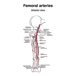
The superficial iliac circumflex artery, the smallest of the cutaneous branches of the femoral artery, arises close to the superficial epigastric artery, and, piercing the fascia lata, runs lateralward, parallel with the inguinal ligament, as far as the crest of the ilium.

The middle cervical ganglion is the smallest of the three cervical sympathetic ganglia. It presumably represents the merging of the sympathetic ganglia of cervical segments C5–C6. It is usually situated at the level of the sixth cervical vertebra.

The cystic veins drain venous blood from the gallbladder and the cystic duct. The cystic veins either drain into various branches and tributaries of the hepatic portal vein.
The left colic vein is a vein that drains the left colic flexure and descending colon. It empties into the inferior mesenteric vein. It accompanies the left colic artery.