Esophageal rupture

Last updated
Boerhaave syndrome
Boorhaave1.JPG
Axial CT image through the upper chest showing extraluminal air (Pneumomediastinum) surrounding the trachea and esophagus
Specialty Gastroenterology
General surgery

Esophageal rupture is a rupture of the esophageal wall. Iatrogenic causes account for approximately 56% of esophageal perforations, usually due to medical instrumentation such as an endoscopy or paraesophageal surgery. [1] The 10% of esophageal perforations caused specifically by vomiting are termed Boerhaave syndrome. [2]

Contents

Spontaneous perforation of the esophagus is most commonly a full-thickness tear in the esophageal wall due to a sudden increase in intraesophageal pressure combined with relatively negative intrathoracic pressure caused by straining or vomiting (effort rupture of the esophagus or Boerhaave's syndrome). Other causes of spontaneous perforation include caustic ingestion, pill esophagitis, Barrett's esophagus, infectious ulcers in patients with AIDS, and following dilation of esophageal strictures.[ citation needed ]

In most cases of Boerhaave's syndrome, the tear occurs at the left postero-lateral aspect of the distal esophagus and extends for several centimeters. The condition is associated with high morbidity and mortality and is fatal without treatment. The occasionally nonspecific nature of the symptoms may contribute to a delay in diagnosis and a poor outcome. Spontaneous effort rupture of the cervical esophagus, leading to localized cervical perforation, may be more common than previously recognized and has a generally benign course. Pre-existing esophageal disease is not a prerequisite for esophageal perforation, but it contributes to increased mortality.[ citation needed ]

This condition was first documented by the 18th-century physician Herman Boerhaave, after whom it is named. [3] [4] A related condition is Mallory-Weiss syndrome which is only a mucosal tear. A common site of iatrogenic perforation is the cervical esophagus just above the upper sphincter, whereas spontaneous rupture as seen in Boerhaave's syndrome perforation commonly occurs in the lower third of the esophagus. [5]

Signs and symptoms

The classic history of esophageal rupture is one of severe retching and vomiting followed by excruciating retrosternal chest and upper abdominal pain. Odynophagia, tachypnea, dyspnea, cyanosis, fever, and shock develop rapidly thereafter. [6]

Physical examination is usually not helpful, particularly early in the course. Subcutaneous emphysema (crepitation) is an important diagnostic finding but is not very sensitive, being present in only 9 of 34 patients (27 percent) in one series. A pleural effusion may be detected. [7]

Mackler's triad includes chest pain, vomiting, and subcutaneous emphysema, and while it is a classical presentation, it is only present in 14% of people. [8]

Pain can occasionally radiate to the left shoulder, causing physicians to confuse an esophageal perforation with a myocardial infarction.[ citation needed ]

It may also be audibly recognized as Hamman's sign. [9]

Pathophysiology

Esophageal rupture, in Boerhaave syndrome, is thought to be the result of a sudden rise in internal esophageal pressure produced during vomiting, as a result of neuromuscular incoordination causing failure of the cricopharyngeus muscle (a sphincter within the pharynx) to relax. As the intra-oesophageal pressure increases, the bolus within the oesophagus has nowhere to go superiorly (as the cricopharyngeus fails to relax) which causes the oesophagus to rupture. The syndrome is commonly associated with the consumption of excessive food and/or alcohol as well as eating disorders such as bulimia.[ citation needed ]

The most common anatomical location of the tear in Boerhaave syndrome is at left posterolateral wall of the lower third of the esophagus, 2–3 cm before the stomach. [10]

Currently, the most common cause of esophageal perforation is iatrogenic. However, iatrogenic perforations, while still constituting a serious medical condition, are easier to treat and less prone to complications, particularly mediastinitis and sepsis. This is because they usually do not involve contamination of the mediastinum with gastric contents.[ citation needed ]

Diagnosis

Upright chest radiography showing mediastinal air adjacent to the aorta and tracking cephalad adjacent to the left common carotid artery. This patient presented to the emergency department with severe chest pain after eating. CXR Pneumomediastinum.jpg
Upright chest radiography showing mediastinal air adjacent to the aorta and tracking cephalad adjacent to the left common carotid artery. This patient presented to the emergency department with severe chest pain after eating.
Sagittal reformatted CT image showing discontinutity in the wall of the posterolateral aspect of the distal esophagus BoorhaaveSag CT.JPG
Sagittal reformatted CT image showing discontinutity in the wall of the posterolateral aspect of the distal esophagus

The diagnosis of Boerhaave's syndrome is suggested on the plain chest radiography and confirmed by chest CT scan. The initial plain chest radiograph is almost always abnormal in patients with Boerhaave's syndrome and usually reveals mediastinal or free peritoneal air as the initial radiologic manifestation. With cervical esophageal perforations, plain films of the neck show air in the soft tissues of the prevertebral space.[ citation needed ]

Hours to days later, pleural effusion(s) with or without pneumothorax, widened mediastinum, and subcutaneous emphysema is typically seen. CT scan may show esophageal wall edema and thickening, extraesophageal air, periesophageal fluid with or without gas bubbles, mediastinal widening, and air and fluid in the pleural spaces, retroperitoneum or lesser sac.[ citation needed ]

The diagnosis of esophageal perforation could also be confirmed by water-soluble contrast esophagram (Gastrografin), which reveals the location and extent of extravasation of contrast material. Although barium is superior in demonstrating small perforations, the spillage of barium sulfate into the mediastinal and pleural cavities can cause an inflammatory response and subsequent fibrosis and is therefore not used as the primary diagnostic study. If, however, the water-soluble study is negative, a barium study should be performed for better definition.[ citation needed ]

Endoscopy has no role in the diagnosis of spontaneous esophageal perforation. Both the endoscope and insufflation of air can extend the perforation and introduce air into the mediastinum.[ citation needed ]

Patients may also have a pleural effusion high in amylase (from saliva), low pH, and may contain particles of food.[ citation needed ]

Differential diagnosis

Common misdiagnoses include myocardial infarction, pancreatitis, lung abscess, pericarditis, and spontaneous pneumothorax. If esophageal perforation is suspected, even in the absence of physical findings, chest xray, water soluble contrast radiographic studies of the esophagus and a CT scan should be promptly obtained. In most cases, non-operative management is administered based on radiological evidence contained in mediastinal collection. [11]

Treatment

With the exception of a few case reports describing survival without surgery, [2] the mortality of untreated Boerhaave syndrome is 100%. [12] With surgical intervention, the mortality rate decreases to 30%. [12] Its treatment includes immediate antibiotic therapy to prevent mediastinitis and sepsis, surgical repair of the perforation, [13] and if there is significant fluid loss it should be replaced with IV fluid therapy since oral rehydration is not possible. Even with early surgical intervention (within 24 hours) the risk of death is 25%. [14]

Related Research Articles

<span class="mw-page-title-main">Pleural cavity</span> Thin fluid-filled space between the two pulmonary pleurae (visceral and parietal) of each lung

The pleural cavity, pleural space, or interpleural space is the potential space between the pleurae of the pleural sac that surrounds each lung. A small amount of serous pleural fluid is maintained in the pleural cavity to enable lubrication between the membranes, and also to create a pressure gradient.

<span class="mw-page-title-main">Hiatal hernia</span> Type of hernia

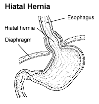
A hiatal hernia or hiatus hernia is a type of hernia in which abdominal organs slip through the diaphragm into the middle compartment of the chest. This may result in gastroesophageal reflux disease (GERD) or laryngopharyngeal reflux (LPR) with symptoms such as a taste of acid in the back of the mouth or heartburn. Other symptoms may include trouble swallowing and chest pains. Complications may include iron deficiency anemia, volvulus, or bowel obstruction.

<span class="mw-page-title-main">Aortic dissection</span> Injury to the innermost layer of the aorta

Aortic dissection (AD) occurs when an injury to the innermost layer of the aorta allows blood to flow between the layers of the aortic wall, forcing the layers apart. In most cases, this is associated with a sudden onset of severe chest or back pain, often described as "tearing" in character. Vomiting, sweating, and lightheadedness may also occur. Damage to other organs may result from the decreased blood supply, such as stroke, lower extremity ischemia, or mesenteric ischemia. Aortic dissection can quickly lead to death from insufficient blood flow to the heart or complete rupture of the aorta.

<span class="mw-page-title-main">Pleural effusion</span> Accumulation of excess fluid in the pleural cavity

A pleural effusion is accumulation of excessive fluid in the pleural space, the potential space that surrounds each lung. Under normal conditions, pleural fluid is secreted by the parietal pleural capillaries at a rate of 0.6 millilitre per kilogram weight per hour, and is cleared by lymphatic absorption leaving behind only 5–15 millilitres of fluid, which helps to maintain a functional vacuum between the parietal and visceral pleurae. Excess fluid within the pleural space can impair inspiration by upsetting the functional vacuum and hydrostatically increasing the resistance against lung expansion, resulting in a fully or partially collapsed lung.

<span class="mw-page-title-main">Mallory–Weiss syndrome</span> Bleeding from a laceration in the mucosa at the junction of the stomach and esophagus

Mallory–Weiss syndrome or gastro-esophageal laceration syndrome refers to bleeding from a laceration in the mucosa at the junction of the stomach and esophagus. This is usually caused by severe vomiting because of alcoholism or bulimia, but can be caused by any condition which causes violent vomiting and retching such as food poisoning. The syndrome presents with hematemesis. The laceration is sometimes referred to as a Mallory–Weiss tear.

<span class="mw-page-title-main">Mediastinum</span> Central part of the thoracic cavity

The mediastinum is the central compartment of the thoracic cavity. Surrounded by loose connective tissue, it is an undelineated region that contains a group of structures within the thorax, namely the heart and its vessels, the esophagus, the trachea, the phrenic and cardiac nerves, the thoracic duct, the thymus and the lymph nodes of the central chest.

<span class="mw-page-title-main">Hemothorax</span> Blood accumulation in the pleural cavity

A hemothorax is an accumulation of blood within the pleural cavity. The symptoms of a hemothorax may include chest pain and difficulty breathing, while the clinical signs may include reduced breath sounds on the affected side and a rapid heart rate. Hemothoraces are usually caused by an injury, but they may occur spontaneously due to cancer invading the pleural cavity, as a result of a blood clotting disorder, as an unusual manifestation of endometriosis, in response to Pneumothorax, or rarely in association with other conditions.

<span class="mw-page-title-main">Chylothorax</span> Medical condition

A chylothorax is an abnormal accumulation of chyle, a type of lipid-rich lymph, in the space surrounding the lung. The lymphatics of the digestive system normally returns lipids absorbed from the small bowel via the thoracic duct, which ascends behind the esophagus to drain into the left brachiocephalic vein. If normal thoracic duct drainage is disrupted, either due to obstruction or rupture, chyle can leak and accumulate within the negative-pressured pleural space. In people on a normal diet, this fluid collection can sometimes be identified by its turbid, milky white appearance, since chyle contains emulsified triglycerides.

<span class="mw-page-title-main">Thoracentesis</span> Removal of fluids/air from the pleural cavity of the lungs

Thoracentesis, also known as thoracocentesis, pleural tap, needle thoracostomy, or needle decompression, is an invasive medical procedure to remove fluid or air from the pleural space for diagnostic or therapeutic purposes. A cannula, or hollow needle, is carefully introduced into the thorax, generally after administration of local anesthesia. The procedure was first performed by Morrill Wyman in 1850 and then described by Henry Ingersoll Bowditch in 1852.

<span class="mw-page-title-main">Pancreatic fistula</span> Medical condition

A pancreatic fistula is an abnormal communication between the pancreas and other organs due to leakage of pancreatic secretions from damaged pancreatic ducts. An external pancreatic fistula is one that communicates with the skin, and is also known as a pancreaticocutaneous fistula, whereas an internal pancreatic fistula communicates with other internal organs or spaces. Pancreatic fistulas can be caused by pancreatic disease, trauma, or surgery.

<span class="mw-page-title-main">Pneumoperitoneum</span> Medical condition

Pneumoperitoneum is pneumatosis in the peritoneal cavity, a potential space within the abdominal cavity. The most common cause is a perforated abdominal organ, generally from a perforated peptic ulcer, although any part of the bowel may perforate from a benign ulcer, tumor or abdominal trauma. A perforated appendix seldom causes a pneumoperitoneum.

<span class="mw-page-title-main">Mediastinitis</span> Inflammatory process affecting the mediastinum

Mediastinitis is inflammation of the tissues in the mid-chest, or mediastinum. It can be either acute or chronic. It is thought to be due to four different etiologies:

<span class="mw-page-title-main">Pneumomediastinum</span> Abnormal presence of gas in the thorax

Pneumomediastinum is pneumatosis in the mediastinum, the central part of the chest cavity. First described in 1819 by René Laennec, the condition can result from physical trauma or other situations that lead to air escaping from the lungs, airways, or bowel into the chest cavity. In underwater divers it is usually the result of pulmonary barotrauma.

<span class="mw-page-title-main">Hamman's syndrome</span> Medical condition

Hamman's syndrome, also known as Macklin's syndrome, is a syndrome of spontaneous subcutaneous emphysema and pneumomediastinum, sometimes associated with pain and, less commonly, dyspnea, dysphonia, and a low-grade fever.

<span class="mw-page-title-main">Mediastinal tumors</span> Medical condition

A mediastinal tumor is a tumor in the mediastinum, the cavity that separates the lungs from the rest of the chest. It contains the heart, esophagus, trachea, thymus, and aorta. The most common mediastinal masses are neurogenic tumors, usually found in the posterior mediastinum, followed by thymoma (15–20%) located in the anterior mediastinum. Lung cancer typically spreads to the lymph nodes in the mediastinum.

<span class="mw-page-title-main">Subcutaneous emphysema</span> Medical condition

Subcutaneous emphysema occurs when gas or air accumulates and seeps under the skin, where normally no gas should be present. Subcutaneous refers to the subcutaneous tissue, and emphysema refers to trapped air pockets. Since the air generally comes from the chest cavity, subcutaneous emphysema usually occurs around the upper torso, such as on the chest, neck, face, axillae and arms, where it is able to travel with little resistance along the loose connective tissue within the superficial fascia. Subcutaneous emphysema has a characteristic crackling-feel to the touch, a sensation that has been described as similar to touching warm Rice Krispies. This sensation of air under the skin is known as subcutaneous crepitation, a form of crepitus.

Pleural disease occurs in the pleural space, which is the thin fluid-filled area in between the two pulmonary pleurae in the human body. There are several disorders and complications that can occur within the pleural area, and the surrounding tissues in the lung.

<span class="mw-page-title-main">Tracheobronchial injury</span> Damage to the tracheobronchial tree

Tracheobronchial injury is damage to the tracheobronchial tree. It can result from blunt or penetrating trauma to the neck or chest, inhalation of harmful fumes or smoke, or aspiration of liquids or objects.

Norman Rupert Barrett CBE FRSA was an Australian-born British thoracic surgeon who is widely yet mistakenly remembered for describing what became known as Barrett's oesophagus.

<span class="mw-page-title-main">Pulmonary pleurae</span> Serous membrane that lines the wall of the thoracic cavity and the surface of the lung

The pulmonary pleurae are the two opposing layers of serous membrane overlying the lungs, mediastinum and the inside surfaces of the surrounding chest walls.

References

  1. Marx, John A.; Hockberger, Robert S.; Walls, Ron M.; Adams, James, eds. (2010). Rosen's emergency medicine: concepts and clinical practice. Vol. 1 (7th ed.). St. Louis: Mosby/Elsevier. ISBN   978-0-323-05472-0.[ page needed ]
  2. 1 2 Boerhaave syndrome at eMedicine
  3. synd/2800 at Who Named It?
  4. Boerhaave, Herman (1724). Atrocis, nec descripti prius, morbis historia: Secundum medicae artis leges conscripta[Bitter or the results were registered before the history of the disease. According to the written laws of medicine] (in Latin). Lugduni Batavorum; Ex officine Boutesteniana. OCLC   952706276.[ page needed ]
  5. Bailey & Love, 25th ed., page 1014[ full citation needed ]
  6. "Esophageal Rupture". MSD Manuals. Retrieved 22 July 2021.
  7. "Pleural Effusion". The Lecturio Medical Concept Library. Retrieved 22 July 2021.
  8. Bautz, B; Schneider, JI (May 2020). "High-Risk Chief Complaints I: Chest Pain-The Big Three (an Update)". Emergency Medicine Clinics of North America. 38 (2): 453–498. doi:10.1016/j.emc.2020.01.009. PMID   32336336. S2CID   216556980.
  9. "Hamman sign" "at Dorland's Medical Dictionary
  10. Korn, Owen; Oñate, Juan C.; López, René (2007). "Anatomy of the Boerhaave syndrome". Surgery. 141 (2): 222–8. doi:10.1016/j.surg.2006.06.034. PMID   17263979.
  11. Wong, Kenneth; Roy, Gerard (2006). "Oesophageal rupture arising as a complication of acute appendicitis in a child". The Medical Journal of Australia. 184 (11): 588. doi:10.5694/j.1326-5377.2006.tb00391.x. PMID   16768671. S2CID   34353120.
  12. 1 2 Curci, Joseph J.; Horman, Marc J. (1976). "Boerhaaveʼs Syndrome. The Importance of Early Diagnosis and Treatment". Annals of Surgery. 183 (4): 401–8. doi:10.1097/00000658-197604000-00013. PMC   1344212 . PMID   1267496.
  13. Matsuda, Akihisa; Miyashita, Masao; Sasajima, Koji; Nomura, Tsutomu; Makino, Hiroshi; Matsutani, Takeshi; Katsuno, Akira; Sasaki, Junpei; Tajiri, Takashi (2006). "Boerhaave Syndrome Treated Conservatively Following Early Endoscopic Diagnosis: A Case Report". Journal of Nippon Medical School. 73 (6): 341–5. doi: 10.1272/jnms.73.341 . PMID   17220586.
  14. Jougon, J; Mcbride, T; Delcambre, F; Minniti, A; Velly, J (2004). "Primary esophageal repair for Boerhaave's syndrome whatever the free interval between perforation and treatment". European Journal of Cardio-Thoracic Surgery. 25 (4): 475–9. doi: 10.1016/j.ejcts.2003.12.029 . PMID   15037257.

Further reading