Hereditary diffuse leukoencephalopathy with spheroids

Last updated
Hereditary diffuse leukoencephalopathy with spheroids (HDLS)
Other namesAdult-onset leukoencephalopathy with axonal spheroids and pigmented glia, Autosomal dominant leukoencephalopathy with neuroaxonal spheroids
Autosomal dominant - en.svg
Hereditary diffuse leukoencephalopathy with spheroids is inherited in an autosomal dominant manner

Hereditary diffuse leukoencephalopathy with spheroids (HDLS) is a rare adult onset autosomal dominant disorder characterized by cerebral white matter degeneration with demyelination and axonal spheroids leading to progressive cognitive and motor dysfunction. Spheroids are axonal swellings with discontinuous or absence of myelin sheaths. It is believed that the disease arises from primary microglial dysfunction that leads to secondary disruption of axonal integrity, neuroaxonal damage, and focal axonal spheroids leading to demyelination. Spheroids in HDLS resemble to some extent those produced by shear stress in a closed head injury with damage to axons, causing them to swell due to blockage of axoplasmic transport. In addition to trauma, axonal spheroids can be found in aged brain, stroke, and in other degenerative diseases. [1] In HDLS, it is uncertain whether demyelination occurs prior to the axonal spheroids or what triggers neurodegeneration after apparently normal brain and white matter development, although genetic deficits suggest that demyelination and axonal pathology may be secondary to microglial dysfunction. [2] The clinical syndrome in patients with HDLS is not specific and it can be mistaken for Alzheimer's disease, frontotemporal dementia, atypical Parkinsonism, multiple sclerosis, or corticobasal degeneration. [3]

Contents

Symptoms

With symptoms of personality changes, behavioral changes, dementia, depression, and epilepsy, HDLS has been commonly misdiagnosed for a number of other diseases. [4] Dementia or frontotemporal behavioral changes, for example, have commonly steered some clinicians to mistakenly consider diagnoses such as Alzheimer's disease, frontotemporal dementia or atypical Parkinsonism. The presence of white matter changes has led to misdiagnosis of multiple sclerosis. HDLS commonly manifests with neuropsychiatric symptoms, progressing to dementia, and after a few years shows motor dysfunction. Eventually patients become reliant on wheelchairs. [3]

White matter degeneration is associated with and makes differential diagnoses out of other adult onset leukodystrophies such as metachromatic leukodystrophy (MLD), Krabbe disease (globoid cell leukodystrophy), and X-linked adrenoleukodystrophy (X-ADL). [2]

DiseaseExclusive Trait
MLDAccumulation of metachromatic material in white matter
Krabbe DiseasePresence of globoid cells derived from microglia which have multiple nuclei
X-ALDPredominant parieto-occipital white matter abnormality
Vanishing White Matter (VWM) Disease
Nasu-Hakola
  • Pain and tenderness of ankles/feet/wrist
  • Cystic bone lesions noticeable on radiological films

[2]

Neuropsychiatric symptoms

Many neuropsychiatric symptoms have been identified in clinical studies of HDLS patients. These include severe depression and anxiety that have been identified in about 70% of HDLS families, verging on suicidal tendencies and substance abuse such as alcoholism. Additionally, patients may exhibit disorientation, confusion, agitation, irritability, aggressiveness, an altered mental state, the loss of the ability to execute learned movements (apraxia), or the inability to speak (mutism). [3]

Motor impairment

Persons with HDLS can develop tremors, decreased body movement, unsteadiness (Parkinsonism, muscles on one side of the body in constant contraction (spastic hemiparesis), impairment in motor and sensory function in the lower extremities (paraparesis), paralysis resulting in partial or total loss of all extremities and torso (tetraparesis), and the lack of voluntary coordination of muscle movements (ataxia). [3]

Causes

The cause of HDLS in most families is mutation in the colony stimulating factor 1 receptor (CSF1R), a growth factor for microglia and monocyte/macrophages, suggesting that microglial dysfunction may be primary in HDLS. [4]

The mutations are concentrated in tyrosine kinase domain (TKD) of the protein. Mutations were mainly found in exons 12-22 of the intracellular TKD, including 10 missense mutations that have a single nucleotide deletion and a single codon deletion that consists of a triplet of nucleotides that have been removed causing a whole amino acid to not be coded. Additionally, three splice site mutations were identified that caused an in-frame deletion of an exon, an expressed nucleotide sequence, leading to the removal of more than 40 amino acids in the TKD. [4]

This determination has based upon genetic studies of 14 HDLS families confirming mutations in this gene. The CSF1 receptor protein primarily functions in regulation, survival, proliferation, and differentiation of microglial cells. [5] The mechanism of microglial dysfunction due to mutations in CSF1R to the myelin loss and axonal spheroid formation remains unknown. Further research is needed to better understand disease pathogenesis.

Pathology

An axial CT scan of a patient presenting a leukoencephalopathy, the base disorder to the family of disorders that HDLS falls under. Leukencephalopathy.PNG
An axial CT scan of a patient presenting a leukoencephalopathy, the base disorder to the family of disorders that HDLS falls under.

In HDLS, there is enlargement of the lateral ventricles and marked thinning or weakening of cerebral white matter. [6] The loss of white matter is caused by myelin loss. These changes are associated with diffuse gliosis, moderate loss of axons and many axonal spheroids. [1]

Activated or ameboid microglia and macrophages that contain myelin debris, lipid droplets and brown autofluorescent pigment granules are found in the areas with demyelination and axonal spheroids. In severely degenerated areas there are many large, reactive astrocytes filled with glial fibrils. [1]

In autopsy cases, it has been shown that white matter abnormalities are relatively confined to the cerebrum while avoiding the cerebellum and many of the major fiber tracts of the nervous system. The exception is the corticospinal tracts(pyramidal tracts) in the brainstem and sometimes spinal cord. [2]

The brain pathology of HDLS resembles that of Nasu-Hakola disease (polycystic lipomembranous osteodysplasia with sclerosing leukoencephalopathy). [7]

Diagnosis

Research as of 2012 includes investigations of microglial function. This work would further clarify whether the disease is primarily a defect in microglia function. For such a study, microglial cells from HDLS kindred can be cultured from autopsy brain and analyzed in comparison to normal microglial cells on the basis of differences in mutation occurrences and growth factor expression. [5]

Differential diagnosis

Related disorders in the same disease spectrum as HDLS include Nasu-Hakola disease (polycystic lipomembranous osteodysplasia with sclerosing leukoencephalopathy), and a type of leukodystrophy with pigment-filled macrophages called pigmentary orthochromatic leukodystrophy (POLD). [3] In addition to white matter disease, Nasu-Hakola causes bone cysts. It is caused by mutations in the genes involved in the same colony stimulating factor (CSF) signaling pathway cascade as identified in HDLS. [8]

Nasu-Hakola disease appears to be caused by mutations in the TYRO protein tyrosine kinase-binding protein (TYROBP - also known as DAP12) or the triggering receptor expressed on myeloid cells 2 (TREM2) protein. While different gene mutations occur within the pathway for Nasu-Hakola and HDLS, both are characterized by white matter degeneration with axonal spheroids. Current researchers in the field believe that more in depth analysis and comparison of the two genetic abnormalities in these disorders could lead to a better understanding of the disease mechanisms in these rare disorders. POLD exhibits noninflammatory demyelination of axons with initial symptoms of euphoria, apathy, headache, and executive dysfunction. While HDLS is autosomal dominant, some families with POLD have features that suggest autosomal recessive inheritance. [9] Nevertheless, POLD has recently been shown to have the same genetic basis as HDLS.

Clinical and genealogic studies

To gain a better understanding of the disease, researchers have retrospectively reviewed medical records of probands and others who were assessed through clinical examinations or questionnaires. Blood samples are collected from the families of the probands for genetic testing. These family members are assessed using their standard medical history, on their progression of Parkinson's like symptoms (Unified Parkinson's Disease Rating Scale), and on their progression of cognitive impairment such as dementia (Folstein Test). [2]

Neuroimaging

Standard MRI scans have been performed on 1.5 Tesla scanners with 5 mm thickness and 5 mm spacing to screen for white matter lesions in identified families. If signal intensities of the MRI scans are higher in white matter regions than in grey matter regions, the patient is considered to be at risk for HDLS, although a number of other disorders can also produce white matter changes and the findings are not diagnostic without genetic testing or pathologic confirmation. [2]

Pathology

Tissue sections from brain biopsies or autopsy brains are commonly embedded in paraffin from which sections are cut and mounted on glass slides for histologic studies. Special stains for myelin and axonal pathology show the abnormal changes that are characteristic of HDLS are identified in white matter of the neocortex, basal ganglia, thalamus, midbrain, pons and spinal cord. [2] [10] In addition to routine histologic methods (H&E staining), samples are evaluated with immunohistochemistry for ubiquitin, amyloid precursor protein, and neurofilament to characterize axonal changes and myelin basic protein for myelin pathology. Immunohistochemical stains for microglia (CD68 or HLA-DR) and astrocytes (GFAP) are also helpful techniques to characterize white matter pathology. [6] With a similar pathology to POLD, HDLS is commonly grouped as adult-onset leukoencephalopathy with axonal spheroids and pigmented glia (ALSP) so as to give these individually under-recognized conditions heightened attention. [3]

Classification

HDLS falls under the category of brain white matter diseases called leukoencephalopathies that are characterized by some degree of white matter dysfunction. HDLS has white matter lesions with abnormalities in myelin sheath around axons, where the causative influences are being continually explored based upon recent genetic findings. Studies by Sundal and colleagues from Sweden showed that a risk allele in Caucasians may be causative because cases identified have thus far been among large Caucasian families. [2]

Management

Epidemiology

An average clinical profile from published studies shows that the median onset age for HDLS patients is 44.3 years with a mean disease duration of 5.8 years and mean age of death at 53.2 years. [2] [11] As of 2012, there have been around 15 cases identified with at least 11 sporadic cases of HDLS. [2] [11] HDLS cases have been located in Germany, Norway, Sweden, and the United States, showing an international distribution focusing between Northern Europe and the United States. [2]

Through the study of numerous kindred, it was found that the disease did not occur among just males or females, but rather was evenly distributed indicative of an autosomal rather than a sex-linked genetic disorder. It was also observed that the HDLS cases did not skip generations as it would occur with a recessive inheritance, and as such has been labeled autosomal dominant. [2]

History

This disease was first described in 1984 by Axelsson et al. in a large Swedish pedigree. [12] It is a disorder better known to neuropathologists than clinicians. A neuropathologist with an interest in HDLS, Dr. Dennis W. Dickson, has identified a number of cases from neuropathology study of brains submitted for investigation of familial adult-onset dementia and movement disorders in New York and later in Florida. Recognition of the importance of this disorder as a cause of adult onset dementia and movement disorders was further heightened in 1997 at the Mayo Clinic when Dr. Zbigniew K. Wszolek identified a family with HDLS that was initially thought to be due to another disease process (FTDP-17), but only an autopsy of one and then other family members revealed it to be HDLS. Wszolek established an international consortium in 2005 to identify other families and to collect DNA or brain samples from family members for neuropathologic confirmation and genetic research at the Mayo Clinic in Florida. [2]

See also

Related Research Articles

<span class="mw-page-title-main">Myelin</span> Fatty substance that surrounds nerve cell axons to insulate them and increase transmission speed

Myelin is a lipid-rich material that surrounds nerve cell axons to insulate them and increase the rate at which electrical impulses pass along the axon. The myelinated axon can be likened to an electrical wire with insulating material (myelin) around it. However, unlike the plastic covering on an electrical wire, myelin does not form a single long sheath over the entire length of the axon. Rather, myelin ensheaths the axon segmentally: in general, each axon is encased in multiple long sheaths with short gaps between, called nodes of Ranvier. At the nodes of Ranvier, which are approximately one thousandth of a mm in length, the axon's membrane is bare of myelin.

Hereditary spastic paraplegia (HSP) is a group of inherited diseases whose main feature is a progressive gait disorder. The disease presents with progressive stiffness (spasticity) and contraction in the lower limbs. HSP is also known as hereditary spastic paraparesis, familial spastic paraplegia, French settlement disease, Strumpell disease, or Strumpell-Lorrain disease. The symptoms are a result of dysfunction of long axons in the spinal cord. The affected cells are the primary motor neurons; therefore, the disease is an upper motor neuron disease. HSP is not a form of cerebral palsy even though it physically may appear and behave much the same as spastic diplegia. The origin of HSP is different from cerebral palsy. Despite this, some of the same anti-spasticity medications used in spastic cerebral palsy are sometimes used to treat HSP symptoms.

<span class="mw-page-title-main">Demyelinating disease</span> Any neurological disease in which the myelin sheath of neurons is damaged

A demyelinating disease refers to any disease affecting the nervous system where the myelin sheath surrounding neurons is damaged. This damage disrupts the transmission of signals through the affected nerves, resulting in a decrease in their conduction ability. Consequently, this reduction in conduction can lead to deficiencies in sensation, movement, cognition, or other functions depending on the nerves affected.

<span class="mw-page-title-main">Alexander disease</span> Rare genetic disorder of the white matter of the brain

Alexander disease is a very rare autosomal dominant leukodystrophy, which are neurological conditions caused by anomalies in the myelin which protects nerve fibers in the brain. The most common type is the infantile form that usually begins during the first two years of life. Symptoms include mental and physical developmental delays, followed by the loss of developmental milestones, an abnormal increase in head size and seizures. The juvenile form of Alexander disease has an onset between the ages of 2 and 13 years. These children may have excessive vomiting, difficulty swallowing and speaking, poor coordination, and loss of motor control. Adult-onset forms of Alexander disease are less common. The symptoms sometimes mimic those of Parkinson’s disease or multiple sclerosis, or may present primarily as a psychiatric disorder.

<span class="mw-page-title-main">CADASIL</span> Medical condition

CADASIL or CADASIL syndrome, involving cerebral autosomal dominant arteriopathy with subcortical infarcts and leukoencephalopathy, is the most common form of hereditary stroke disorder, and is thought to be caused by mutations of the Notch 3 gene on chromosome 19. The disease belongs to a family of disorders called the leukodystrophies. The most common clinical manifestations are migraine headaches and transient ischemic attacks or strokes, which usually occur between 40 and 50 years of age, although MRI is able to detect signs of the disease years prior to clinical manifestation of disease.

<span class="mw-page-title-main">Krabbe disease</span> Medical condition

Krabbe disease (KD) is a rare and often fatal lysosomal storage disease that results in progressive damage to the nervous system. KD involves dysfunctional metabolism of sphingolipids and is inherited in an autosomal recessive pattern. The disease is named after the Danish neurologist Knud Krabbe (1885–1961).

<span class="mw-page-title-main">Leukodystrophy</span> Group of disorders characterised by degeneration of white matter in the brain

Leukodystrophies are a group of, usually, inherited disorders, characterized by degeneration of the white matter in the brain. The word leukodystrophy comes from the Greek roots leuko, "white", dys, "abnormal" and troph, "growth". The leukodystrophies are caused by imperfect growth or development of the glial cells which produce the myelin sheath, the fatty insulating covering around nerve fibers. Leukodystrophies may be classified as hypomyelinating or demyelinating diseases, respectively, depending on whether the damage is present before birth or occurs after. Other demyelinating diseases are usually not congenital and have a toxic or autoimmune cause.

<span class="mw-page-title-main">Lesional demyelinations of the central nervous system</span>

Multiple sclerosis and other demyelinating diseases of the central nervous system (CNS) produce lesions and glial scars or scleroses. They present different shapes and histological findings according to the underlying condition that produces them.

Leukoencephalopathy is a term that describes all of the brain white matter diseases, whether their molecular cause is known or unknown. It can refer specifically to any of these diseases:

<span class="mw-page-title-main">Leukoencephalopathy with vanishing white matter</span> Neurological disease

Leukoencephalopathy with vanishing white matter is an autosomal recessive neurological disease. The cause of the disease are mutations in any of the 5 genes encoding subunits of the translation initiation factor eIF2B: EIF2B1, EIF2B2, EIF2B3, EIF2B4, or EIF2B5. The disease belongs to a family of conditions called the Leukodystrophies.

<span class="mw-page-title-main">Colony stimulating factor 1 receptor</span> Protein found in humans

Colony stimulating factor 1 receptor (CSF1R), also known as macrophage colony-stimulating factor receptor (M-CSFR), and CD115, is a cell-surface protein encoded by the human CSF1R gene. CSF1R is a receptor that can be activated by two ligands: colony stimulating factor 1 (CSF-1) and interleukin-34 (IL-34). CSF1R is highly expressed in myeloid cells, and CSF1R signaling is necessary for the survival, proliferation, and differentiation of many myeloid cell types in vivo and in vitro. CSF1R signaling is involved in many diseases and is targeted in therapies for cancer, neurodegeneration, and inflammatory bone diseases.

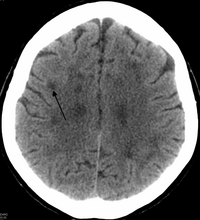
Leukoencephalopathy with neuroaxonal spheroids (LENAS) is an extremely rare kind of leukoencephalopathy and is classified as a neurodegenerative disease. LENAS is a cause of severe and subacute dementia that results from damage to certain areas of the brain. This damage is to a type of brain tissue called white matter and axon damage due to swellings which are termed spheroids.

<span class="mw-page-title-main">Hyperintensity</span> High intensity on MRI brain scans

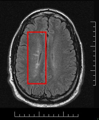
A hyperintensity or T2 hyperintensity is an area of high intensity on types of magnetic resonance imaging (MRI) scans of the brain of a human or of another mammal that reflect lesions produced largely by demyelination and axonal loss. These small regions of high intensity are observed on T2 weighted MRI images within cerebral white matter or subcortical gray matter. The volume and frequency is strongly associated with increasing age. They are also seen in a number of neurological disorders and psychiatric illnesses. For example, deep white matter hyperintensities are 2.5 to 3 times more likely to occur in bipolar disorder and major depressive disorder than control subjects. WMH volume, calculated as a potential diagnostic measure, has been shown to correlate to certain cognitive factors. Hyperintensities appear as "bright signals" on an MRI image and the term "bright signal" is occasionally used as a synonym for a hyperintensity.

Megalencephalic leukoencephalopathy with subcortical cysts is a form of hereditary CNS demyelinating disease. It belongs to a group of disorders called leukodystrophies. It is characterized by early-onset enlargement of the head (macrocephaly) as well as delayed-onset neurological deterioration to include spasticity, epilepsy, and lack of muscular coordination. MLC does not appear to be a disease that is fatal at birth or early in life despite its symptoms, although the number of patients throughout history known to have the disease is fairly limited.

Grinker's myelinopathy, also known as anoxic leukoencephalopathy, is a rare disease of the central nervous system. The disease is characterized by a delayed leukoencephalopathy after a hypoxic episode. It is typically, though not necessarily, related to carbon monoxide poisoning or heroin overdose. It occurs in roughly 2.8% of those who experience an acute hypoxic/anoxic episode. Because of the wide range of symptoms and the delay in onset, it is often misdiagnosed as other neuropathologies. Grinker's myelinopathy was originally characterized by Roy R. Grinker in 1925 or 1926, depending on the source.

<span class="mw-page-title-main">Cerebral autosomal recessive arteriopathy with subcortical infarcts and leukoencephalopathy</span> Medical condition

Cerebral autosomal recessive arteriopathy with subcortical infarcts and leukoencephalopathy (CARASIL) is disease of the arteries in the brain, which causes tissue loss in the subcortical region of the brain and the destruction of myelin in the CNS. CARASIL is characterized by symptoms such as gait disturbances, hair loss, low back pain, dementia, and stroke. CARASIL is a rare disease, having only been diagnosed in about 50 patients, of which ten have been genetically confirmed. Most cases have been reported in Japan, but Chinese and caucasian individuals have also been diagnosed with the disease. CARASIL is inherited in an autosomal recessive pattern. There is currently no cure for CARASIL. Other names for CARASIL include familial young-adult-onset arteriosclerotic leukoencephalopathy with alopecia and lumbago without arterial hypertension, Nemoto disease and Maeda syndrome.

Nasu–Hakola disease also known as polycystic lipomembranous osteodysplasia with sclerosing leukoencephalopathy is a rare disease characterised by early-onset dementia and multifocal bone cysts. It is caused by autosomal recessive loss of function mutations in either the TREM2 or TYROBP gene that are found most frequently in the Finnish and Japanese populations.

<span class="mw-page-title-main">Hypomyelination-congenital cataract syndrome</span> Medical condition

Hypomyelination-congenital cataract syndrome is a rare autosomal recessive hereditary disorder that affects the brain's white matter and is characterized by congenital cataract, psychomotor development delays, and moderate intellectual disabilities. It is a type of leukoencephalopathy.

<span class="mw-page-title-main">Autosomal dominant leukodystrophy with autonomic disease</span> Medical condition

Autosomal dominant leukodystrophy with autonomic disease is a rare neurological condition of genetic origin which is characterized by gradual demyelination of the central nervous system which results in various impairments, including ataxia, mild cognitive disability and autonomic dysfunction. It is part of a group of disorders called "leukodystrophies".

References

  1. 1 2 3 Lin, W. L., Wszolek, Z. K., & Dickson, D. W. (2010). Hereditary diffuse leukoencephalopathy with spheroids: ultrastructural and immunoelectron microscopic studies. Int J Clin Exp Pathol, 3(7), 665-674.
  2. 1 2 3 4 5 6 7 8 9 10 11 12 13 Sundal, C., Lash, J., Aasly, J., Oygarden, S., Roeber, S., Kretzschman, H., . . . Wszolek, Z. K. (2012). Hereditary diffuse leukoencephalopathy with axonal spheroids (HDLS): a misdiagnosed disease entity. J Neurol Sci, 314(1-2), 130-137. doi : 10.1016/j.jns.2011.10.006
  3. 1 2 3 4 5 6 Wider, C., Van Gerpen, J. A., DeArmond, S., Shuster, E. A., Dickson, D. W., & Wszolek, Z. K. (2009). Leukoencephalopathy with spheroids (HDLS) and pigmentary leukodystrophy (POLD): a single entity? Neurology, 72(22), 1953–1959. doi : 10.1212/WNL.0b013e3181a826c0
  4. 1 2 3 Rademakers, R., Baker, M., Nicholson, A., Rutherford, N., Finch, N., Soto-Ortolaza, A., . . . Wszolek, Z. (2012). Mutations in the colony stimulating factor 1 receptor (CSF1R) cause hereditary diffuse leukoencephalopathy with spheroids. Movement Disorders, 27, S399-S400.
  5. 1 2 Kinoshita, M., Yoshida, K., Oyanagi, K., Hashimoto, T., & Ikeda, S. (2012). Hereditary diffuse leukoencephalopathy with axonal spheroids caused by R782H mutation in CSF1R: Case report. Journal of the Neurological Sciences, 318(1-2), 115-118. doi : 10.1016/j.jns.2012.03.012
  6. 1 2 Baba, Y., Ghetti, B., Baker, M. C., Uitti, R. J., Hutton, M. L., Yamaguchi, K., . . . Wszolek, Z. K. (2006). Hereditary diffuse leukoencephalopathy with spheroids: clinical, pathologic and genetic studies of a new kindred. Acta Neuropathol, 111(4), 300-311. doi : 10.1007/s00401-006-0046-z
  7. Hancock, N., Poon, M., Taylor, B., & McLean, C. (2003). Hereditary diffuse leucoencephalopathy with spheroids. J Neurol Neurosurg Psychiatry, 74(9), 1345–1347.
  8. Paloneva, J., Mandelin, J., Kiialainen, A., Böhling, T., Prudlo, J., Hakola, P., . . . Peltonen, L. (2003). DAP12/TREM2 deficiency results in impaired osteoclast differentiation and osteoporotic features. The Journal of experimental medicine, 198(4), 669-675.
  9. Knaap, Marjo S., & Valk, Jaap. (2005). Pigmentary Orthochromatic Leukodystrophy Magnetic Resonance of Myelination and Myelin Disorders (pp. 557-558): Springer Berlin Heidelberg.
  10. Van Gerpen, J. A., Wider, C., Broderick, D. F., Dickson, D. W., Brown, L. A., & Wszolek, Z. K. (2008). Insights into the dynamics of hereditary diffuse leukoencephalopathy with axonal spheroids. Neurology, 71(12), 925-929. doi : 10.1212/01.wnl.0000325916.30701.21
  11. 1 2 Sundal, C., Van Gerpen, J. A., Nicholson, A. M., Wider, C., Shuster, E. A., Aasly, J., . . . Wszolek, Z. K. (2012). MRI characteristics and scoring in HDLS due to CSF1R gene mutations. Neurology, 79(6), 566-574. doi : 10.1212/WNL.0b013e318263575a
  12. Axelsson, R., Roytta, M., Sourander, P., Akesson, H. O., & Andersen, O. (1984). Hereditary diffuse leucoencephalopathy with spheroids. Acta Psychiatr Scand Suppl , 314, 1-65.