Demyelinating disease

Last updated
Demyelinating disease
MS Demyelinisation CD68 10xv2.jpg
Photomicrograph of a demyelinating MS-lesion: Immunohistochemical staining for CD68 highlights numerous macrophages (brown). Original magnification 10×.
Specialty Neurology   OOjs UI icon edit-ltr-progressive.svg

A demyelinating disease refers to any disease affecting the nervous system where the myelin sheath surrounding neurons is damaged. [1] This damage disrupts the transmission of signals through the affected nerves, resulting in a decrease in their conduction ability. Consequently, this reduction in conduction can lead to deficiencies in sensation, movement, cognition, or other functions depending on the nerves affected.

Contents

Various factors can contribute to the development of demyelinating diseases, including genetic predisposition, infectious agents, autoimmune reactions, and other unknown factors. Proposed causes of demyelination include genetic predisposition, environmental factors such as viral infections or exposure to certain chemicals. Additionally, exposure to commercial insecticides like sheep dip, weed killers, and flea treatment preparations for pets, which contain organophosphates, can also lead to nerve demyelination. [2] Chronic exposure to neuroleptic medications may also cause demyelination. [3] Furthermore, deficiencies in vitamin B12 can result in dysmyelination. [4] [5]

Demyelinating diseases are traditionally classified into two types: demyelinating myelinoclastic diseases and demyelinating leukodystrophic diseases. In the first group, a healthy and normal myelin is destroyed by toxic substances, chemicals, or autoimmune reactions. In the second group, the myelin is inherently abnormal and undergoes degeneration. [6] The Poser criteria named this second group dysmyelinating diseases. [7]

In the most well-known demyelinating disease, multiple sclerosis, evidence suggests that the body's immune system plays a significant role. Acquired immune system cells, specifically T-cells, are found at the site of lesions. Other immune system cells, such as macrophages (and possibly mast cells), also contribute to the damage. [8]

Signs and symptoms

Symptoms and signs that present in demyelinating diseases are different for each condition. These symptoms and signs can present in a person with a demyelinating disease: [9]

Evolutionary considerations

The role of prolonged cortical myelination in human evolution has been implicated as a contributing factor in some cases of demyelinating disease. Unlike other primates, humans exhibit a unique pattern of postpubertal myelination, which may contribute to the development of psychiatric disorders and neurodegenerative diseases that present in early adulthood and beyond. The extended period of cortical myelination in humans may allow greater opportunities for disruption in myelination, resulting in the onset of demyelinating disease. [10] Furthermore, humans have significantly greater prefrontal white matter volume than other primate species, which implies greater myelin density. [11] Increased myelin density in humans as a result of a prolonged myelination may, therefore, structure risk for myelin degeneration and dysfunction. Evolutionary considerations for the role of prolonged cortical myelination as a risk factor for demyelinating disease are particularly pertinent given that genetics and autoimmune deficiency hypotheses fail to explain many cases of demyelinating disease. As has been argued, diseases such as multiple sclerosis cannot be accounted for by autoimmune deficiency alone, but strongly imply the influence of flawed developmental processes in disease pathogenesis. [12] Therefore, the role of the human-specific prolonged period of cortical myelination is an important evolutionary consideration in the pathogenesis of demyelinating disease.[ citation needed ]

Diagnosis

Various methods/techniques are used to diagnose demyelinating diseases:

Types

Demyelinating diseases can be divided in those affecting the central nervous system (CNS) and those affecting the peripheral nervous system (PNS). They can also be classified by the presence or absence of inflammation. Finally, a division may be made based on the underlying cause of demyelination: the disease process can be demyelinating myelinoclastic, wherein myelin is destroyed; or dysmyelinating leukodystrophic, wherein myelin is abnormal and degenerative.

CNS

The demyelinating disorders of the central nervous system include:[ citation needed ]

The myelinoclastic disorders are typically associated with symptoms such as optic neuritis and transverse myelitis, because the demyelinating inflammation can affect the optic nerve or spinal cord. Many are idiopathic. Both myelinoclastic and leukodystrophic modes of disease may result in lesional demyelinations of the central nervous system.

PNS

Guillain-Barre syndrome - demyelination Guillain-barre syndrome - Nerve Damage.gif
Guillain–Barré syndrome – demyelination

The demyelinating diseases of the peripheral nervous system include:[ citation needed ]

Treatment

Treatments are patient-specific and depend on the symptoms that present with the disorder, as well as the progression of the condition. Improvements to the patient's life may be accomplished through the management of symptoms or slowing of the rate of demyelination. Treatment can include medication, lifestyle changes (i.e. smoking cessation, increased rest, and dietary changes), counselling, relaxation, physical exercise, patient education, and in some cases, deep brain thalamic stimulation (to ameliorate tremors). [13] :227–248

Prognosis

Prognosis depends on the condition itself. Some conditions such as MS depend on the subtype of the disease and various attributes of the patient such as age, sex, initial symptoms, and the degree of disability the patient experiences. [14] Life expectancy in MS patients is 5 to 10 years lower than unaffected people. [15] MS is an inflammatory demyelinating disease of the central nervous system (CNS) that develops in genetically susceptible individuals after exposure to unknown environmental trigger(s). The bases for MS are unknown but are strongly suspected to involve immune reactions against autoantigens, particularly myelin proteins. The most accepted hypothesis is that dialogue between T-cell receptors and myelin antigens leads to an immune attack on the myelin-oligodendrocyte complex. These interactions between active T cells and myelin antigens provoke a massive destructive inflammatory response and promote continuing proliferation of T and B cells and macrophage activation, which sustains secretion of inflammatory mediators. [16] Other conditions such as central pontine myelinolysis have about a third of patients recover and the other two-thirds experience varying degrees of disability. [17] In some cases, such as transverse myelitis, the patient can begin recovery as early as 2 to 12 weeks after the onset of the condition.[ citation needed ]

Epidemiology

Incidence of demyelinating diseases varies by disorder. Some conditions, such as tabes dorsalis appear predominantly in males and begin in midlife. Optic neuritis, though, occurs preferentially in females typically between the ages of 30 and 35. [18] Other conditions such as multiple sclerosis vary in prevalence depending on the country and population. [19] This condition can appear in children and adults. [15]

Research

Much of the research conducted on demyelinating diseases is targeted towards discovering the mechanisms by which these disorders function in an attempt to develop therapies and treatments for individuals affected by these conditions. For example, proteomics has revealed several proteins which contribute to the pathophysiology of demyelinating diseases. [20] For example, COX-2 has been implicated in oligodendrocyte death in animal models of demyelination. [21] The presence of myelin debris has been correlated with damaging inflammation as well as poor regeneration, due to the presence of inhibitory myelin components. [22] [23]

N-cadherin is expressed in regions of active remyelination and may play an important role in generating a local environment conducive to remyelination. [24] N-cadherin agonists have been identified and observed to stimulate neurite growth and cell migration, key aspects of promoting axon growth and remyelination after injury or disease. [25]

Immunomodulatory drugs such as fingolimod have been shown to reduce immune-mediated damage to the CNS, preventing further damage in patients with MS. The drug targets the role of macrophages in disease progression. [26] [27]

Manipulating thyroid hormone levels may become a viable strategy to promote remyelination and prevent irreversible damage in MS patients. [28] It has also been shown that intranasal administration of apotransferrin (aTf) can protect myelin and induce remyelination. [29] Finally, electrical stimulation which activates neural stem cells may provide a method by which regions of demyelination can be repaired. [30]

In other animals

Demyelinating diseases/disorders have been found worldwide in various animals. Some of these animals include mice, pigs, cattle, hamsters, rats, sheep, Siamese kittens, and a number of dog breeds (including Chow Chow, Springer Spaniel, Dalmatian, Samoyed, Golden Retriever, Lurcher, Bernese Mountain Dog, Vizsla, Weimaraner, Australian Silky Terrier, and mixed breeds). [31] [32]

See also

Related Research Articles

<span class="mw-page-title-main">Myelin</span> Fatty substance that surrounds nerve cell axons to insulate them and increase transmission speed

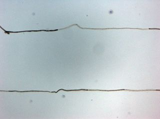
Myelin is a lipid-rich material that surrounds nerve cell axons to insulate them and increase the rate at which electrical impulses pass along the axon. The myelinated axon can be likened to an electrical wire with insulating material (myelin) around it. However, unlike the plastic covering on an electrical wire, myelin does not form a single long sheath over the entire length of the axon. Rather, myelin ensheaths the axon segmentally: in general, each axon is encased in multiple long sheaths with short gaps between, called nodes of Ranvier. At the nodes of Ranvier, which are approximately one thousandth of a mm in length, the axon's membrane is bare of myelin.

<span class="mw-page-title-main">Optic neuritis</span> Medical condition

Optic neuritis describes any condition that causes inflammation of the optic nerve; it may be associated with demyelinating diseases, or infectious or inflammatory processes.

<span class="mw-page-title-main">Multiple sclerosis</span> Disease that damages the myelin sheaths around nerves

Multiple sclerosis (MS) is an autoimmune disease in which the insulating covers of nerve cells in the brain and spinal cord are damaged. This damage disrupts the ability of parts of the nervous system to transmit signals, resulting in a range of signs and symptoms, including physical, mental, and sometimes psychiatric problems. Specific symptoms can include double vision, vision loss, eye pain, muscle weakness, and loss of sensation or coordination. MS takes several forms, with new symptoms either occurring in isolated attacks or building up over time. In the relapsing forms of MS, between attacks, symptoms may disappear completely, although some permanent neurological problems often remain, especially as the disease advances. In the progressive forms of MS, bodily function slowly deteriorates and disability worsens once symptoms manifest and will steadily continue to do so if the disease is left untreated.

Myelitis is inflammation of the spinal cord which can disrupt the normal responses from the brain to the rest of the body, and from the rest of the body to the brain. Inflammation in the spinal cord can cause the myelin and axon to be damaged resulting in symptoms such as paralysis and sensory loss. Myelitis is classified to several categories depending on the area or the cause of the lesion; however, any inflammatory attack on the spinal cord is often referred to as transverse myelitis.

<span class="mw-page-title-main">Oligodendrocyte</span> Neural cell type

Oligodendrocytes, also known as oligodendroglia, are a type of neuroglia whose main functions are to provide support and insulation to axons within the central nervous system (CNS) of jawed vertebrates. Their function is similar to that of Schwann cells, which perform the same task in the peripheral nervous system (PNS). Oligodendrocytes accomplish this by forming the myelin sheath around axons. Unlike Schwann cells, a single oligodendrocyte can extend its processes to cover around 50 axons, with each axon being wrapped in approximately 1 μm of myelin sheath. Furthermore, an oligodendrocyte can provide myelin segments for multiple adjacent axons.

Oligodendrocyte progenitor cells (OPCs), also known as oligodendrocyte precursor cells, NG2-glia, O2A cells, or polydendrocytes, are a subtype of glia in the central nervous system named for their essential role as precursors to oligodendrocytes. They are typically identified in the human by co-expression of PDGFRA and CSPG4.

Experimental autoimmune encephalomyelitis, sometimes experimental allergic encephalomyelitis (EAE), is an animal model of brain inflammation. It is an inflammatory demyelinating disease of the central nervous system (CNS). It is mostly used with rodents and is widely studied as an animal model of the human CNS demyelinating diseases, including multiple sclerosis (MS) and acute disseminated encephalomyelitis (ADEM). EAE is also the prototype for T-cell-mediated autoimmune disease in general.

<span class="mw-page-title-main">Chronic inflammatory demyelinating polyneuropathy</span> Medical condition

Chronic inflammatory demyelinating polyneuropathy (CIDP) is an acquired autoimmune disease of the peripheral nervous system characterized by progressive weakness and impaired sensory function in the legs and arms. The disorder is sometimes called chronic relapsing polyneuropathy (CRP) or chronic inflammatory demyelinating polyradiculoneuropathy. CIDP is closely related to Guillain–Barré syndrome and it is considered the chronic counterpart of that acute disease. Its symptoms are also similar to progressive inflammatory neuropathy. It is one of several types of neuropathy.

<span class="mw-page-title-main">Pathophysiology of multiple sclerosis</span>

Multiple sclerosis is an inflammatory demyelinating disease of the CNS in which activated immune cells invade the central nervous system and cause inflammation, neurodegeneration, and tissue damage. The underlying cause is currently unknown. Current research in neuropathology, neuroimmunology, neurobiology, and neuroimaging, together with clinical neurology, provide support for the notion that MS is not a single disease but rather a spectrum.

<span class="mw-page-title-main">Lesional demyelinations of the central nervous system</span>

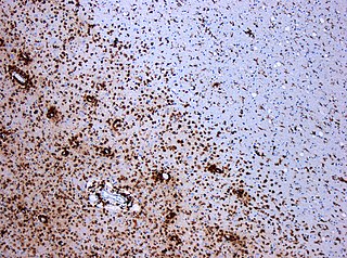
Multiple sclerosis and other demyelinating diseases of the central nervous system (CNS) produce lesions and glial scars or scleroses. They present different shapes and histological findings according to the underlying condition that produces them.

Remyelination is the process of propagating oligodendrocyte precursor cells to form oligodendrocytes to create new myelin sheaths on demyelinated axons in the CNS. This is a process naturally regulated in the body and tends to be very efficient in a healthy CNS. The process creates a thinner myelin sheath than normal, but it helps to protect the axon from further damage, from overall degeneration, and proves to increase conductance once again. The processes underlying remyelination are under investigation in the hope of finding treatments for demyelinating diseases, such as multiple sclerosis.

Inflammatory demyelinating diseases (IDDs), sometimes called Idiopathic (IIDDs) due to the unknown etiology of some of them, are a heterogenous group of demyelinating diseases - conditions that cause damage to myelin, the protective sheath of nerve fibers - that occur against the background of an acute or chronic inflammatory process. IDDs share characteristics with and are often grouped together under Multiple Sclerosis. They are sometimes considered different diseases from Multiple Sclerosis, but considered by others to form a spectrum differing only in terms of chronicity, severity, and clinical course.

<span class="mw-page-title-main">Balo concentric sclerosis</span> Medical condition

Baló's concentric sclerosis is a disease in which the white matter of the brain appears damaged in concentric layers, leaving the axis cylinder intact. It was described by József Mátyás Baló who initially named it "leuko-encephalitis periaxialis concentrica" from the previous definition, and it is currently considered one of the borderline forms of multiple sclerosis.

Myelinogenesis is the formation and development of myelin sheaths in the nervous system, typically initiated in late prenatal neurodevelopment and continuing throughout postnatal development. Myelinogenesis continues throughout the lifespan to support learning and memory via neural circuit plasticity as well as remyelination following injury. Successful myelination of axons increases action potential speed by enabling saltatory conduction, which is essential for timely signal conduction between spatially separate brain regions, as well as provides metabolic support to neurons.

<span class="mw-page-title-main">GJB1</span> Protein-coding gene in humans

Gap junction beta-1 protein (GJB1), also known as connexin 32 (Cx32), is a transmembrane protein that in humans is encoded by the GJB1 gene. Gap junction beta-1 protein is a member of the gap junction connexin family of proteins that regulates and controls the transfer of communication signals across cell membranes, primarily in the liver and peripheral nervous system. However, the protein is expressed in multiple organs, including in oligodendrocytes in the central nervous system.

<span class="mw-page-title-main">Tumefactive multiple sclerosis</span> Medical condition

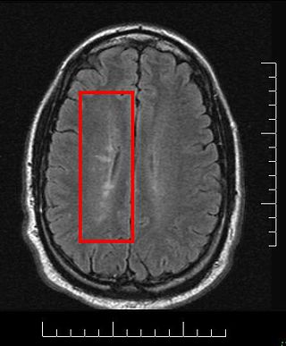
Tumefactive multiple sclerosis is a condition in which the central nervous system of a person has multiple demyelinating lesions with atypical characteristics for those of standard multiple sclerosis (MS). It is called tumefactive as the lesions are "tumor-like" and they mimic tumors clinically, radiologically and sometimes pathologically.

Anti-MAG peripheral neuropathy is a specific type of peripheral neuropathy in which the person's own immune system attacks cells that are specific in maintaining a healthy nervous system. As these cells are destroyed by antibodies, the nerve cells in the surrounding region begin to lose function and create many problems in both sensory and motor function. Specifically, antibodies against myelin-associated glycoprotein (MAG) damage Schwann cells. While the disorder occurs in only 10% of those afflicted with peripheral neuropathy, people afflicted have symptoms such as muscle weakness, sensory problems, and other motor deficits usually starting in the form of a tremor of the hands or trouble walking. There are, however, multiple treatments that range from simple exercises in order to build strength to targeted drug treatments that have been shown to improve function in people with this type of peripheral neuropathy.

<span class="mw-page-title-main">Pathology of multiple sclerosis</span> Pathologic overview

Multiple sclerosis (MS) can be pathologically defined as the presence of distributed glial scars (scleroses) in the central nervous system that must show dissemination in time (DIT) and in space (DIS) to be considered MS lesions.

MOG antibody disease (MOGAD) or MOG antibody-associated encephalomyelitis (MOG-EM) is an inflammatory demyelinating disease of the central nervous system. Serum anti-myelin oligodendrocyte glycoprotein antibodies are present in up to half of patients with an acquired demyelinating syndrome and have been described in association with a range of phenotypic presentations, including acute disseminated encephalomyelitis, optic neuritis, transverse myelitis, and neuromyelitis optica.

Anti-neurofascin demyelinating diseases refers to health conditions engendered by auto-antibodies against neurofascins, which can produce both central and peripheral demyelination. Some cases of combined central and peripheral demyelination (CCPD) could be produced by them.

References

  1. "demyelinating disease" "at Dorland's Medical Dictionary
  2. Lotti M, Moretto A (2005). "Organophosphate-induced delayed polyneuropathy". Toxicol Rev. 24 (1): 37–49. doi:10.2165/00139709-200524010-00003. PMID   16042503. S2CID   29313644.
  3. Konopaske GT, Dorph-Petersen KA, Sweet RA, Pierri JN, Zhang W, Sampson AR, Lewis DA (April 2008). "Effect of chronic antipsychotic exposure on astrocyte and oligodendrocyte numbers in macaque monkeys". Biol Psychiatry. 63 (8): 759–65. doi:10.1016/j.biopsych.2007.08.018. PMC   2386415 . PMID   17945195.
  4. Agadi S, Quach MM, Haneef Z (2013). "Vitamin-responsive epileptic encephalopathies in children". Epilepsy Res Treat. 2013: 510529. doi: 10.1155/2013/510529 . PMC   3745849 . PMID   23984056.
  5. Yoganathan S, Varman M, Oommen SP, Thomas M (2017). "A Tale of Treatable Infantile Neuroregression and Diagnostic Dilemma with Glutaric Aciduria Type I". J Pediatr Neurosci. 12 (4): 356–359. doi: 10.4103/jpn.JPN_35_17 . PMC   5890558 . PMID   29675077.
  6. Fernández O, Fernández VE, Guerrero M (2015). "Demyelinating diseases of the central nervous system". Medicine. 11 (77): 4601–4609. doi:10.1016/j.med.2015.04.001.
  7. POSER CM (March 1961). "Leukodystrophy and the concept of dysmyelination". Arch Neurol. 4 (3): 323–32. doi:10.1001/archneur.1961.00450090089013. PMID   13737358.
  8. Laetoli (January 2008). "Demyelination". Archived from the original on 2012-07-28.
  9. "Symptoms of Demyelinating Disorders - Right Diagnosis." Right Diagnosis. Right Diagnosis, 01 Feb 2012. Web. 24 Sep 2012
  10. Miller DJ, Duka T, Stimpson CD, Schapiro SJ, Baze WB, McArthur MJ, Fobbs AJ, Sousa AM, Sestan N, Wildman DE, Lipovich L, Kuzawa CW, Hof PR, Sherwood CC (October 2012). "Prolonged myelination in human neocortical evolution". Proc Natl Acad Sci U S A. 109 (41): 16480–5. Bibcode:2012PNAS..10916480M. doi: 10.1073/pnas.1117943109 . PMC   3478650 . PMID   23012402.
  11. Schoenemann PT, Sheehan MJ, Glotzer LD (February 2005). "Prefrontal white matter volume is disproportionately larger in humans than in other primates". Nat Neurosci. 8 (2): 242–52. doi:10.1038/nn1394. PMID   15665874. S2CID   205430527.
  12. Chaudhuri A (October 2013). "Multiple sclerosis is primarily a neurodegenerative disease". J Neural Transm (Vienna). 120 (10): 1463–6. doi:10.1007/s00702-013-1080-3. PMID   23982272. S2CID   8575687.
  13. 1 2 3 4 5 6 7 Freedman MS (2005). Advances in Neurology Volume 98: Multiple Sclerosis and Demyelinating Diseases. Philadelphia: Lippincott Williams & Wilkins. p. 112. ISBN   0781751705.
  14. Weinshenker BG (1994). "Natural history of multiple sclerosis". Ann Neurol. 36 Suppl: S6–11. doi:10.1002/ana.410360704. PMID   8017890. S2CID   7140070.
  15. 1 2 Compston A, Coles A (October 2008). "Multiple sclerosis". Lancet. 372 (9648): 1502–17. doi:10.1016/S0140-6736(08)61620-7. PMID   18970977. S2CID   195686659.
  16. Minagar A, Alexander JS (December 2003). "Blood-brain barrier disruption in multiple sclerosis". Mult Scler. 9 (6): 540–9. doi:10.1191/1352458503ms965oa. PMID   14664465. S2CID   10189144.
  17. Abbott R, Silber E, Felber J, Ekpo E (October 2005). "Osmotic demyelination syndrome". BMJ. 331 (7520): 829–30. doi:10.1136/bmj.331.7520.829. PMC   1246086 . PMID   16210283.
  18. Rodriguez M, Siva A, Cross SA, O'Brien PC, Kurland LT (February 1995). "Optic neuritis: a population-based study in Olmsted County, Minnesota". Neurology. 45 (2): 244–50. doi:10.1212/wnl.45.2.244. PMID   7854520. S2CID   25800388.
  19. Rosati G (April 2001). "The prevalence of multiple sclerosis in the world: an update". Neurol Sci. 22 (2): 117–39. doi:10.1007/s100720170011. PMID   11603614. S2CID   207051545.
  20. Newcombe J, Eriksson B, Ottervald J, Yang Y, Franzén B (February 2005). "Extraction and proteomic analysis of proteins from normal and multiple sclerosis postmortem brain". J Chromatogr B Analyt Technol Biomed Life Sci. 815 (1–2): 191–202. doi:10.1016/j.jchromb.2004.10.073. PMID   15652809.
  21. Palumbo S, Toscano CD, Parente L, Weigert R, Bosetti F (May 2012). "The cyclooxygenase-2 pathway via the PGE₂ EP2 receptor contributes to oligodendrocytes apoptosis in cuprizone-induced demyelination". J Neurochem. 121 (3): 418–27. doi:10.1111/j.1471-4159.2011.07363.x. PMC   3220805 . PMID   21699540.
  22. Clarner T, Diederichs F, Berger K, Denecke B, Gan L, van der Valk P, Beyer C, Amor S, Kipp M (October 2012). "Myelin debris regulates inflammatory responses in an experimental demyelination animal model and multiple sclerosis lesions". Glia. 60 (10): 1468–80. doi:10.1002/glia.22367. PMID   22689449. S2CID   205834726.
  23. Podbielska M, Banik NL, Kurowska E, Hogan EL (August 2013). "Myelin recovery in multiple sclerosis: the challenge of remyelination". Brain Sci. 3 (4): 1282–324. doi: 10.3390/brainsci3031282 . PMC   4061877 . PMID   24961530.
  24. Hochmeister S, Romauch M, Bauer J, Seifert-Held T, Weissert R, Linington C, Hartung HP, Fazekas F, Storch MK (September 2012). "Re-expression of N-cadherin in remyelinating lesions of experimental inflammatory demyelination". Exp Neurol. 237 (1): 70–7. doi:10.1016/j.expneurol.2012.06.010. PMID   22735489. S2CID   33883037.
  25. Burden-Gulley SM, Gates TJ, Craig SE, Gupta M, Brady-Kalnay SM (May 2010). "Stimulation of N-cadherin-dependent neurite outgrowth by small molecule peptide mimetic agonists of the N-cadherin HAV motif". Peptides. 31 (5): 842–9. doi:10.1016/j.peptides.2010.02.002. PMID   20153391. S2CID   207357858.
  26. Gasperini C, Ruggieri S (2012). "Development of oral agent in the treatment of multiple sclerosis: how the first available oral therapy, fingolimod will change therapeutic paradigm approach". Drug Des Devel Ther. 6: 175–86. doi: 10.2147/DDDT.S8927 . PMC   3414371 . PMID   22888218.
  27. Ransohoff RM, Howe CL, Rodriguez M (November 2002). "Growth factor treatment of demyelinating disease: at last, a leap into the light". Trends Immunol. 23 (11): 512–6. doi:10.1016/s1471-4906(02)02321-9. PMID   12401395.
  28. Silvestroff L, Bartucci S, Pasquini J, Franco P (May 2012). "Cuprizone-induced demyelination in the rat cerebral cortex and thyroid hormone effects on cortical remyelination". Exp Neurol. 235 (1): 357–67. doi:10.1016/j.expneurol.2012.02.018. PMID   22421533. S2CID   1534460.
  29. Guardia Clausi M, Paez PM, Campagnoni AT, Pasquini LA, Pasquini JM (October 2012). "Intranasal administration of aTf protects and repairs the neonatal white matter after a cerebral hypoxic-ischemic event". Glia. 60 (10): 1540–54. doi:10.1002/glia.22374. hdl: 11336/67318 . PMID   22736466. S2CID   28658807.
  30. Sherafat MA, Heibatollahi M, Mongabadi S, Moradi F, Javan M, Ahmadiani A (September 2012). "Electromagnetic field stimulation potentiates endogenous myelin repair by recruiting subventricular neural stem cells in an experimental model of white matter demyelination". J Mol Neurosci. 48 (1): 144–53. doi:10.1007/s12031-012-9791-8. PMID   22588976. S2CID   15779187.
  31. "Merck Veterinary Manual – Demyelinating Disorders: Introduction". Archived from the original on 2010-12-19. Retrieved 2012-10-30.
  32. Johnson RT (2004). "Demyelinating Diseases". The Infectious Etiology of Chronic Diseases: Defining the Relationship, Enhancing the Research, and Mitigating the Effects: Workshop Summary. NIH.