Hemiballismus

Last updated
Hemiballismus
Other namesBallismus or Ballism
Specialty Neurology   OOjs UI icon edit-ltr-progressive.svg

Hemiballismus or hemiballism is a basal ganglia syndrome resulting from damage to the subthalamic nucleus in the basal ganglia. [1] Hemiballismus is a rare hyperkinetic movement disorder, [2] that is characterized by violent involuntary limb movements, [1] [3] on one side of the body, [4] and can cause significant disability. [5] Ballismus affects both sides of the body and is much rarer. [4] Symptoms can decrease during sleep. [6]

Contents

Hemiballismus differs from chorea in that the movements occur in the proximal limbs whereas in chorea the limb movements are in the distal limbs. [4] Also in chorea the movements are more dance-like, flowing from one region to another. [7]

Symptoms

Ballism was defined by Meyers in 1968 [8] as "Repetitive, but constantly varying, large amplitude involuntary movements of the proximal parts of the limbs. This activity is almost ceaseless and movements are often complex and combined". Hemiballismus is usually characterized by involuntary flinging motions of the extremities. [2] The movements are often violent and have wide amplitudes of motion. [9] They are continuous and random and can involve proximal or distal muscles on one side of the body. Some cases even include the facial muscles. [6] It is common for arms and legs to move together. The more a patient is active, the more the movements increase. With relaxation comes a decrease in movements. [10] Physicians can measure the severity of the disorder by having the patient perform a series of basic, predetermined tasks and counting the hemiballistic movements during a set time session. [11] The physicians then rate the patient on a severity scale. This scale gives scientists and clinicians a way to compare patients and determine the range of the disorder.[ citation needed ]

The name hemiballismus literally means "half ballistic", referring to the violent, flailing movements observed on one side of the body.

Causes

In examining the causes of hemiballismus, it is important to remember that this disorder is extremely rare. While hemiballismus can result from the following list, just because a patient has one of these disorders does not mean they will also experience hemiballismus.[ citation needed ]

Stroke

Hemisballismus as a result of stroke occurs in only about 0.45 cases per hundred thousand stroke patients. [2] Even at such a small rate, stroke is by far the most common cause of hemiballismus. [12] A stroke causes tissue to die due to a lack of oxygen resulting from an impaired blood supply. In the basal ganglia, this can result in the death of tissue that helps to control movement. As a result, the brain is left with damaged tissue that sends damaged signals to the skeletal muscles in the body. The result is occasionally a patient with hemiballismus.[ citation needed ]

Traumatic brain injury

Hemiballismus can also occur as a result of a traumatic brain injury. There are cases in which survivors of assault or other forms of violence have developed hemiballismus. [5] Through these acts of violence, the survivor's brain has been damaged and the hemiballistic movements have developed.[ citation needed ]

Amyotrophic lateral sclerosis

This disease causes neuronal loss and gliosis, which can include the subthalamic nucleus and other areas of the brain. [13] Essentially any disorder that causes some form of neuronal loss or gliosis in the basal ganglia has the potential to cause hemiballismus.[ citation needed ]

Hyperglycemia-induced involuntary movements (in this case, not hemiballismus, but hemichorea (chorea of one side of the body) and bilateral dystonia) in a 62-year-old Japanese woman with type 1 diabetes.

Nonketotic hyperglycemia

Patients with nonketotic hyperglycemia can develop hemiballismus as a complication to the disease through the development of a subthalamic nucleus lesion. [14] This is the second most common reported cause of hemiballismus. It can be found primarily in the elderly and many of the reported cases have come from East Asian origin, which suggests that there may be some genetic disposition to development of hemiballismus as a result of hyperglycemia. Hemiballistic movements appear when blood glucose levels get too high and then subside once glucose levels return to normal. This time scale for this is usually several hours. In patients with this type of hemiballismus, imaging reveals abnormalities in the putamen contralateral to the movements as well as the globus pallidus and caudate nucleus. While the hyperglycemia itself is not the cause of the hemiballistic movements, it has been suggested that petechial hemorrhage or a decreased production of GABA and acetylcholine could result secondary to the hyperglycemia. One of these issues could be responsible for the hemiballistic movements. [10]

Neoplasms

A neoplasm is an abnormal growth of cells. Cases have shown that if this occurs somewhere in the basal ganglia, hemiballismus can result. [10]

Vascular malformations

Vascular malformations can cause abnormal blood flow to areas of the brain. If too little blood is delivered to the basal ganglia, a stroke can occur. [10]

Tuberculomas

This is another form of tumor that can result in the brain as a result of a tuberculous meningitis infection. This type of tumor can also damage parts of the basal ganglia, sometimes resulting in hemiballismus. [10]

Demyelinating plaques

Demyelinating plaques attack the myelin sheaths on neurons. This decreases the conduction velocity of the neurons, making the signals received by the basal ganglia garbled and incomplete. This disorganized signal can also cause the chaotic movements characterized by hemiballismus. [10]

Complications from HIV infection

Patients with HIV often have complications that arise along with AIDS. Hypoglycemia due to pentamidine use in patients with AIDS has been known to cause hemiballismus. In some patients, hemiballismus has been the only visible symptom to alert the physician that the patients may have AIDS. It is typically a result of a secondary infection that occurs due to the compromised immune system and the most common infection causing hemiballismus is cerebral toxoplasmosis. Most of the lesions that result from this infection are found in the basal ganglia. As long as the diagnosis is not missed, this type of hemiballismus can be treated just as well as in patients without HIV. [10]

Anatomy

Basal ganglia

The basal ganglia are a collection of nuclei that connects to several other areas of the brain. Due to the diverse nuclei that they contain, the basal ganglia are involved in numerous functions, including motor control. It is within this structure that hemiballismus primarily occurs in the brain.[ citation needed ]

Subthalamic nucleus

This structure within the basal ganglia innervates other structures, including a very important connection to the internal globus pallidus. The subthalamic nucleus essentially provides the excitement needed to drive the globus pallidus. [10] Injury to this area or its efferent or afferent connections can induce this disorder contralateral to the side of the lesion. [9] The structure itself is a regulator of motor function and is also involved in associative and limbic functions. [15] It was traditionally thought that the disorder was only caused by injury to the subthalamic nucleus, but later studies have shown that damage to other basal ganglia regions can also be responsible for causing this disorder. [12] Hemiballismus caused by lesions in the subthalamic nucleus is more severe than other forms of the disorder. [10]

Globus pallidus

From recent studies, it is now thought that hemiballismus can be associated with a decreased output of the globus pallidus. This is because studies have shown that firing rates decrease from 70/s to 40/s. [10] In addition to a decreased firing rate, degenerative neurological disorders that cause patients to exhibit hemiballistic movements show a marked decrease in the globus pallidus mass as well. [13] Increases in activity in this area causes there to be an inhibition of the motor thalamus. This causes cortical activation and thus a movement inhibition. In the case of hemiballismus, the opposite occurs, leading to the characteristic large, irregular movements. [10]

Putamen

The putamen is also part of the basal ganglia and can be involved in hemiballismus due to the fact that it projects to the premotor cortex through the globus pallidus. As a result, damage to this area can also cause hemiballistic movements to be seen as it is also part of the chain in movement. [6]

Caudate nucleus

The caudate nucleus is the portion of the basal ganglia that helps control voluntary movement. Damage to this area can also result in hemiballismus as it is directly related to voluntary movement. [6]

Cortical structures

While the majority of damage that causes hemiballismus occurs within the basal ganglia, there are still cases that have been documented on which damage to cortical structures has caused hemiballistic movements. [10]

Diagnosis

Diagnosis of hemiballismus is a clinical one, made with observation during clinical examination. Hemiballismus is a clinical sign with a number of different causes. Therefore, a diagnosis underlying this clinical sign should be sought. The observer should note sudden, flinging movements of a limb(s) and occasionally the face. This is commonly unilateral ("Hemiballismus"). The movements must be distinguished from other hyperkinetic movement disorders such as tremor (generally more rhythmic, and smaller amplitude) and chorea, akathisia and athetosis (all are often of lower amplitude and less violent).[ citation needed ]

Treatments

When treating hemiballismus, it is first important to treat whatever may be causing the manifestation of this disorder. This could be hyperglycemia, infections, or neoplastic lesions. Some patients may not even need treatment because the disorder is not severe and can be self-limited. [10]

Dopamine blockers

When pharmacological treatment is necessary, the most standard type of drug to use is an antidopaminergic drug. Blocking dopamine is effective in about ninety percent of patients. Perphenazine, pimozide, haloperidol, and chlorpromazine are standard choices for treatment. Scientists are still unsure as to why this form of treatment works, as dopamine has not been directly linked to hemiballismus. [10]

Anticonvulsants

An anticonvulsant called topiramate has helped patients in three cases and may be a viable treatment for the future. [16]

ITB therapy

Intrathecal baclofen (ITB) therapy is used to treat a variety of movement disorders such as cerebral palsy and multiple sclerosis. It can also be a possibility to help treat hemiballismus. [5] In one case, before ITB the patient had an average of 10–12 ballism episodes of the right lower limb per hour. During episodes, the right hip would flex up to about 90 degrees, with a fully extended knee. After an ITB pump was implanted and the correct dosage was found, the frequency of ballistic right leg movements decreased to about three per day, and the right hip flexed to only 30 degrees. The patient was also able to better isolate individual distal joint movements in the right lower limb. The patient currently receives 202.4 microg/day of ITB and continues to benefit almost 6 years after the ITB pump was implanted. [5]

Botulinum injections

New uses for botulinum toxin have included treatment of hemiballismus. However, this is still in the early stages of testing. This treatment deals with the muscular manifestations of hemiballismus as opposed to the neurological causes. [5] [10]

Tetrabenazine

Tetrabenazine has been used to treat other movement disorders, but is now being used to treat hemiballismus. Patients using this medication have had a dramatic response. However, lowering the dosage leads to a return of symptoms. This drug works by depleting dopamine. [6]

Antipsychotics

In one case, a patient had not been responding to haloperidol, thus the physician tried olanzapine. The patient made a significant recovery. More research is being performed on the use of these types of drugs in treating hemiballismus. [11]

Functional neurosurgery

Surgery as a treatment should only be used on patients with severe hemiballismus that has not responded to treatment. Lesioning of the globus pallidus or deep brain stimulation of the globus pallidus are procedures that can be used on humans. Usually, lesioning is favored over deep brain stimulation because of the maintenance required to continue stimulating the brain correctly and effectively. [10]

Prognosis

In the past, the prognosis for patients with this disease had been very poor; with many patients experiencing severe disability or death. Now, patients are responding remarkably well to current treatments and the majority of patients go into spontaneous remission. For those that do not go into remission, the symptoms of hemiballismus can generally be very well controlled with medication. [10] Due to the rarity of this disorder, scientists know very little about the details of hemiballismus.

History

The work of J.R. Whittier, F.A. Mettler, and M.B. Carpenter in the mid-1900s helped scientists and clinicians form a more complete picture of hemiballismus. In their experiments, several lesions were made in the basal ganglia structures in monkeys and then they monitored the results. They noticed that the majority of the time, the monkeys did not have any unusual movements. However, when at least twenty percent of the subthalamic nucleus was damaged, abnormal movements were seen in the limbs opposite to the side of the brain that was damaged. This observation caused scientists to believe that hemiballismus outside the subthalamic nucleus did not occur. It was not until much later that this classical model began to expand to include other areas of the basal ganglia and even some cortical structures. They also noticed that unlike human patients, the unusual movements in the monkeys were mainly in the lower extremities. In about half of the monkeys, the hemiballismus continued until the monkey died. [10]

Other scientists have also worked on this perplexing disorder and have found that the symptoms can be induced by injecting kainic acid or ibotenic acid into the subthalamic nucleus. I. Hamada and M.R. DeLong found that by using these chemicals, they could destroy only four percent of the subthalamic nucleus and still see hemiballistic movements. However, the abnormal movements would usually disappear within four to five hours even though it did not appear as though the damaged tissue had healed. This suggests that the subthalamic nucleus is plastic enough to adapt to small amounts of damage in order to resume normal function. [10]

See also

Related Research Articles

<span class="mw-page-title-main">Putamen</span> Round structure at the base of the forebrain

The putamen is a round structure located at the base of the forebrain (telencephalon). The putamen and caudate nucleus together form the dorsal striatum. It is also one of the structures that compose the basal nuclei. Through various pathways, the putamen is connected to the substantia nigra, the globus pallidus, the claustrum, and the thalamus, in addition to many regions of the cerebral cortex. A primary function of the putamen is to regulate movements at various stages and influence various types of learning. It employs GABA, acetylcholine, and enkephalin to perform its functions. The putamen also plays a role in degenerative neurological disorders, such as Parkinson's disease.

<span class="mw-page-title-main">Substantia nigra</span> Structure in the basal ganglia of the brain

The substantia nigra (SN) is a basal ganglia structure located in the midbrain that plays an important role in reward and movement. Substantia nigra is Latin for "black substance", reflecting the fact that parts of the substantia nigra appear darker than neighboring areas due to high levels of neuromelanin in dopaminergic neurons. Parkinson's disease is characterized by the loss of dopaminergic neurons in the substantia nigra pars compacta.

<span class="mw-page-title-main">Basal ganglia</span> Group of subcortical nuclei involved in the motor and reward systems

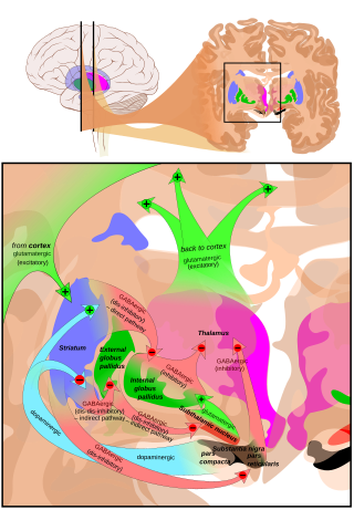
The basal ganglia (BG), or basal nuclei, are a group of subcortical nuclei found in the brains of vertebrates. In humans and some primates, differences exist, primarily in the division of the globus pallidus into external and internal regions, and in the division of the striatum. Positioned at the base of the forebrain and the top of the midbrain, they have strong connections with the cerebral cortex, thalamus, brainstem and other brain areas. The basal ganglia are associated with a variety of functions, including regulating voluntary motor movements, procedural learning, habit formation, conditional learning, eye movements, cognition, and emotion.

<span class="mw-page-title-main">Deep brain stimulation</span> Neurosurgical treatment involving implantation of a brain pacemaker

Deep brain stimulation (DBS) is a surgical procedure that implants a neurostimulator and electrodes which sends electrical impulses to specified targets in the brain responsible for movement control. The treatment is designed for a range of movement disorders such as Parkinson's disease, essential tremor, and dystonia, as well as for certain neuropsychiatric conditions like obsessive-compulsive disorder (OCD) and epilepsy. The exact mechanisms of DBS are complex and not entirely clear, but it is known to modify brain activity in a structured way.

<span class="mw-page-title-main">Globus pallidus</span> Structure of the basal ganglia of the brain

The globus pallidus (GP), also known as paleostriatum or dorsal pallidum, is a subcortical structure of the brain. It consists of two adjacent segments, one external, known in rodents simply as the globus pallidus, and one internal, known in rodents as the entopeduncular nucleus. It is part of the telencephalon, but retains close functional ties with the subthalamus in the diencephalon – both of which are part of the extrapyramidal motor system. The globus pallidus is a major component of the basal ganglia, with principal inputs from the striatum, and principal direct outputs to the thalamus and the substantia nigra. The latter is made up of similar neuronal elements, has similar afferents from the striatum, similar projections to the thalamus, and has a similar synaptology. Neither receives direct cortical afferents, and both receive substantial additional inputs from the intralaminar thalamus.

<span class="mw-page-title-main">Athetosis</span> Medical condition

Athetosis is a symptom characterized by slow, involuntary, convoluted, writhing movements of the fingers, hands, toes, and feet and in some cases, arms, legs, neck and tongue. Movements typical of athetosis are sometimes called athetoid movements. Lesions to the brain are most often the direct cause of the symptoms, particularly to the corpus striatum. This symptom does not occur alone and is often accompanied by the symptoms of cerebral palsy, as it is often a result of this physical disability. Treatments for athetosis are not very effective, and in most cases are simply aimed at managing the uncontrollable movement, rather than the cause itself.

<span class="mw-page-title-main">Indirect pathway</span> Neuronal circuit that suppresses unwanted movements

The indirect pathway, sometimes known as the indirect pathway of movement, is a neuronal circuit through the basal ganglia and several associated nuclei within the central nervous system (CNS) which helps to prevent unwanted muscle contractions from competing with voluntary movements. It operates in conjunction with the direct pathway.

<span class="mw-page-title-main">Subthalamic nucleus</span> Small lens-shaped nucleus in the brain

The subthalamic nucleus (STN) is a small lens-shaped nucleus in the brain where it is, from a functional point of view, part of the basal ganglia system. In terms of anatomy, it is the major part of the subthalamus. As suggested by its name, the subthalamic nucleus is located ventral to the thalamus. It is also dorsal to the substantia nigra and medial to the internal capsule. It was first described by Jules Bernard Luys in 1865, and the term corpus Luysi or Luys' body is still sometimes used.

<span class="mw-page-title-main">Hyperkinesia</span> Excessive movements due to basal ganglia dysfunction

Hyperkinesia refers to an increase in muscular activity that can result in excessive abnormal movements, excessive normal movements, or a combination of both. Hyperkinesia is a state of excessive restlessness which is featured in a large variety of disorders that affect the ability to control motor movement, such as Huntington's disease. It is the opposite of hypokinesia, which refers to decreased bodily movement, as commonly manifested in Parkinson's disease.

Hypokinesia is one of the classifications of movement disorders, and refers to decreased bodily movement. Hypokinesia is characterized by a partial or complete loss of muscle movement due to a disruption in the basal ganglia. Hypokinesia is a symptom of Parkinson's disease shown as muscle rigidity and an inability to produce movement. It is also associated with mental health disorders and prolonged inactivity due to illness, amongst other diseases.

<span class="mw-page-title-main">Spasmodic torticollis</span> Medical condition

Spasmodic torticollis is an extremely painful chronic neurological movement disorder causing the neck to involuntarily turn to the left, right, upwards, and/or downwards. The condition is also referred to as "cervical dystonia". Both agonist and antagonist muscles contract simultaneously during dystonic movement. Causes of the disorder are predominantly idiopathic. A small number of patients develop the disorder as a result of another disorder or disease. Most patients first experience symptoms midlife. The most common treatment for spasmodic torticollis is the use of botulinum toxin type A.

<span class="mw-page-title-main">Pallidotomy</span> Surgical procedure to damage the globus pallidus

Pallidotomy is a neurosurgical procedure. It is used to treat Parkinson's disease and some other conditions, often as an alternative to deep brain stimulation. It involves placing a tiny electrical probe in the globus pallidus, one of the basal ganglia of the brain, to damage it. Unilateral pallidotomy can cause side effects including problems with language learning, visuospatial constructional ability, and executive functions. Bilateral pallidotomy is not effective, with many severe side effects.

<span class="mw-page-title-main">External globus pallidus</span> Part of the globus pallidus

The external globus pallidus combines with the internal globus pallidus (GPi) to form the globus pallidus, an anatomical subset of the basal ganglia. Globus pallidus means "pale globe" in Latin, indicating its appearance. The external globus pallidus is the segment of the globus pallidus that is relatively further (lateral) from the midline of the brain.

<span class="mw-page-title-main">Internal globus pallidus</span>

The internal globus pallidus and the external globus pallidus (GPe) make up the globus pallidus. The GPi is one of the output nuclei of the basal ganglia. The GABAergic neurons of the GPi send their axons to the ventral anterior nucleus (VA) and the ventral lateral nucleus (VL) in the dorsal thalamus, to the centromedian complex, and to the pedunculopontine complex.

Ablative brain surgery is the surgical ablation by various methods of brain tissue to treat neurological or psychological disorders. The word "Ablation" stems from the Latin word Ablatus meaning "carried away". In most cases, however, ablative brain surgery does not involve removing brain tissue, but rather destroying tissue and leaving it in place. The lesions it causes are irreversible. There are some target nuclei for ablative surgery and deep brain stimulation. Those nuclei are the motor thalamus, the globus pallidus, and the subthalamic nucleus.

<span class="mw-page-title-main">Basal ganglia disease</span> Group of physical problems resulting from basal ganglia dysfunction

Basal ganglia disease is a group of physical problems that occur when the group of nuclei in the brain known as the basal ganglia fail to properly suppress unwanted movements or to properly prime upper motor neuron circuits to initiate motor function. Research indicates that increased output of the basal ganglia inhibits thalamocortical projection neurons. Proper activation or deactivation of these neurons is an integral component for proper movement. If something causes too much basal ganglia output, then the ventral anterior (VA) and ventral lateral (VL) thalamocortical projection neurons become too inhibited, and one cannot initiate voluntary movement. These disorders are known as hypokinetic disorders. However, a disorder leading to abnormally low output of the basal ganglia leads to reduced inhibition, and thus excitation, of the thalamocortical projection neurons which synapse onto the cortex. This situation leads to an inability to suppress unwanted movements. These disorders are known as hyperkinetic disorders.

<span class="mw-page-title-main">Blocq's disease</span> Loss of memory of specialized movements causing the inability to maintain an upright posture

Blocq's disease was first considered by Paul Blocq (1860–1896), who described this phenomenon as the loss of memory of specialized movements causing the inability to maintain an upright posture, despite normal function of the legs in the bed. The patient is able to stand up, but as soon as the feet are on the ground, the patient cannot hold himself upright nor walk; however when lying down, the subject conserved the integrity of muscular force and the precision of movements of the lower limbs. The motivation of this study came when a fellow student Georges Marinesco (1864) and Paul published a case of parkinsonian tremor (1893) due to a tumor located in the substantia nigra.

<span class="mw-page-title-main">Camptocormia</span> Symptom of a multitude of diseases, most commonly seen in the elderly

Camptocormia, also known as bent spine syndrome (BSS), is a symptom of a multitude of diseases that is most commonly seen in the elderly. It is identified by an abnormal thoracolumbar spinal flexion, which is a forward bending of the lower joints of the spine, occurring in a standing position. In order to be classified as BSS, the anterior flexion must be of 45 degrees anteriorly. This classification differentiates it from a similar syndrome known as kyphosis. Although camptocormia is a symptom of many diseases, there are two common origins: neurological and muscular. Camptocormia is treated by alleviating the underlying condition causing it through therapeutic measures or lifestyle changes.

<span class="mw-page-title-main">Neuroferritinopathy</span> Medical condition

Neuroferritinopathy is a genetic neurodegenerative disorder characterized by the accumulation of iron in the basal ganglia, cerebellum, and motor cortex of the human brain. Symptoms, which are extrapyramidal in nature, progress slowly and generally do not become apparent until adulthood. These symptoms include chorea, dystonia, and cognitive deficits which worsen with age.

<span class="mw-page-title-main">Athetoid cerebral palsy</span> Type of cerebral palsy associated with basal ganglia damage

Athetoid cerebral palsy, or dyskinetic cerebral palsy, is a type of cerebral palsy primarily associated with damage, like other forms of CP, to the basal ganglia in the form of lesions that occur during brain development due to bilirubin encephalopathy and hypoxic–ischemic brain injury. Unlike spastic or ataxic cerebral palsies, ADCP is characterized by both hypertonia and hypotonia, due to the affected individual's inability to control muscle tone. Clinical diagnosis of ADCP typically occurs within 18 months of birth and is primarily based upon motor function and neuroimaging techniques. While there are no cures for ADCP, some drug therapies as well as speech, occupational therapy, and physical therapy have shown capacity for treating the symptoms.

References

  1. 1 2 Purves, Dale (2012). Neuroscience (5th ed.). Sunderland, Mass. pp. 411–412. ISBN   9780878936953.{{cite book}}: CS1 maint: location missing publisher (link)
  2. 1 2 3 Das RR, Romero JR, Mandel A (2005). "Hemiballismus in a patient with Contralateral Carotid Artery Occlusion". Journal of the Neurological Sciences. 238: S392. doi:10.1016/S0022-510X(05)81507-2. S2CID   54398493.
  3. Gale J. T., Amirnovin R., Wiliams Z., Flaherty A. W. & Eskandar, E. N. (2008). "Symphony to cacophony: Pathophysiology of the human Basal Ganglia in Parkinson disease". Neuroscience and Biobehavioral Reviews. 32 (3): 378–387. doi:10.1016/j.neubiorev.2006.11.005. PMID   17466375. S2CID   14612243.{{cite journal}}: CS1 maint: multiple names: authors list (link)
  4. 1 2 3 "Chorea, Athetosis, and Hemiballismus - Neurologic Disorders". MSD Manual Professional Edition. Retrieved 9 October 2020.
  5. 1 2 3 4 5 Francisco GE (2006). "Successful treatment of posttraumatic hemiballismus with intrathecal baclofen therapy". American Journal of Physical Medicine & Rehabilitation. 85 (9): 779–782. doi:10.1097/01.phm.0000233173.32432.6f. PMID   16924190.
  6. 1 2 3 4 5 Sitburana O, Ondo W (2006). "Tetrabenazine in hyperglycemic-induced hemichorea-hemiballismus". Movement Disorders. 21 (11): S353–S354. doi:10.1002/mds.21100. PMID   16986158. S2CID   26271552.
  7. Haines, Duane; Mihailoff, Gregory (2018). Fundamental neuroscience for basic and clinical applications (Fifth ed.). Philadelphia, PA: Elsevier. p. 387. ISBN   9780323396325.
  8. Meyer, R. (1968) Ballismus. In: Vinken, P.J. and Bruyn, G.W. (Eds.), Handbook of Clinical Neurology, Vol. 6, North-Holland Publishing Co., Amsterdam, pp. 476-490.
  9. 1 2 Gimenez-Munoz A, Alarcia R, Ledesma L, Ara JR (2008). "Pseudoballism secondary to spinal trauma". Neurologia. 23 (5): 315–318. PMID   18247185.
  10. 1 2 3 4 5 6 7 8 9 10 11 12 13 14 15 16 17 18 19 20 21 22 23 Postuma RB, Lang AE (2003). "Hemiballism: revisiting a classic disorder". Lancet Neurology. 2 (11): 661–668. doi:10.1016/S1474-4422(03)00554-4. PMID   14572734. S2CID   33892692.
  11. 1 2 Mukand JA, Fitzsimmons C, Wennemer HK, Carrillo A, Cai CB, Bailey KM (2005). "Olanzapine for the treatment of hemiballismus: A case report". Archives of Physical Medicine and Rehabilitation. 86 (3): 587–590. doi:10.1016/j.apmr.2004.05.012. PMID   15759249.
  12. 1 2 Grandas, F (2011). "Hemiballismus". Hyperkinetic Movement Disorders. Handbook of Clinical Neurology. Vol. 100. pp. 249–60. doi:10.1016/B978-0-444-52014-2.00017-3. ISBN   9780444520142. PMID   21496584.
  13. 1 2 Gamez J, Corbera-Bellalta M, Mila M, Lopez-Lisbona R, Boluda S, Ferrer I (2008). "Chorea-ballism associated with familial amyotrophic lateral sclerosis. A clinical, genetic, and neuropathological study". Movement Disorders. 23 (3): 434–438. doi:10.1002/mds.21856. PMID   18072201. S2CID   20844407.
  14. Kim HJ, Moon WJ, Oh J, Lee IK, Kim HY, Han SH (2008). "Subthalamic lesion on MR imaging in a patient with nonketotic hyperglycemia-induced hemiballism". American Journal of Neuroradiology. 29 (3): 526–527. doi: 10.3174/ajnr.A0927 . PMC   8118868 . PMID   18184834.
  15. Temel Y, Blokland A, Steinbusch HW, Visser-Vandewalle V (2005). "The functional role of the subthalamic nucleus in cognitive and limbic circuits". Progress in Neurobiology. 76 (6): 393–413. doi:10.1016/j.pneurobio.2005.09.005. PMID   16249050. S2CID   12222747.
  16. Driver-Dunckley E, Evidente VG (2005). "Hemichorea-hemiballismus may respond to topiramate". Clinical Neuropharmacology. 28 (3): 142–144. doi:10.1097/01.wnf.0000164160.71206.a3. PMID   15965315.