Posterior cortical atrophy

Last updated
Posterior cortical atrophy
Other namesBiparietal Alzheimer disease
LobesCaptsLateral.png
Lobes of the human brain
Specialty Neurology

Posterior cortical atrophy (PCA), also called Benson's syndrome, is a rare form of dementia which is considered a visual variant or an atypical variant of Alzheimer's disease (AD). [1] [2] [3] The disease causes atrophy of the posterior part of the cerebral cortex, resulting in the progressive disruption of complex visual processing. [4] PCA was first described by D. Frank Benson in 1988. [5] [6]

Contents

PCA usually affects people at an earlier age than typical cases of Alzheimer's disease, with initial symptoms often experienced in people in their mid-fifties or early sixties. [4] This was the case with writer Terry Pratchett (1948–2015), who went public in 2007 about being diagnosed with PCA. [7] In rare cases, PCA can be caused by dementia with Lewy bodies and Creutzfeldt–Jakob disease. [6] [4]

Symptoms

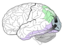
The main symptom resulting from posterior cortical atrophy is a decrease in visuospatial and visuoperceptual capabilities, since the area of atrophy involves the occipital lobe responsible for visual processing. [8] The atrophy is progressive; early symptoms include difficulty reading, blurred vision, light sensitivity, issues with depth perception, and trouble navigating through space. [9] [10] Additional symptoms include apraxia, a disorder of movement planning, alexia, an impaired ability to read, and visual agnosia, an object recognition disorder. [11] In the two-streams hypothesis, damage to the ventral, or "what" stream, of the visual system, located in the temporal lobe, leads to the symptoms related to general vision and object recognition deficits; damage to the dorsal, or "where/how" stream, located in the parietal lobe, leads to PCA symptoms related to impaired movements in response to visual stimuli, such as navigation and apraxia. [11] [10]

The dorsal stream (green) runs through the parietal lobe, and the ventral stream (purple) runs through the temporal lobe. Both streams originate in the occipital lobe (blue) located posteriorly. Ventral-dorsal streams.svg
The dorsal stream (green) runs through the parietal lobe, and the ventral stream (purple) runs through the temporal lobe. Both streams originate in the occipital lobe (blue) located posteriorly.

As neurodegeneration spreads, more severe symptoms emerge, including the inability to recognize familiar people and objects, trouble navigating familiar places, and sometimes visual hallucinations. [8] [9] In addition, difficulty may be experienced in making guiding movements towards objects, and a decline in literacy skills including reading, writing, and spelling may develop. [9] [12] [13] Furthermore, if neural death spreads into other anterior cortical regions, symptoms similar to Alzheimer's disease, such as memory loss, may result. [9] [12] In PCA where there is significant atrophy in one hemisphere of the brain hemispatial neglect may result – the inability to see stimuli on one half of the visual field. [10] Anxiety and depression are also common symptoms. [14]

Connection to Alzheimer's disease

Studies have shown that PCA may be a variant of Alzheimer's disease (AD), with an emphasis on visual deficits. [2] [11] Although in primarily different, but sometimes overlapping, brain regions, both involve progressive neural degeneration, as shown by the loss of neurons and synapses, and the presence of neurofibrillary tangles and senile plaques in affected brain regions; this eventually leads to dementia in both diseases. [15] [16] In PCA there is more cortical damage and gray matter (cell body) loss in posterior regions, especially in the occipital, parietal, and temporal lobes, whereas in Alzheimer's there is typically more damage in the prefrontal cortex and hippocampus. [12] [15] [17] PCA tends to impair working memory and anterograde memory, while leaving episodic memory intact, whereas in AD there is typically impaired episodic memory, suggesting some differences still lie in the primary areas of cortical damage. [9] [15]

Over time, however, atrophy in PCA may spread to regions that are commonly damaged in AD, leading to shared AD symptoms such as deficits in memory, language, learning, and cognition. [11] [12] [15] [16] Although PCA has an earlier onset, a diagnosis with Alzheimer's is often made, suggesting that the degeneration has simply migrated anteriorly to other cortical brain regions. [8] [11]

There is no standard definition of PCA and no established diagnostic criteria, so it is not possible to know how many people have the condition. Some studies have found that about 5 percent of people diagnosed with Alzheimer's disease have PCA. However, because PCA often goes unrecognized, the true percentage may be as high as 15 percent. Researchers and physicians are working to establish a standard definition and diagnostic criteria for PCA. [18]

PCA may also be correlated with Lewy body disease, Creutzfeldt–Jakob disease, Bálint's syndrome, and Gerstmann syndrome. [9] [10] [19] In addition, PCA may result in part from mutations in the presenilin 1 gene (PSEN1). [10]

Diagnosis

The cause of PCA is unknown, and there are no fully accepted diagnostic criteria for the disease. [2] [10] This is partially due to the gradual onset of PCA symptoms, their variety, the rare nature of the disease, and the younger age of onset typically 50–60 years. [20] In 2012, the first international conference on PCA was held in Vancouver, Canada. Continued research and testing will hopefully result in accepted and standardized criteria for diagnosis. [10]

PCA is often initially misdiagnosed as an anxiety disorder or depression. It has been suggested that depression or anxiety may result from the symptoms of decreased visual function, and the progressive nature of the disease. Early visual impairments have often led to a referral to an ophthalmologist, which can result in unnecessary cataract surgery. [20]

Due to the lack of biomarkers for PCA, neuropsychological examinations are advised. [21] Neuroimaging can also assist in the diagnosis of PCA. [20] For PCA and AD neuroimaging is carried out using MRI scans, single-photon emission computed tomography, and positron emission tomography (PET scans). [22] Neuroimages are often compared to those of people with AD to assist diagnosis. Due to the early onset of PCA in comparison to AD, images taken at the early stages of the disease will vary from brain images in AD. At this early stage brain atrophy will be shown to be more centrally located in the right posterior lobe and occipital gyrus, while AD brain images show the majority of atrophy in the medial temporal cortex. This variation within the images will assist in early diagnosis of PCA; however, as the years go on the images will become increasingly similar, due to the majority of PCA also developing to AD later in life because of continued brain atrophy. [10] [23] A key aspect found through brain imaging of PCA patients is a loss of grey matter (collections of neuronal cell bodies) in the posterior and occipital temporal cortices within the right hemisphere. [24]

For some people with PCA, neuroimaging may not give a clear diagnosis; therefore, careful observation in relation to PCA symptoms can also assist in the diagnosis. [20] The variation and lack of organized clinical testing has led to continued difficulties and delays in the diagnosis of PCA. [10]

Treatment

Specific and accepted treatment for PCA has yet to be discovered; this may be due to the rarity and variations of the disease. [10] [25] At times people with PCA are treated with AD treatments, such as cholinesterase inhibitors: donepezil, rivastigmine, galantamine, and also memantine. [10] Antidepressant drugs have also provided some positive effects. [20]

Other treatments such as occupational therapy, or help with adapting to visual changes may help. [10] [20] People with PCA and their caregivers are likely to have different needs than the more typical cases of Alzheimer's disease, and may benefit from specialized support groups, or other groups for young people with dementia. No study to date has been definitive to provide accepted conclusive analysis on treatment options. [20]

Related Research Articles

<span class="mw-page-title-main">Dementia with Lewy bodies</span> Type of progressive dementia

Dementia with Lewy bodies (DLB) is a type of dementia characterized by changes in sleep, behavior, cognition, movement, and regulation of automatic bodily functions. Memory loss is not always an early symptom. The disease worsens over time and is usually diagnosed when cognitive impairment interferes with normal daily functioning. Together with Parkinson's disease dementia, DLB is one of the two Lewy body dementias. It is a common form of dementia, but the prevalence is not known accurately and many diagnoses are missed. The disease was first described by Kenji Kosaka in 1976.

<span class="mw-page-title-main">Binswanger's disease</span> Medical condition

Binswanger's disease, also known as subcortical leukoencephalopathy and subcortical arteriosclerotic encephalopathy, is a form of small-vessel vascular dementia caused by damage to the white brain matter. White matter atrophy can be caused by many circumstances including chronic hypertension as well as old age. This disease is characterized by loss of memory and intellectual function and by changes in mood. These changes encompass what are known as executive functions of the brain. It usually presents between 54 and 66 years of age, and the first symptoms are usually mental deterioration or stroke.

<span class="mw-page-title-main">Precuneus</span> Region of the parietal lobe of the brain

In neuroanatomy, the precuneus is the portion of the superior parietal lobule on the medial surface of each brain hemisphere. It is located in front of the cuneus. The precuneus is bounded in front by the marginal branch of the cingulate sulcus, at the rear by the parieto-occipital sulcus, and underneath by the subparietal sulcus. It is involved with episodic memory, visuospatial processing, reflections upon self, and aspects of consciousness.

<span class="mw-page-title-main">Occipital lobe</span> Part of the brain at the back of the head

The occipital lobe is one of the four major lobes of the cerebral cortex in the brain of mammals. The name derives from its position at the back of the head, from the Latin ob, 'behind', and caput, 'head'.

<span class="mw-page-title-main">Frontotemporal dementia</span> Types of dementia involving the frontal or temporal lobes

Frontotemporal dementia (FTD), frontotemporal degeneration disease, or frontotemporal neurocognitive disorder, encompasses several types of dementia involving the progressive degeneration of the brain's frontal and temporal lobes. FTDs broadly present as behavioral or language disorders with gradual onsets. Common signs and symptoms include significant changes in social and personal behavior, apathy, blunting of emotions, and deficits in both expressive and receptive language. Signs and symptoms tend to appear in late adulthood, typically between the ages of 45 and 65. Men and women appear to be equally affected. FTD is the second most prevalent type of early onset dementia after Alzheimer's disease. Currently, there is no cure, but there are treatments that help alleviate symptoms.

Cerebral atrophy is a common feature of many of the diseases that affect the brain. Atrophy of any tissue means a decrement in the size of the cell, which can be due to progressive loss of cytoplasmic proteins. In brain tissue, atrophy describes a loss of neurons and the connections between them. Brain atrophy can be classified into two main categories: generalized and focal atrophy. Generalized atrophy occurs across the entire brain whereas focal atrophy affects cells in a specific location. If the cerebral hemispheres are affected, conscious thought and voluntary processes may be impaired.

Semantic dementia (SD), also known as semantic variant primary progressive aphasia (svPPA), is a progressive neurodegenerative disorder characterized by loss of semantic memory in both the verbal and non-verbal domains. However, the most common presenting symptoms are in the verbal domain. Semantic dementia is a disorder of semantic memory that causes patients to lose the ability to match words or images to their meanings. However, it is fairly rare for patients with semantic dementia to develop category specific impairments, though there have been documented cases of it occurring. Typically, a more generalized semantic impairment results from dimmed semantic representations in the brain.

Imaging genetics refers to the use of anatomical or physiological imaging technologies as phenotypic assays to evaluate genetic variation. Scientists that first used the term imaging genetics were interested in how genes influence psychopathology and used functional neuroimaging to investigate genes that are expressed in the brain.

<span class="mw-page-title-main">Corticobasal degeneration</span> Rare neurodegenerative disease

Corticobasal degeneration (CBD) is a rare neurodegenerative disease involving the cerebral cortex and the basal ganglia. CBD symptoms typically begin in people from 50 to 70 years of age, and typical survival before death is eight years. It is characterized by marked disorders in movement and cognition, and is classified as one of the Parkinson plus syndromes. Diagnosis is difficult, as symptoms are often similar to those of other disorders, such as Parkinson's disease, progressive supranuclear palsy, and dementia with Lewy bodies, and a definitive diagnosis of CBD can only be made upon neuropathologic examination.

Cortical blindness is the total or partial loss of vision in a normal-appearing eye caused by damage to the brain's occipital cortex. Cortical blindness can be acquired or congenital, and may also be transient in certain instances. Acquired cortical blindness is most often caused by loss of blood flow to the occipital cortex from either unilateral or bilateral posterior cerebral artery blockage and by cardiac surgery. In most cases, the complete loss of vision is not permanent and the patient may recover some of their vision. Congenital cortical blindness is most often caused by perinatal ischemic stroke, encephalitis, and meningitis. Rarely, a patient with acquired cortical blindness may have little or no insight that they have lost vision, a phenomenon known as Anton–Babinski syndrome.

<span class="mw-page-title-main">Posterior cerebral artery</span> Artery which supplies blood to the occipital lobe of the brain

The posterior cerebral artery (PCA) is one of a pair of cerebral arteries that supply oxygenated blood to the occipital lobe, part of the back of the human brain. The two arteries originate from the distal end of the basilar artery, where it bifurcates into the left and right posterior cerebral arteries. These anastomose with the middle cerebral arteries and internal carotid arteries via the posterior communicating arteries.

Psychoorganic syndrome (POS), also known as organic psychosyndrome, is a progressive disease comparable to presenile dementia. It consists of psychopathological complex of symptoms that are caused by organic brain disorders that involve a reduction in memory and intellect. Psychoorganic syndrome is often accompanied by asthenia.

<span class="mw-page-title-main">Inferior temporal gyrus</span> One of three gyri of the temporal lobe of the brain

The inferior temporal gyrus is one of three gyri of the temporal lobe and is located below the middle temporal gyrus, connected behind with the inferior occipital gyrus; it also extends around the infero-lateral border on to the inferior surface of the temporal lobe, where it is limited by the inferior sulcus. This region is one of the higher levels of the ventral stream of visual processing, associated with the representation of objects, places, faces, and colors. It may also be involved in face perception, and in the recognition of numbers and words.

<span class="mw-page-title-main">Posterior cingulate cortex</span> Caudal part of the cingulate cortex of the brain

The posterior cingulate cortex (PCC) is the caudal part of the cingulate cortex, located posterior to the anterior cingulate cortex. This is the upper part of the "limbic lobe". The cingulate cortex is made up of an area around the midline of the brain. Surrounding areas include the retrosplenial cortex and the precuneus.

<span class="mw-page-title-main">Posterior cerebral artery syndrome</span> Medical condition

Posterior cerebral artery syndrome is a condition whereby the blood supply from the posterior cerebral artery (PCA) is restricted, leading to a reduction of the function of the portions of the brain supplied by that vessel: the occipital lobe, the inferomedial temporal lobe, a large portion of the thalamus, and the upper brainstem and midbrain.

Phonagnosia is a type of agnosia, or loss of knowledge, that involves a disturbance in the recognition of familiar voices and the impairment of voice discrimination abilities in which the affected individual does not suffer from comprehension deficits. Phonagnosia is an auditory agnosia, an acquired auditory processing disorder resulting from brain damage, other auditory agnosias include cortical deafness and auditory verbal agnosia also known as pure word deafness.

Alzheimer's Disease Neuroimaging Initiative (ADNI) is a multisite study that aims to improve clinical trials for the prevention and treatment of Alzheimer's disease (AD). This cooperative study combines expertise and funding from the private and public sector to study subjects with AD, as well as those who may develop AD and controls with no signs of cognitive impairment. Researchers at 63 sites in the US and Canada track the progression of AD in the human brain with neuroimaging, biochemical, and genetic biological markers. This knowledge helps to find better clinical trials for the prevention and treatment of AD. ADNI has made a global impact, firstly by developing a set of standardized protocols to allow the comparison of results from multiple centers, and secondly by its data-sharing policy which makes available all at the data without embargo to qualified researchers worldwide. To date, over 1000 scientific publications have used ADNI data. A number of other initiatives related to AD and other diseases have been designed and implemented using ADNI as a model. ADNI has been running since 2004 and is currently funded until 2021.

<span class="mw-page-title-main">Ulegyria</span> Type of cortical scarring deep in the sulci

Ulegyria is a diagnosis used to describe a specific type of cortical scarring in the deep regions of the sulcus that leads to distortion of the gyri. Ulegyria is identified by its characteristic "mushroom-shaped" gyri, in which scarring causes shrinkage and atrophy in the deep sulcal regions while the surface gyri are spared. This condition is most often caused by hypoxic-ischemic brain injury in the perinatal period. The effects of ulegyria can range in severity, although it is most commonly associated with cerebral palsy, mental retardation and epilepsy. N.C. Bresler was the first to view ulegyria in 1899 and described this abnormal morphology in the brain as “mushroom-gyri." Although ulegyria was first identified in 1899, there is still limited information known or reported about the condition.

Parkinson's disease dementia (PDD) is dementia that is associated with Parkinson's disease (PD). Together with dementia with Lewy bodies (DLB), it is one of the Lewy body dementias characterized by abnormal deposits of Lewy bodies in the brain.

The occipital face area (OFA) is a region of the human cerebral cortex which is specialised for face perception. The OFA is located on the lateral surface of the occipital lobe adjacent to the inferior occipital gyrus. The OFA comprises a network of brain regions including the fusiform face area (FFA) and posterior superior temporal sulcus (STS) which support facial processing.

References

  1. Schott JM, Crutch SJ (February 2019). "Posterior Cortical Atrophy". Continuum (Minneap Minn). 25 (1): 52–75. doi:10.1212/CON.0000000000000696. PMC   6548537 . PMID   30707187.
  2. 1 2 3 "Visual variant of Alzheimer's disease - EyeWiki". eyewiki.aao.org. Retrieved 2017-11-07.
  3. Nestor PJ, Caine D, Fryer TD, Clarke J, Hodges JR (2003). "The topography of metabolic deficits in posterior cortical atrophy (the visual variant of Alzheimer's disease) with FDG-PET". J Neurol Neurosurg Psychiatry. 74 (11): 1521–1529. doi:10.1136/jnnp.74.11.1521. PMC   1738241 . PMID   14617709.
  4. 1 2 3 "Posterior Cortical Atrophy". UCSF Memory and Aging Center. University of California, San Francisco . Retrieved 2011-10-22.
  5. Benson DF, Davis RJ, Snyder BD (July 1988). "Posterior Cortical Atrophy". Archives of Neurology . 45 (7): 789–793. doi:10.1001/archneur.1988.00520310107024. PMID   3390033.
  6. 1 2 "Posterior Cortical Atrophy". Martinos Center for Biomedical Imaging. Harvard University. 2009-01-19. Retrieved 2011-10-22.
  7. "Terry Pratchett pledges $1 million to Alzheimer's research". Alzheimer's Research Trust. 2011-07-29. Archived from the original on 2013-01-14. Retrieved 2011-07-29.
  8. 1 2 3 Mendez, Mario; Mehdi Ghajarania; Kent Perryman (14 June 2002). "Posterior cortical atrophy: Clinical characteristics and differences compared to Alzheimer's disease". Dementia and Geriatric Cognitive Disorders. 14 (1): 33–40. doi:10.1159/000058331. PMID   12053130. S2CID   12291848.
  9. 1 2 3 4 5 6 Crutch, Sebastian; Manja Lehmann; Jonathan Schott; Gil Rabinovici; Martin Rosser; Nick Fox (February 2012). "Posterior Cortical Atrophy" (PDF). The Lancet Neurology. 11 (2): 170–178. doi:10.1016/s1474-4422(11)70289-7. PMC   3740271 . PMID   22265212.
  10. 1 2 3 4 5 6 7 8 9 10 11 12 Borruat, François-Xavier (18 October 2013). "Posterior Cortical Atrophy: Review of the Recent Literature" (PDF). Neuro-Ophthalmology. 13 (12): 406. doi:10.1007/s11910-013-0406-8. PMID   24136454. S2CID   45579994.
  11. 1 2 3 4 5 Goenthals, Maartin; Patrick Santens (20 February 2001). "Posterior cortical atrophy. Two case reports and a review of the literature". Clinical Neurology and Neurosurgery. 103 (2): 115–119. doi:10.1016/s0303-8467(01)00114-7. PMID   11516556. S2CID   46582748.
  12. 1 2 3 4 Crutch SJ, Schott JM, Rabinovici GD, Boeve BF, Cappa SF, Dickerson BC, Dubois B, Graff-Radford NR, Krolak-Salmon P, Lehmann M, Mendez MF, Pijnenburg Y, Ryan NS, Scheltens P, Shakespeare T, Tang-Wai DF, van der Flier WM, Bain L, Carrillo MC, Fox NC (Jul 2013). "Shining a light on posterior cortical atrophy". Alzheimer's & Dementia. 9 (4): 463–5. doi:10.1016/j.jalz.2012.11.004. PMID   23274153. S2CID   16404876.
  13. Tsunoda, Ayami; Shuji Iritani; Norio Ozaki (17 March 2011). "Presenile dementia diagnosed as posterior cortical atrophy". Psychogeriatrics. 11 (3): 171–176. doi:10.1111/j.1479-8301.2011.00366.x. PMID   21951958.
  14. Crutch, Sebastian; Manja Lehmann; Jonathan Schott; Gil Rabinovici; Martin Rosser; Nick Fox (February 2012). "Posterior cortical atrophy". The Lancet Neurology. 11 (2): 170–178. doi:10.1016/s1474-4422(11)70289-7. PMC   3740271 . PMID   22265212.
  15. 1 2 3 4 Kennedy, Jonathan; Manja Lehmann; Magdalena J. Sokolska; Hilary Archer; Elizabeth K. Warrington; Nick C. Fox; Sebastian J. Crutch (25 October 2011). "Visualizing the emergence of posterior cortical atrophy". Neurocase: The Neural Basis of Cognition. 18 (3): 248–257. doi:10.1080/13554794.2011.588180. PMID   22026812. S2CID   20617012.
  16. 1 2 Hof, Patrick; Brent Vogt; Constantin Bouras; John Morrison (December 1997). "Atypical Form of Alzheimer's Disease with Prominent Posterior Cortical Atrophy: a Review of Lesion Distribution and Circuit Disconnection in Cortical Visual Pathways". Vision Res. 37 (24): 3609–3625. doi: 10.1016/s0042-6989(96)00240-4 . PMID   9425534.
  17. Tsunoda, Ayami; Shuji Iritani; Norio Ozaki (17 March 2011). "Presenile dementia diagnosed as posterior cortical atrophy". Psychogeriatrics. 11 (171–176): 171–176. doi:10.1111/j.1479-8301.2011.00366.x. PMID   21951958.
  18. "Posterior Cortical Atrophy | Signs, Symptoms, & Diagnosis". Dementia. Retrieved 2016-07-25.
  19. Nagaratnam, Nages; Kujan Nagaratnam; Daniel Jolley; Allan Ting (6 June 2001). "Dementia following posterior cortical atrophy—a descriptive clinical case report". Archives of Gerontology and Geriatrics. 33 (2): 179–190. doi:10.1016/s0167-4943(01)00179-0. PMID   15374033.
  20. 1 2 3 4 5 6 7 Crutch, Sebastian J.; Manja Lehmann; Jonathan M. Schott; Gil D. Rabinovici; Martin N. Rossor; Nick C. Fox (February 2012). "Posterior Cortical Atrophy". The Lancet Neurology. 11 (2): 170–178. doi:10.1016/s1474-4422(11)70289-7. PMC   3740271 . PMID   22265212.
  21. Croisile, MD. Bernard; Alexis Brice (September 2004). "Benson's syndrome or Posterior Cortical Atrophy" (PDF). Orphanet Encyclopedia. Archived from the original (PDF) on 12 November 2013. Retrieved 11 November 2013.
  22. Goldstein, Martin A.; Iliyan Ivanov; Michael E. Silverman (May 2011). "Posterior Cortical Atrophy: An Exemplar for Renovating Diagnostic Formulation in Neurosychiaty". Comprehensive Psychiatry. 52 (3): 326–333. doi:10.1016/j.comppsych.2010.06.013. PMID   21497228.
  23. Möller C, van der Flier WM, Versteeg A, Benedictus MR, Wattjes MP, Koedam EL, Scheltens P, Barkhof F, Vrenken H (Feb 2014). "Quantitative Regional Validation of the Visual Rating Scale for Posterior Cortical Atrophy". European Radiology. 24 (2): 397–404. doi:10.1007/s00330-013-3025-5. PMID   24092044. S2CID   14187936.
  24. Migliaccio R, Agosta F, Toba MN, Samri D, Corlier F, de Souza LC, Chupin M, Sharman M, Gorno-Tempini ML, Dubois B, Filippi M, Bartolomeo P (November 2012). "Brain Networks in Posterior Cortical Atrophy: A Single Case Tractography Study and Literature Review". Cortex. 48 (10): 1298–1309. doi:10.1016/j.cortex.2011.10.002. PMC   4813795 . PMID   22099855.
  25. Caine, Diana (2004). "Posterior Cortical Atrophy: A Review of the Literature". Neurocase. 10 (5): 382–385. doi:10.1080/13554790490892239. PMID   15788276. S2CID   45491962.