Cerebral herniation | |
---|---|
MRI showing injury due to brain herniation | |
Specialty | Neurology, neurosurgery |
Brain herniation is a potentially deadly side effect of very high pressure within the skull that occurs when a part of the brain is squeezed across structures within the skull. The brain can shift across such structures as the falx cerebri, the tentorium cerebelli, and even through the foramen magnum (the hole in the base of the skull through which the spinal cord connects with the brain). Herniation can be caused by a number of factors that cause a mass effect and increase intracranial pressure (ICP): these include traumatic brain injury, intracranial hemorrhage, or brain tumor. [1]
Herniation can also occur in the absence of high ICP when mass lesions such as hematomas occur at the borders of brain compartments. In such cases local pressure is increased at the place where the herniation occurs, but this pressure is not transmitted to the rest of the brain, and therefore does not register as an increase in ICP. [2]
Because herniation puts extreme pressure on parts of the brain and thereby cuts off the blood supply to various parts of the brain, it is often fatal. Therefore, extreme measures are taken in hospital settings to prevent the condition by reducing intracranial pressure, or decompressing (draining) a hematoma which is putting local pressure on a part of the brain.
Brain herniation frequently presents with abnormal posturing, [2] a characteristic positioning of the limbs indicative of severe brain damage. These patients have a lowered level of consciousness, with Glasgow Coma Scores of three to five. [2] One or both pupils may be dilated and fail to constrict in response to light. [2] Vomiting can also occur due to compression of the vomiting center in the medulla oblongata. Severe headaches and seizures as a result of increased intracranial pressure are not uncommon. Cardiovascular and pulmonary symptoms may also be present as the brain loses function, but might also be associated with bleeding. These symptoms are known as Cushing's Triad: hypertension (with widening pulse pressure), irregular respirations (commonly Cheyne-Stokes), bradycardia and in severe cases cardiac arrest. [3] [4]
Causes of brain herniation include: [5]
The tentorium is an extension of the dura mater that separates the cerebellum from the cerebrum. There are two major classes of herniation: supratentorial and infratentorial. Supratentorial refers to herniation of structures normally found above the tentorial notch, and infratentorial refers to structures normally found below it. [7]
1) Uncal (transtentorial)
2) Central
3) Cingulate (subfalcine or transfalcine)
5) Tectal (posterior)
6) Upward (upward cerebellar or upward transtentorial)
7) Tonsillar (downward cerebellar)
In uncal herniation, a common subtype of transtentorial herniation, the innermost part of the temporal lobe, the uncus, can be squeezed so much that it moves towards the tentorium and puts pressure on the brainstem, most notably the midbrain. [8] The tentorium is a structure within the skull formed by the dura mater of the meninges. Tissue may be stripped from the cerebral cortex in a process called decortication. [9]
The uncus can squeeze the oculomotor nerve (a.k.a. CN III), which may affect the parasympathetic input to the eye on the side of the affected nerve, causing the pupil of the affected eye to dilate and fail to constrict in response to light as it should. Pupillary dilation often precedes the somatic motor effects of CN III compression called oculomotor nerve palsy or third nerve palsy. This palsy presents as deviation of the eye to a "down and out" position due to loss of innervation to all ocular motility muscles except for the lateral rectus (innervated by abducens nerve (a.k.a. CN VI) and the superior oblique (innervated by trochlear nerve a.k.a. CN IV). The symptoms occur in this order because the parasympathetic fibers surround the motor fibers of CN III and are hence compressed first.[ citation needed ]
Compression of the ipsilateral posterior cerebral artery will result in ischemia of the ipsilateral primary visual cortex and contralateral visual field deficits in both eyes (contralateral homonymous hemianopsia).
Another important finding is a false localizing sign, the so-called Kernohan's notch, which results from compression of the contralateral [10] cerebral crus containing descending corticospinal and some corticobulbar tract fibers. This leads to Ipsilateral hemiparesis in reference to the herniation and contralateral hemiparesis with reference to the cerebral crus.[ citation needed ]
With increasing pressure and progression of the hernia there will be distortion of the brainstem leading to Duret hemorrhages (tearing of small vessels in the parenchyma) in the median and paramedian zones of the mesencephalon and pons. The rupture of these vessels leads to linear or flamed shaped hemorrhages. The disrupted brainstem can lead to decorticate posture, respiratory center depression and death. Other possibilities resulting from brain stem distortion include lethargy, slow heart rate, and pupil dilation. [9]
Uncal herniation may advance to central herniation. [7] The sliding uncus syndrome represents uncal herniation without alteration in the level of consciousness and other sequelae mentioned above.
In central herniation, the diencephalon and parts of the temporal lobes of both of the cerebral hemispheres are squeezed through a notch in the tentorium cerebelli. [8] [11] Transtentorial herniation can occur when the brain moves either up or down across the tentorium, called ascending and descending transtentorial herniation respectively; however descending herniation is much more common. [1] Downward herniation can stretch branches of the basilar artery (pontine arteries), causing them to tear and bleed, known as a Duret hemorrhage. The result is usually fatal. [11] Other symptoms of this type of herniation include small, fixed pupils with [12] paralysis of upward eye movement giving the characteristic appearance of "sunset eyes". Also found in these patients, often as a terminal complication is the development of diabetes insipidus due to the compression of the pituitary stalk. Radiographically, downward herniation is characterized by obliteration of the suprasellar cistern from temporal lobe herniation into the tentorial hiatus with associated compression on the cerebral peduncles. Upwards herniation, on the other hand, can be radiographically characterized by obliteration of the quadrigeminal cistern. Intracranial hypotension syndrome has been known to mimic downwards transtentorial herniation.
In cingulate or subfalcine herniation, the most common type, the innermost part of the frontal lobe is scraped under part of the falx cerebri, the dura mater at the top of the head between the two hemispheres of the brain. [8] [13] Cingulate herniation can be caused when one hemisphere swells and pushes the cingulate gyrus by the falx cerebri. [7] This does not put as much pressure on the brainstem as the other types of herniation, but it may interfere with blood vessels in the frontal lobes that are close to the site of injury (anterior cerebral artery), or it may progress to central herniation. [8] Interference with the blood supply can cause dangerous increases in ICP that can lead to more dangerous forms of herniation. [14] Symptoms for cingulate herniation are not well defined. [14] Usually occurring in addition to uncal herniation, cingulate herniation may present with abnormal posturing and coma. [7] Cingulate herniation is frequently believed to be a precursor to other types of herniation. [14]
In transcalvarial herniation, the brain squeezes through a fracture or a surgical site in the skull. [7] Also called "external herniation", this type of herniation may occur during craniectomy, surgery in which a flap of skull is removed, the protruding brain region preventing the piece of skull from being replaced during the operation. [1]
Increased pressure in the posterior fossa can cause the cerebellum to move up through the tentorial opening in upward, or cerebellar herniation. [8] The midbrain is pushed through the tentorial notch upward. This is also known as ascending transtentorial herniation since it occurs across the tentorium cerebelli.
In tonsillar herniation, also called downward cerebellar herniation, [7] transforaminal herniation, or "coning", the cerebellar tonsils move downward through the foramen magnum possibly causing compression of the lower brainstem and upper cervical spinal cord as they pass through the foramen magnum. [8] Increased pressure on the brainstem can result in dysfunction of the centers in the brain responsible for controlling respiratory and cardiac function. The most common signs are intractable headache, head tilt, and neck stiffness due to tonsillar impaction. The level of consciousness may decrease and also give rise to flaccid paralysis. Blood pressure instability is also evident in these patients. [12]
Congenital tonsillar herniation of the cerebellum is also known as a Chiari malformation (CM), or previously an Arnold-Chiari malformation (ACM). There are four types of Chiari malformation, and they represent very different disease processes with different symptoms and prognosis. These conditions can be found in asymptomatic patients as an incidental finding, or can be so severe as to be life-threatening. This condition is now being diagnosed more frequently by radiologists, as more patients undergo MRI scans of their heads, especially upright MRI, which is more than twice as sensitive for detecting this condition. [15] Cerebellar tonsillar ectopia (CTE) is a term used by radiologists to describe cerebellar tonsils that are "low lying" but that do not meet the radiographic criteria for definition as a Chiari malformation. The currently accepted radiographic definition for a Chiari malformation is that cerebellar tonsils lie at least 5mm below the level of the foramen magnum. Some clinicians have reported that some patients appear to experience symptoms consistent with a Chiari malformation without radiographic evidence of tonsillar herniation. Sometimes these patients are described as having a 'Chiari [type] 0'.
There are many suspected causes of tonsillar herniation including: decreased or malformed posterior fossa (the lower, back part of the skull) not providing enough room for the cerebellum; hydrocephalus or abnormal CSF volume pushing the tonsils out; or dural tension pulling the brain caudally. Connective tissue disorders, such as Ehlers Danlos syndrome, can be associated.
For further evaluation of tonsillar herniation, CINE flow studies are used. This type of MRI examines flow of CSF at the cranio-cervical joint. For persons experiencing symptoms but without clear MRI evidence, especially if the symptoms are better in the supine position and worse upon standing/upright, an upright MRI may be useful. [2]
Treatment involves removal of the etiologic mass and decompressive craniectomy. Brain herniation can cause severe disability or death. In fact, when herniation is visible on a CT scan, the prognosis for a meaningful recovery of neurological function is poor. [2] The patient may become paralyzed on the same side as the lesion causing the pressure, or damage to parts of the brain caused by herniation may cause paralysis on the side opposite the lesion. [11] Damage to the midbrain, which contains the reticular activating network which regulates consciousness, will result in coma. [11] Damage to the cardio-respiratory centers in the medulla oblongata will cause respiratory arrest and (secondarily) cardiac arrest. [11] Investigation is underway regarding the use of neuroprotective agents during the prolonged post-traumatic period of brain hypersensitivity associated with the syndrome. [17]
A cerebral arteriovenous malformation is an abnormal connection between the arteries and veins in the brain—specifically, an arteriovenous malformation in the cerebrum.
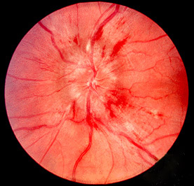
Papilledema or papilloedema is optic disc swelling that is caused by increased intracranial pressure due to any cause. The swelling is usually bilateral and can occur over a period of hours to weeks. Unilateral presentation is extremely rare.
Hydrocephalus is a condition in which an accumulation of cerebrospinal fluid (CSF) occurs within the brain. This typically causes increased pressure inside the skull. Older people may have headaches, double vision, poor balance, urinary incontinence, personality changes, or mental impairment. In babies, it may be seen as a rapid increase in head size. Other symptoms may include vomiting, sleepiness, seizures, and downward pointing of the eyes.
Chiari malformation (CM) is a structural defect in the cerebellum, characterized by a downward displacement of one or both cerebellar tonsils through the foramen magnum. CMs can cause headaches, difficulty swallowing, vomiting, dizziness, neck pain, unsteady gait, poor hand coordination, numbness and tingling of the hands and feet, and speech problems. Less often, people may experience ringing or buzzing in the ears, weakness, slow heart rhythm, or fast heart rhythm, curvature of the spine (scoliosis) related to spinal cord impairment, abnormal breathing, such as central sleep apnea, characterized by periods of breathing cessation during sleep, and, in severe cases, paralysis.
The trochlear nerve, also known as the fourth cranial nerve, cranial nerve IV, or CN IV, is a cranial nerve that innervates a single muscle - the superior oblique muscle of the eye. Unlike most other cranial nerves, the trochlear nerve is exclusively a motor nerve.
In neuroanatomy, dura mater is a thick membrane made of dense irregular connective tissue that surrounds the brain and spinal cord. It is the outermost of the three layers of membrane called the meninges that protect the central nervous system. The other two meningeal layers are the arachnoid mater and the pia mater. It envelops the arachnoid mater, which is responsible for keeping in the cerebrospinal fluid. It is derived primarily from the neural crest cell population, with postnatal contributions of the paraxial mesoderm.
Intracranial hemorrhage (ICH), also known as intracranial bleed, is bleeding within the skull. Subtypes are intracerebral bleeds, subarachnoid bleeds, epidural bleeds, and subdural bleeds. More often than not it ends in death.
Epidural hematoma is when bleeding occurs between the tough outer membrane covering the brain and the skull. Often there is loss of consciousness following a head injury, a brief regaining of consciousness, and then loss of consciousness again. Other symptoms may include headache, confusion, vomiting, and an inability to move parts of the body. Complications may include seizures.
The falx cerebri is a large, crescent-shaped fold of dura mater that descends vertically into the longitudinal fissure between the cerebral hemispheres of the human brain, separating the two hemispheres and supporting dural sinuses that provide venous and CSF drainage to the brain. It is attached to the crista galli anteriorly, and blends with the tentorium cerebelli posteriorly.
The cerebellar tentorium or tentorium cerebelli is an extension of the dura mater between the inferior aspect of the occipital lobes and the superior aspect of the cerebellum. The free border of the tentorium gives passage to the midbrain.
Abnormal posturing is an involuntary flexion or extension of the arms and legs, indicating severe brain injury. It occurs when one set of muscles becomes incapacitated while the opposing set is not, and an external stimulus such as pain causes the working set of muscles to contract. The posturing may also occur without a stimulus. Since posturing is an important indicator of the amount of damage that has occurred to the brain, it is used by medical professionals to measure the severity of a coma with the Glasgow Coma Scale and the Pediatric Glasgow Coma Scale.
Dandy–Walker malformation (DWM), also known as Dandy–Walker syndrome (DWS), is a rare congenital brain malformation in which the part joining the two hemispheres of the cerebellum does not fully form, and the fourth ventricle and space behind the cerebellum are enlarged with cerebrospinal fluid. Most of those affected develop hydrocephalus within the first year of life, which can present as increasing head size, vomiting, excessive sleepiness, irritability, downward deviation of the eyes and seizures. Other, less common symptoms are generally associated with comorbid genetic conditions and can include congenital heart defects, eye abnormalities, intellectual disability, congenital tumours, other brain defects such as agenesis of the corpus callosum, skeletal abnormalities, an occipital encephalocele or underdeveloped genitalia or kidneys. It is sometimes discovered in adolescents or adults due to mental health problems.
Duret haemorrhages are small linear areas of bleeding in the midbrain and upper pons of the brainstem. They are caused by a traumatic downward displacement of the brainstem.
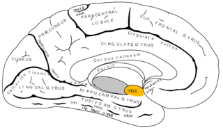
The uncus is an anterior extremity of the parahippocampal gyrus. It is separated from the apex of the temporal lobe by a slight fissure called the incisura temporalis.
The cerebellar tonsil is a rounded lobule on the undersurface of each cerebellar hemisphere, continuous medially with the uvula of the cerebellar vermis and superiorly by the flocculonodular lobe. Synonyms include: tonsilla cerebelli, amygdala cerebelli, the latter of which is not to be confused with the cerebral tonsils or amygdala nuclei located deep within the medial temporal lobes of the cerebral cortex.
Kernohan's notch is a cerebral peduncle indentation associated with some forms of transtentorial herniation. It is a secondary condition caused by a primary injury on the opposite hemisphere of the brain. Kernohan's notch is an ipsilateral condition, in that a left-sided primary lesion evokes motor impairment in the left side of the body and a right-sided primary injury evokes motor impairment in the right side of the body. The seriousness of Kernohan's notch varies depending on the primary problem causing it, which may range from benign brain tumors to advanced subdural hematoma.
A cerebrospinal fluid leak is a medical condition where the cerebrospinal fluid (CSF) surrounding the brain or spinal cord leaks out of one or more holes or tears in the dura mater. A cerebrospinal fluid leak can be either cranial or spinal, and these are two different disorders. A spinal CSF leak can be caused by one or more meningeal diverticula or CSF-venous fistulas not associated with an epidural leak.
Basilar invagination is invagination (infolding) of the base of the skull that occurs when the top of the C2 vertebra migrates upward. It can cause narrowing of the foramen magnum. It also may press on the lower brainstem.
Macrocephaly-capillary malformation (M-CM) is a multiple malformation syndrome causing abnormal body and head overgrowth and cutaneous, vascular, neurologic, and limb abnormalities. Though not every patient has all features, commonly found signs include macrocephaly, congenital macrosomia, extensive cutaneous capillary malformation, body asymmetry, polydactyly or syndactyly of the hands and feet, lax joints, doughy skin, variable developmental delay and other neurologic problems such as seizures and low muscle tone.
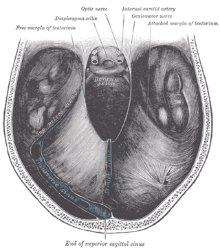
The tentorial notch refers to the anterior opening between the free edge of the cerebellar tentorium and the clivus for the passage of the brainstem.