Kaposi's sarcoma

Last updated
Kaposi's sarcoma, multiple haemorrhagic sarcoma
Other namesKaposi sarcoma
Kaposis sarcoma 01.jpg
Kaposi sarcoma. Characteristic purple lesions on the nose in an HIV-positive female. [1]
Pronunciation
Specialty Oncology
Symptoms Purple colored skin lesions [4]
TypesClassic, endemic, immunosuppression therapy-related, epidemic [4] [5]
Risk factors Human herpesvirus 8 (HHV8), poor immune function [4] [6]
Diagnostic method Tissue biopsy, medical imaging [4] [6]
Differential diagnosis Blue rubber bleb nevus syndrome, pyogenic granuloma, melanocytic nevi, melanoma [6]
TreatmentSurgery, chemotherapy, radiation therapy, biologic therapy [4]
Frequency42,000 (new cases, 2018) [7]
Deaths20,000 (2018) [7]

Kaposi's sarcoma (KS) is a type of cancer that can form masses in the skin, in lymph nodes, in the mouth, or in other organs. [4] [6] The skin lesions are usually painless, purple and may be flat or raised. [6] [8] Lesions can occur singly, multiply in a limited area, or may be widespread. [6] Depending on the sub-type of disease and level of immune suppression, KS may worsen either gradually or quickly. [6] Except for Classical KS where there is generally no immune suppression, KS is caused by a combination of immune suppression (such as due to HIV/AIDS) and infection by Human herpesvirus 8 (HHV8 – also called KS-associated herpesvirus (KSHV)). [8]

Contents

Classic, endemic, immunosuppression therapy-related (also known as iatrogenic), and epidemic (also known as AIDS-related) sub-types are all described. [8] Classic KS tends to affect older men in regions where KSHV is highly prevalent (Mediterranean, Eastern Europe, Middle East), is usually slow-growing, and most often affects only the legs. [8] Endemic KS is most common in Sub-Saharan Africa and is more aggressive in children, while older adults present similarly to classic KS. [8] Immunosuppression therapy-related KS generally occurs in people following organ transplantation and mostly affects the skin. [8] Epidemic KS occurs in people with AIDS and many parts of the body can be affected. [8] KS is diagnosed by tissue biopsy, while the extent of disease may be determined by medical imaging. [4] [6] [8]

Treatment is based on the sub-type, whether the condition is localized or widespread, and the person's immune function. [6] Localized skin lesions may be treated by surgery, injections of chemotherapy into the lesion, or radiation therapy. [6] Widespread disease may be treated with chemotherapy or biologic therapy. [4] [6] In those with HIV/AIDS, highly active antiretroviral therapy (HAART) prevents and often treats KS. [8] [9] In certain cases the addition of chemotherapy may be required. [9] With widespread disease, death may occur. [6]

The condition is relatively common in people with HIV/AIDS and following organ transplant. [6] [8] [9] Over 35% of people with AIDS may be affected. [10] KS was first described by Moritz Kaposi in 1872. [11] [12] It became more widely known as one of the AIDS-defining illnesses in the 1980s. [11] KSHV was discovered as a causative agent in 1994. [11] [13]

Signs and symptoms

KS lesions are nodules or blotches that may be red, purple, brown, or black, and are usually papular.[ citation needed ]

They are typically found on the skin, but spread elsewhere is common, especially the mouth, gastrointestinal tract and respiratory tract. Growth can range from very slow to explosively fast, and is associated with significant mortality and morbidity. [14]

The lesions are painless, but become cosmetically disfiguring or interruptive to organs. [15]

Skin

An example of Kaposi's sarcoma Kaposis Sarcoma Lesions.jpg
An example of Kaposi's sarcoma
Patch stage Kaposi's sarcoma. Red to brownish irregularly shaped macules and plaques. Patch stage Kaposi's sarcoma.jpg
Patch stage Kaposi's sarcoma. Red to brownish irregularly shaped macules and plaques.

Commonly affected areas include the lower limbs, back, face, mouth, and genitalia. The lesions are usually as described above, but may occasionally be plaque-like (often on the soles of the feet) or even involved in skin breakdown with resulting fungating lesions. Associated swelling may be from either local inflammation or lymphoedema (obstruction of local lymphatic vessels by the lesion). Kaposi's sarcoma skin lesions may be psychologically distressing. [17] [18]

Mouth

An HIV-positive person presenting with a Kaposi's sarcoma lesion with an overlying candidiasis infection in their mouth Kaposi's sarcoma intraoral AIDS 072 lores.jpg
An HIV-positive person presenting with a Kaposi's sarcoma lesion with an overlying candidiasis infection in their mouth

The mouth is involved in about 30% of cases, and is the initial site in 15% of AIDS-related KS. In the mouth, the hard palate is most frequently affected, followed by the gums. [19] Lesions in the mouth may be easily damaged by chewing and bleed or develop secondary infection, and even interfere with eating or speaking.[ citation needed ]

Gastrointestinal tract

Involvement can be common in those with transplant-related or AIDS-related KS, and it may occur in the absence of skin involvement. The gastrointestinal lesions may be silent or cause weight loss, pain, nausea/vomiting, diarrhea, bleeding (either vomiting blood or passing it with bowel movements), malabsorption, or intestinal obstruction. [20]

Respiratory tract

Involvement of the airway can present with shortness of breath, fever, cough, coughing up blood or chest pain, or as an incidental finding on chest x-ray. [21] The diagnosis is usually confirmed by bronchoscopy, when the lesions are directly seen and often biopsied. Kaposi's sarcoma of the lung has a poor prognosis.[ citation needed ]

Cause

Kaposi's sarcoma-associated herpesvirus (KSHV), also called HHV-8, is present in almost 100% of Kaposi sarcoma lesions, whether HIV-related, classic, endemic, or iatrogenic. [22] KSHV encodes oncogenes, microRNAs and circular RNAs that promote cancer cell proliferation and escape from the immune system. [23]

Transmission

In Europe and North America, KSHV is transmitted through saliva. Thus, kissing is a risk factor for transmission. Higher rates of transmission among gay and bisexual men have been attributed to "deep kissing" sexual partners with KSHV. [24] Another alternative theory suggests that use of saliva as a sexual lubricant might be a major mode for transmission. Prudent advice is to use commercial lubricants when needed and avoid deep kissing with partners with KSHV infection or whose status is unknown.[ citation needed ]

KSHV is also transmissible via organ transplantation [25] and blood transfusion. [26] Testing for the virus before these procedures is likely to effectively limit iatrogenic transmission.[ citation needed ]

Pathology

Micrograph of a Kaposi sarcoma showing its typical features. Histopathology of Kaposi's sarcoma.png
Micrograph of a Kaposi sarcoma showing its typical features.

Despite its name, in general it is not considered a true sarcoma, [27] [28] which is a tumor arising from mesenchymal tissue. The histogenesis of KS remains controversial. [29] KS may arise as a cancer of lymphatic endothelium [30] and forms vascular channels that fill with blood cells, giving the tumor its characteristic bruise-like appearance. KSHV proteins are uniformly detected in KS cancer cells.[ citation needed ]

KS lesions contain tumor cells with a characteristic abnormal elongated shape, called spindle cells. The most typical feature of Kaposi sarcoma is the presence of spindle cells forming slits containing red blood cells. Mitotic activity is only moderate and pleomorphism is usually absent. [31] The tumor is highly vascular, containing abnormally dense and irregular blood vessels, which leak red blood cells into the surrounding tissue and give the tumor its dark color. Inflammation around the tumor may produce swelling and pain. Variously sized PAS positive hyaline bodies are often seen in the cytoplasm or sometimes extracellularly.[ citation needed ]

The spindle cells of Kaposi sarcoma differentiate toward endothelial cells, probably of lymph vessel rather than blood vessel origin. [32] The consistent immunoreactivity for podoplanin supports the lymphatic nature of the lesion.[ citation needed ]

Diagnosis

Although KS may be suspected from the appearance of lesions and the patient's risk factors, a definite diagnosis can be made only by biopsy and microscopic examination. Detection of the KSHV protein LANA in tumor cells confirms the diagnosis.[ citation needed ]

In differential diagnosis, arteriovenous malformations, pyogenic granuloma and other vascular proliferations can be microscopically confused with KS. [33]

Differential diagnosis of Kaposi's sarcoma

Source: [34]

  1. Naevus
  2. Histiocytoma
  3. Cryptococcosis
  4. Histoplasmosis
  5. Leishmaniasis
  6. Pneumocystis lesions
  7. Dermatophytosis
  8. Angioma
  9. Bacillary angiomatosis
  10. Pyogenic granuloma
  11. Melanoma

Classification

HHV-8 is responsible for all varieties of KS. Since Moritz Kaposi first described the cancer, the disease has been reported in five separate clinical settings, with different presentations, epidemiology, and prognoses. [35] :599 All of the forms are infected with KSHV and are different manifestations of the same disease but have differences in clinical aggressiveness, prognosis, and treatment.

Prevention

Blood tests to detect antibodies against KSHV have been developed and can be used to determine whether a person is at risk for transmitting the infection to their sexual partner, or whether an organ is infected before transplantation. However, these tests are not available except as research tools, and, thus, there is little screening for persons at risk for becoming infected with KSHV, such as people following a transplant.[ citation needed ]

Treatment

Kaposi sarcoma is not curable, but it can often be treatable for many years. In KS associated with immunodeficiency or immunosuppression, treating the cause of the immune system dysfunction can slow or stop the progression of KS. In 40% or more of patients with AIDS-associated Kaposi sarcoma, the Kaposi lesions will shrink upon first starting highly active antiretroviral therapy (HAART). Therefore, HAART is considered the cornerstone of therapy in AIDS-associated Kaposi sarcoma. However, in a certain percentage[ vague ] of such people, Kaposi sarcoma may recur after many years on HAART, especially if HIV is not completely suppressed.

People with a few local lesions can often be treated with local measures such as radiation therapy or cryosurgery. [48] [49] Weak evidence suggests that antiretroviral therapy in combination with chemotherapy is more effective than either of those two therapies individually. [50] Limited basic and clinical evidence suggest that topical beta-blockers, such as timolol, may induce regression of localized lesions in classic as well as HIV-associated Kaposi sarcoma. [51] [52] In general, surgery is not recommended, as Kaposi sarcoma can appear in wound edges. In general, more widespread disease, or disease affecting internal organs, is treated with systemic therapy with interferon alpha, liposomal anthracyclines (such as liposomal doxorubicin or daunorubicin), thalidomide, or paclitaxel. [53] [54]

Alitretinoin, applied to the lesion, may be used when the lesion is not getting better with standard treatment of HIV/AIDS and chemotherapy or radiation therapy cannot be used. [55]

Epidemiology

With the decrease in the death rate among people with HIV/AIDS receiving new treatments in the 1990s, the rates and severity of epidemic KS also decreased. However, the number of people living with HIV/AIDS is increasing in the United States, and it is possible that the number of people with AIDS-associated Kaposi sarcoma will again rise as these people live longer with HIV infection.[ citation needed ]

Society

Because of their highly visible nature, external lesions are sometimes the presenting symptom of AIDS. Kaposi sarcoma entered the awareness of the general public with the release of the film Philadelphia , in which the main character was fired after his employers found out he was HIV-positive due to visible lesions. By the time KS lesions appear, likely, the immune system has already been severely weakened.[ citation needed ] It has been reported that only 6% of men who have sex with men are aware that KS is caused by a virus different from HIV. [56] Thus, there is little community effort to prevent KSHV infection. Likewise, no systematic screening of organ donations is in place.

In people with AIDS, Kaposi sarcoma is considered an opportunistic infection, a disease that can gain a foothold in the body because the immune system has been weakened. With the rise of HIV/AIDS in Africa, where KSHV is widespread, KS has become the most frequently reported cancer in some countries.

Related Research Articles

<span class="mw-page-title-main">Castleman disease</span> Group of lymphoproliferative disorders

Castlemandisease (CD) describes a group of rare lymphoproliferative disorders that involve enlarged lymph nodes, and a broad range of inflammatory symptoms and laboratory abnormalities. Whether Castleman disease should be considered an autoimmune disease, cancer, or infectious disease is currently unknown.

AIDS-related lymphoma describes lymphomas occurring in patients with acquired immunodeficiency syndrome (AIDS).

Rhadinovirus is a genus of viruses in the order Herpesvirales, in the family Herpesviridae, in the subfamily Gammaherpesvirinae. Humans and other mammals serve as natural hosts. There are 12 species in this genus. Diseases associated with this genus include: Kaposi's sarcoma, primary effusion lymphoma and multicentric Castleman's disease, caused by Human gammaherpesvirus 8 (HHV-8), also known as Kaposi's sarcoma-associated herpesvirus (KSHV). The term rhadino comes from the Latin fragile, referring to the tendency of the viral genome to break apart when it is isolated.

<span class="mw-page-title-main">Kaposi's sarcoma-associated herpesvirus</span> Species of virus

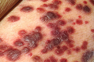
Kaposi's sarcoma-associated herpesvirus (KSHV) is the ninth known human herpesvirus; its formal name according to the International Committee on Taxonomy of Viruses (ICTV) is Human gammaherpesvirus 8, or HHV-8 in short. Like other herpesviruses, its informal names are used interchangeably with its formal ICTV name. This virus causes Kaposi's sarcoma, a cancer commonly occurring in AIDS patients, as well as primary effusion lymphoma, HHV-8-associated multicentric Castleman's disease and KSHV inflammatory cytokine syndrome. It is one of seven currently known human cancer viruses, or oncoviruses. Even after many years since the discovery of KSHV/HHV8, there is no known cure for KSHV associated tumorigenesis.

<span class="mw-page-title-main">Oncovirus</span> Viruses that can cause cancer

An oncovirus or oncogenic virus is a virus that can cause cancer. This term originated from studies of acutely transforming retroviruses in the 1950–60s, when the term "oncornaviruses" was used to denote their RNA virus origin. With the letters "RNA" removed, it now refers to any virus with a DNA or RNA genome causing cancer and is synonymous with "tumor virus" or "cancer virus". The vast majority of human and animal viruses do not cause cancer, probably because of longstanding co-evolution between the virus and its host. Oncoviruses have been important not only in epidemiology, but also in investigations of cell cycle control mechanisms such as the retinoblastoma protein.

<span class="mw-page-title-main">Opportunistic infection</span> Infection caused by pathogens that take advantage of an opportunity not normally available

An opportunistic infection is an infection caused by pathogens that take advantage of an opportunity not normally available. These opportunities can stem from a variety of sources, such as a weakened immune system, an altered microbiome, or breached integumentary barriers. Many of these pathogens do not necessarily cause disease in a healthy host that has a non-compromised immune system, and can, in some cases, act as commensals until the balance of the immune system is disrupted. Opportunistic infections can also be attributed to pathogens which cause mild illness in healthy individuals but lead to more serious illness when given the opportunity to take advantage of an immunocompromised host.

AIDS-defining clinical conditions is the list of diseases published by the Centers for Disease Control and Prevention (CDC) that are associated with AIDS and used worldwide as a guideline for AIDS diagnosis. CDC exclusively uses the term AIDS-defining clinical conditions, but the other terms remain in common use.

<span class="mw-page-title-main">Primary effusion lymphoma</span> Medical condition

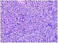
Primary effusion lymphoma (PEL) is classified as a diffuse large B cell lymphoma. It is a rare malignancy of plasmablastic cells that occurs in individuals that are infected with the Kaposi's sarcoma-associated herpesvirus. Plasmablasts are immature plasma cells, i.e. lymphocytes of the B-cell type that have differentiated into plasmablasts but because of their malignant nature do not differentiate into mature plasma cells but rather proliferate excessively and thereby cause life-threatening disease. In PEL, the proliferating plasmablastoid cells commonly accumulate within body cavities to produce effusions, primarily in the pleural, pericardial, or peritoneal cavities, without forming a contiguous tumor mass. In rare cases of these cavitary forms of PEL, the effusions develop in joints, the epidural space surrounding the brain and spinal cord, and underneath the capsule which forms around breast implants. Less frequently, individuals present with extracavitary primary effusion lymphomas, i.e., solid tumor masses not accompanied by effusions. The extracavitary tumors may develop in lymph nodes, bone, bone marrow, the gastrointestinal tract, skin, spleen, liver, lungs, central nervous system, testes, paranasal sinuses, muscle, and, rarely, inside the vasculature and sinuses of lymph nodes. As their disease progresses, however, individuals with the classical effusion-form of PEL may develop extracavitary tumors and individuals with extracavitary PEL may develop cavitary effusions.

<span class="mw-page-title-main">Robert Yarchoan</span>

Robert Yarchoan is a medical researcher who played an important role in the development of the first effective drugs for AIDS. He is the Chief of the HIV and AIDS Malignancy Branch in the NCI and also coordinates HIV/AIDS malignancy research throughout the NCI as director of the Office of HIV and AIDS Malignancy (OHAM).

<span class="mw-page-title-main">Patrick S. Moore</span> Irish and American virologist and epidemiologist

Patrick S. Moore is an American virologist and epidemiologist who co-discovered together with his wife, Yuan Chang, two different human viruses causing the AIDS-related cancer Kaposi's sarcoma and the skin cancer Merkel cell carcinoma. Moore and Chang have discovered two of the seven known human viruses causing cancer. The couple met while in medical school together and were married in 1989 while they pursued fellowships at different universities.

<span class="mw-page-title-main">Yuan Chang</span> American virologist and pathologist

Yuan Chang is a Taiwanese-born American virologist and pathologist who co-discovered together with her husband, Patrick S. Moore, the Kaposi's sarcoma-associated herpesvirus (KSHV) and Merkel cell polyomavirus, two of the seven known human oncoviruses.

<span class="mw-page-title-main">Human herpesvirus 6</span> Informal grouping of viruses which caused human herpesvirus 6 Infection

Human herpesvirus 6 (HHV-6) is the common collective name for human betaherpesvirus 6A (HHV-6A) and human betaherpesvirus 6B (HHV-6B). These closely related viruses are two of the nine known herpesviruses that have humans as their primary host.

Merkel cell polyomavirus was first described in January 2008 in Pittsburgh, Pennsylvania. It was the first example of a human viral pathogen discovered using unbiased metagenomic next-generation sequencing with a technique called digital transcriptome subtraction. MCV is one of seven currently known human oncoviruses. It is suspected to cause the majority of cases of Merkel cell carcinoma, a rare but aggressive form of skin cancer. Approximately 80% of Merkel cell carcinoma (MCC) tumors have been found to be infected with MCV. MCV appears to be a common—if not universal—infection of older children and adults. It is found in respiratory secretions, suggesting that it might be transmitted via a respiratory route. However, it has also been found elsewhere, such as in shedded healthy skin and gastrointestinal tract tissues, thus its precise mode of transmission remains unknown. In addition, recent studies suggest that this virus may latently infect the human sera and peripheral blood mononuclear cells.

The latency-associated nuclear antigen (LANA-1) or latent nuclear antigen is a Kaposi's sarcoma-associated herpesvirus (KSHV) latent protein initially found by Moore and colleagues as a speckled nuclear antigen present in primary effusion lymphoma cells that reacts with antibodies from patients with KS. It is the most immunodominant KSHV protein identified by Western-blotting as 222–234 kDa double bands migrate slower than the predicted molecular weight. LANA has been suspected of playing a crucial role in modulating viral and cellular gene expression. It is commonly used as an antigen in blood tests to detect antibodies in persons that have been exposed to KSHV.

Oral pigmentation is asymptomatic and does not usually cause any alteration to the texture or thickness of the affected area. The colour can be uniform or speckled and can appear solitary or as multiple lesions. Depending on the site, depth, and quantity of pigment, the appearance can vary considerably.

<span class="mw-page-title-main">Signs and symptoms of HIV/AIDS</span>

The stages of HIV infection are acute infection, latency, and AIDS. Acute infection lasts for several weeks and may include symptoms such as fever, swollen lymph nodes, inflammation of the throat, rash, muscle pain, malaise, and mouth and esophageal sores. The latency stage involves few or no symptoms and can last anywhere from two weeks to twenty years or more, depending on the individual. AIDS, the final stage of HIV infection, is defined by low CD4+ T cell counts, various opportunistic infections, cancers, and other conditions.

<span class="mw-page-title-main">Plasmablastic lymphoma</span> Type of large B-cell lymphoma

Plasmablastic lymphoma (PBL) is a type of large B-cell lymphoma recognized by the World Health Organization (WHO) in 2017 as belonging to a subgroup of lymphomas termed lymphoid neoplasms with plasmablastic differentiation. The other lymphoid neoplasms within this subgroup are: plasmablastic plasma cell lymphoma ; primary effusion lymphoma that is Kaposi's sarcoma-associated herpesvirus positive or Kaposi's sarcoma-associated Herpesvirus negative; anaplastic lymphoma kinase-positive large B-cell lymphoma; and human herpesvirus 8-positive diffuse large B-cell lymphoma, not otherwise specified. All of these lymphomas are malignancies of plasmablasts, i.e. B-cells that have differentiated into plasmablasts but because of their malignant nature: fail to differentiate further into mature plasma cells; proliferate excessively; and accumulate in and injure various tissues and organs.

Large B-cell lymphoma arising in HHV8-associated multicentric Castleman's disease is a type of large B-cell lymphoma, recognized in the WHO 2008 classification. It is sometimes called the plasmablastic form of multicentric Castleman disease. It has sometimes been confused with plasmablastic lymphoma in the literature, although that is a dissimilar specific entity. It has variable CD20 expression and unmutated immunoglobulin variable region genes.

<span class="mw-page-title-main">HHV-8-associated MCD</span> Disease subtype

Human herpesvirus 8 associated multicentric Castleman disease is a subtype of Castleman disease, a group of rare lymphoproliferative disorders characterized by lymph node enlargement, characteristic features on microscopic analysis of enlarged lymph node tissue, and a range of symptoms and clinical findings.

Epstein–Barr virus–associated lymphoproliferative diseases are a group of disorders in which one or more types of lymphoid cells, i.e. B cells, T cells, NK cells, and histiocytic-dendritic cells, are infected with the Epstein–Barr virus (EBV). This causes the infected cells to divide excessively, and is associated with the development of various non-cancerous, pre-cancerous, and cancerous lymphoproliferative disorders (LPDs). These LPDs include the well-known disorder occurring during the initial infection with the EBV, infectious mononucleosis, and the large number of subsequent disorders that may occur thereafter. The virus is usually involved in the development and/or progression of these LPDs although in some cases it may be an "innocent" bystander, i.e. present in, but not contributing to, the disease.

References

  1. Sand M, Sand D, Thrandorf C, Paech V, Altmeyer P, Bechara FG (June 2010). "Cutaneous lesions of the nose". Head & Face Medicine. 6: 7. doi: 10.1186/1746-160X-6-7 . PMC   2903548 . PMID   20525327.
  2. Collins English Dictionary – Complete and Unabridged, 12th Edition 2014. S.v. "Kaposi's sarcoma." Retrieved August 15, 2017 from http://www.thefreedictionary.com/Kaposi's+sarcoma
  3. Random House Kernerman Webster's College Dictionary. S.v. "Kaposi's sarcoma." Retrieved August 15, 2017 from http://www.thefreedictionary.com/Kaposi's+sarcoma
  4. 1 2 3 4 5 6 7 8 "Kaposi Sarcoma Treatment". National Cancer Institute. 16 June 2017.
  5. Schneider JW, Dittmer DP (August 2017). "Diagnosis and Treatment of Kaposi Sarcoma". American Journal of Clinical Dermatology. 18 (4): 529–539. doi:10.1007/s40257-017-0270-4. PMC   5509489 . PMID   28324233.
  6. 1 2 3 4 5 6 7 8 9 10 11 12 13 Schwartz RA, Micali G, Nasca MR, Scuderi L (August 2008). "Kaposi sarcoma: a continuing conundrum". Journal of the American Academy of Dermatology. 59 (2): 179–206, quiz 207–8. doi:10.1016/j.jaad.2008.05.001. PMID   18638627.
  7. 1 2 The Global Cancer Observatory (2019). "Kaposi sarcoma" (PDF). Retrieved 14 March 2020.
  8. 1 2 3 4 5 6 7 8 9 10 Cesarman E, Damania B, Krown SE, Martin J, Bower M, Whitby D (January 2019). "Kaposi sarcoma". Nature Reviews. Disease Primers. 5 (1): 9. doi:10.1038/s41572-019-0060-9. PMC   6685213 . PMID   30705286.
  9. 1 2 3 Hoffmann C, Sabranski M, Esser S (2017). "HIV-Associated Kaposi's Sarcoma". Oncology Research and Treatment. 40 (3): 94–98. doi: 10.1159/000455971 . PMID   28259888. S2CID   9700628.
  10. Ferri FF (2017). Ferri's Clinical Advisor 2018 E-Book: 5 Books in 1. Elsevier Health Sciences. p. 735. ISBN   9780323529570.
  11. 1 2 3 "Kaposi Sarcoma Treatment". National Cancer Institute. 1 October 2015. Retrieved 18 December 2017.
  12. Kaposi M (1872). "Idiopathisches multiples Pigmentsarkom der Haut". Archiv für Dermatologie und Syphilis. 4 (2): 265–273. doi:10.1007/BF01830024. S2CID   31438763.
  13. Chang Y, Cesarman E, Pessin MS, Lee F, Culpepper J, Knowles DM, Moore PS (December 1994). "Identification of herpesvirus-like DNA sequences in AIDS-associated Kaposi's sarcoma". Science. 266 (5192): 1865–1869. Bibcode:1994Sci...266.1865C. doi:10.1126/science.7997879. PMID   7997879. S2CID   29977325.
  14. Dezube BJ (October 1996). "Clinical presentation and natural history of AIDS--related Kaposi's sarcoma". Hematology/Oncology Clinics of North America. 10 (5): 1023–9. doi:10.1016/S0889-8588(05)70382-8. PMID   8880194.
  15. "Kaposi's Sarcoma". Taber's Cyclopedic Medical Dictionary. Vol. 21. F. A. Davis Company. 2009. p. 1256.
  16. 1 2 3 4 Jakob L, Metzler G, Chen KM, Garbe C (April 2011). "Non-AIDS associated Kaposi's sarcoma: clinical features and treatment outcome". PLOS ONE. 6 (4): e18397. Bibcode:2011PLoSO...618397J. doi: 10.1371/journal.pone.0018397 . PMC   3075253 . PMID   21533260.
  17. Holland, J. C.; Tross, S. (June 1987). "Psychosocial considerations in the therapy of epidemic Kaposi's sarcoma". Seminars in Oncology. 14 (2 Suppl 3): 48–53. ISSN   0093-7754. PMID   3603057.
  18. França, Katlein; Jafferany, Mohammad (16 December 2016). Stress and Skin Disorders: Basic and Clinical Aspects. Springer. p. 216. ISBN   978-3-319-46352-0.
  19. Nichols CM, Flaitz CM, Hicks MJ (November 1993). "Treating Kaposi's lesions in the HIV-infected patient". Journal of the American Dental Association. 124 (11): 78–84. doi:10.14219/jada.archive.1993.0231. PMID   8227776. Archived from the original on 2007-09-29. Retrieved 2007-06-11.
  20. Danzig JB, Brandt LJ, Reinus JF, Klein RS (June 1991). "Gastrointestinal malignancy in patients with AIDS". The American Journal of Gastroenterology. 86 (6): 715–8. PMID   2038993.
  21. Garay SM, Belenko M, Fazzini E, Schinella R (January 1987). "Pulmonary manifestations of Kaposi's sarcoma". Chest. 91 (1): 39–43. doi:10.1378/chest.91.1.39. PMID   3792084. S2CID   41623543.
  22. Ablashi DV, Chatlynne LG, Whitman JE, Cesarman E (July 2002). "Spectrum of Kaposi's sarcoma-associated herpesvirus, or human herpesvirus 8, diseases". Clinical Microbiology Reviews. 15 (3): 439–464. doi:10.1128/cmr.15.3.439-464.2002. PMC   118087 . PMID   12097251.
  23. Liang C, Lee JS, Jung JU (December 2008). "Immune evasion in Kaposi's sarcoma-associated herpes virus associated oncogenesis". Seminars in Cancer Biology. Manipulation of the immune response by Epstein-Barr virus and Kaposi's sarcoma-associated herpes virus: Consequences for tumor development. 18 (6): 423–436. doi:10.1016/j.semcancer.2008.09.003. PMC   7386567 . PMID   18948197.
  24. Pauk J, Huang ML, Brodie SJ, Wald A, Koelle DM, Schacker T, Celum C, Selke S, Corey L (November 2000). "Mucosal shedding of human herpesvirus 8 in men". The New England Journal of Medicine. 343 (19): 1369–77. doi: 10.1056/NEJM200011093431904 . PMID   11070101.
  25. Parravicini C, Olsen SJ, Capra M, Poli F, Sirchia G, Gao SJ, Berti E, Nocera A, Rossi E, Bestetti G, Pizzuto M, Galli M, Moroni M, Moore PS, Corbellino M (October 1997). "Risk of Kaposi's sarcoma-associated herpes virus transmission from donor allografts among Italian posttransplant Kaposi's sarcoma patients". Blood. 90 (7): 2826–9. PMID   9326251.
  26. Hladik W, Dollard SC, Mermin J, Fowlkes AL, Downing R, Amin MM, Banage F, Nzaro E, Kataaha P, Dondero TJ, Pellett PE, Lackritz EM (September 2006). "Transmission of human herpesvirus 8 by blood transfusion". The New England Journal of Medicine. 355 (13): 1331–8. doi: 10.1056/NEJMoa055009 . PMID   17005950.
  27. Coffin JM, Hughes SH, Varmus HE (1997). Retroviruses. Cold Spring Harbor, NY: Cold Spring Harbor Laboratory Press. ISBN   978-0-87969-571-2.
  28. Ensoli B, Sirianni MC (1998). "Kaposi's sarcoma pathogenesis: a link between immunology and tumor biology". Critical Reviews in Oncogenesis. 9 (2): 107–24. doi:10.1615/CritRevOncog.v9.i2.20. PMID   9973245.
  29. Gurzu S, Ciortea D, Munteanu T, Kezdi-Zaharia I, Jung I (2008). "Mesenchymal-to-endothelial transition in Kaposi sarcoma: a histogenetic hypothesis based on a case series and literature review". PLOS ONE. 8 (8): e71530. Bibcode:2013PLoSO...871530G. doi: 10.1371/journal.pone.0071530 . PMC   3735554 . PMID   23936513.
  30. Beckstead JH, Wood GS, Fletcher V (May 1985). "Evidence for the origin of Kaposi's sarcoma from lymphatic endothelium". The American Journal of Pathology. 119 (2): 294–300. PMC   1887903 . PMID   2986460.
  31. Rosai J (2011). Rosai and Ackerman's Surgical Pathology (10th ed.). Mosby. ISBN   978-0-8089-2433-3.
  32. Weninger W, Partanen TA, Breiteneder-Geleff S, Mayer C, Kowalski H, Mildner M, et al. (February 1999). "Expression of vascular endothelial growth factor receptor-3 and podoplanin suggests a lymphatic endothelial cell origin of Kaposi's sarcoma tumor cells". Laboratory Investigation; A Journal of Technical Methods and Pathology. 79 (2): 243–251. PMID   10068212.
  33. Blumenfeld W, Egbert BM, Sagebiel RW (February 1985). "Differential diagnosis of Kaposi's sarcoma". Archives of Pathology & Laboratory Medicine. 109 (2): 123–127. PMID   2983633.
  34. Griffiths C, Barker J, Bleiker TO, Chalmers R, Creamer D (4 April 2016). Rook's textbook of dermatology (Ninth ed.). p. 31.29. ISBN   9781118441190.
  35. 1 2 3 4 5 6 James W, Berger T, Elston D (2005). Andrews' Diseases of the Skin: Clinical Dermatology (10th ed.). Saunders. ISBN   978-0-7216-2921-6..
  36. Kumar P (2011). "Classic Kaposi's sarcoma in Arabs--widening ethnic involvement". Journal of Cancer Research and Therapeutics. 7 (1): 92–4. doi: 10.4103/0973-1482.80456 . PMID   21546753.
  37. Iscovich J, Boffetta P, Winkelmann R, Brennan P, Azizi E (October 1998). "Classic Kaposi's sarcoma in Jews living in Israel, 1961-1989: a population-based incidence study". AIDS. 12 (15): 2067–72. doi: 10.1097/00002030-199815000-00019 . PMID   9814876. S2CID   23848900.
  38. Fenig E, Brenner B, Rakowsky E, Lapidoth M, Katz A, Sulkes A (October 1998). "Classic Kaposi sarcoma: experience at Rabin Medical Center in Israel". American Journal of Clinical Oncology. 21 (5): 498–500. doi:10.1097/00000421-199810000-00016. PMID   9781608.
  39. 1 2 Cook-Mozaffari P, Newton R, Beral V, Burkitt DP (December 1998). "The geographical distribution of Kaposi's sarcoma and of lymphomas in Africa before the AIDS epidemic". British Journal of Cancer. 78 (11): 1521–8. doi:10.1038/bjc.1998.717. PMC   2063225 . PMID   9836488.
  40. Olsen SJ, Chang Y, Moore PS, Biggar RJ, Melbye M (October 1998). "Increasing Kaposi's sarcoma-associated herpesvirus seroprevalence with age in a highly Kaposi's sarcoma endemic region, Zambia in 1985". AIDS. 12 (14): 1921–5. doi: 10.1097/00002030-199814000-00024 . PMID   9792393. S2CID   1734745.
  41. Olsen SJ, Chang Y, Moore PS, Biggar RJ, Melbye M (October 1998). "Increasing Kaposi's sarcoma-associated herpesvirus seroprevalence with age in a highly Kaposi's sarcoma endemic region, Zambia in 1985". AIDS. 12 (14): 1921–5. doi: 10.1097/00002030-199814000-00024 . PMID   9792393. S2CID   1734745.
  42. Qunibi W, Al-Furayh O, Almeshari K, Lin SF, Sun R, Heston L, Ross D, Rigsby M, Miller G (February 1998). "Serologic association of human herpesvirus eight with posttransplant Kaposi's sarcoma in Saudi Arabia". Transplantation. 65 (4): 583–5. doi: 10.1097/00007890-199802270-00024 . PMID   9500639.
  43. Luppi M, Barozzi P, Schulz TF, Setti G, Staskus K, Trovato R, Narni F, Donelli A, Maiorana A, Marasca R, Sandrini S, Torelli G (November 2000). "Bone marrow failure associated with human herpesvirus 8 infection after transplantation". The New England Journal of Medicine. 343 (19): 1378–85. doi: 10.1056/NEJM200011093431905 . PMID   11070102.
  44. Borkovic SP, Schwartz RA (December 1981). "Kaposi's sarcoma presenting in the homosexual man -- a new and striking phenomenon!". Arizona Medicine. 38 (12): 902–4. PMID   7332494.
  45. Hausen HZ (2006). "Rhadinoviruses". Infections Causing Human Cancer. Weinheim: Wiley-VCH.
  46. Drabell FG (2006). "Kaposi's Sarcoma and Renal Diseases". New Topics in Cancer Research. New York: Nova Biomedical Books.
  47. Beral V, Peterman TA, Berkelman RL, Jaffe HW (January 1990). "Kaposi's sarcoma among persons with AIDS: a sexually transmitted infection?". Lancet. 335 (8682): 123–8. doi:10.1016/0140-6736(90)90001-L. PMID   1967430. S2CID   35639169.
  48. Tappero JW, Berger TG, Kaplan LD, Volberding PA, Kahn JO (1991). "Cryotherapy for cutaneous Kaposi's sarcoma (KS) associated with acquired immune deficiency syndrome (AIDS): a phase II trial". Journal of Acquired Immune Deficiency Syndromes. 4 (9): 839–46. doi:10.1097/00126334-199109000-00002. PMID   1895204. S2CID   19909703.
  49. Zimmerman EE, Crawford P (December 2012). "Cutaneous cryosurgery". American Family Physician. 86 (12): 1118–24. PMID   23316984.
  50. Anglemyer A, Agrawal AK, Rutherford GW (January 2014). "Treatment of Kaposi sarcoma in children with HIV-1 infection". The Cochrane Database of Systematic Reviews. 1 (1): CD009826. doi:10.1002/14651858.CD009826.pub2. PMID   24464843.
  51. McAllister SC, Hanson RS, Manion RD (November 2015). "Propranolol Decreases Proliferation of Endothelial Cells Transformed by Kaposi's Sarcoma-Associated Herpesvirus and Induces Lytic Viral Gene Expression". Journal of Virology. 89 (21): 11144–9. doi:10.1128/JVI.01569-15. PMC   4621132 . PMID   26269192.
  52. Abdelmaksoud A, Filoni A, Giudice G, Vestita M (January 2017). "Classic and HIV-related Kaposi sarcoma treated with 0.1% topical timolol gel". Journal of the American Academy of Dermatology. 76 (1): 153–155. doi: 10.1016/j.jaad.2016.08.041 . PMID   27986137.
  53. Gill PS, Tulpule A, Espina BM, Cabriales S, Bresnahan J, Ilaw M, Louie S, Gustafson NF, Brown MA, Orcutt C, Winograd B, Scadden DT (June 1999). "Paclitaxel is safe and effective in the treatment of advanced AIDS-related Kaposi's sarcoma". Journal of Clinical Oncology. 17 (6): 1876–83. doi:10.1200/jco.1999.17.6.1876. PMID   10561228.
  54. Sgadari C, Toschi E, Palladino C, Barillari G, Carlei D, Cereseto A, Ciccolella C, Yarchoan R, Monini P, Stürzl M, Ensoli B (July 2000). "Mechanism of paclitaxel activity in Kaposi's sarcoma". Journal of Immunology. 165 (1): 509–17. doi: 10.4049/jimmunol.165.1.509 . PMID   10861090.
  55. "Summary of Product Characteristics" (PDF). EMA. Retrieved 14 March 2020.
  56. Phillips AM, Jones AG, Osmond DH, Pollack LM, Catania JA, Martin JN (December 2008). "Awareness of Kaposi's sarcoma-associated herpesvirus among men who have sex with men". Sexually Transmitted Diseases. 35 (12): 1011–4. doi:10.1097/OLQ.0b013e318182c91f. PMC   2593118 . PMID   18665016.