Neuropathic arthropathy

Last updated
Neuropathic joint disease
Neuropathic heel ulcer.jpg
A 68-year-old diabetic female on dialysis presented with a chronic right heel ulcer (3.4 cm X 3.1 cm) of greater than three months' duration. Photograph of the wound after thorough wound bed preparation over the course of 2 weeks.
Specialty Rheumatology   OOjs UI icon edit-ltr-progressive.svg

Neuropathic arthropathy (or neuropathic osteoarthropathy), also known as Charcot joint (often Charcot foot) after the first to describe it, Jean-Martin Charcot, refers to progressive degeneration of a weight-bearing joint, a process marked by bony destruction, bone resorption, and eventual deformity due to loss of sensation. Onset is usually insidious.

Contents

If this pathological process continues unchecked, it can result in joint deformity, ulceration and/or superinfection, loss of function, and in the worst-case scenario, amputation or death. Early identification of joint changes is the best way to limit morbidity.

Symptoms and signs

Oblique view X-ray in a 45-year-old male diabetic revealed a divergent, Lisfranc dislocation of the first metatarsal with associated lesser metatarsal fractures. Charcot arthropathy X-ray.jpg
Oblique view X-ray in a 45-year-old male diabetic revealed a divergent, Lisfranc dislocation of the first metatarsal with associated lesser metatarsal fractures.
The same 45-year-old man with diabetes mellitus presented with a diffusely swollen, warm and non-tender left foot due to Charcot arthropathy. There are no changes to the skin itself. Charcot arthropathy clinical examination.jpg
The same 45-year-old man with diabetes mellitus presented with a diffusely swollen, warm and non-tender left foot due to Charcot arthropathy. There are no changes to the skin itself.

The clinical presentation varies depending on the stage of the disease from mild swelling to severe swelling and moderate deformity. Inflammation, erythema, pain and increased skin temperature (3–7 degrees Celsius) around the joint may be noticeable on examination. X-rays may reveal bone resorption and degenerative changes in the joint. These findings in the presence of intact skin and loss of protective sensation are pathognomonic of acute Charcot arthropathy.

Roughly 75% of patients experience pain, but it is less than what would be expected based on the severity of the clinical and radiographic findings.

Pathogenesis

Any condition resulting in decreased peripheral sensation, proprioception, and fine motor control:

Underlying mechanisms

In reality, both of these mechanisms probably play a role in the development of a Charcot joint.

Joint involvement

Diabetes is the foremost cause in America today for neuropathic joint disease, [1] and the foot is the most affected region. In those with foot deformity, approximately 60% are in the tarsometatarsal joints (medial joints affected more than lateral), 30% metatarsophalangeal joints, and 10% have ankle disease. Over half of diabetic patients with neuropathic joints can recall some kind of precipitating trauma, usually minor.

Patients with neurosyphilis tend to have knee involvement, and patients with syringomyelia of the spinal cord may demonstrate shoulder deformity. [2]

Hip joint destruction is also seen in neuropathic patients.

Diagnosis

Clinical findings

Clinical findings include erythema, edema and increased temperature in the affected joint. In neuropathic foot joints, plantar ulcers may be present. It is often difficult to differentiate osteomyelitis from a Charcot joint, as they may have similar tagged WBC scan and MRI features (joint destruction, dislocation, edema). Definitive diagnosis may require bone or synovial biopsy.

Radiologic findings

First, it is important to recognize that two types of abnormality may be detected. One is termed atrophic, in which there is osteolysis of the distal metatarsals in the forefoot. The more common form of destruction is hypertrophic joint disease, characterized by acute peri-articular fracture and joint dislocation. According to Yochum and Rowe, the "6 D's" of hypertrophy are:

  1. Distended joint
  2. Density increase
  3. Debris production
  4. Dislocation
  5. Disorganization
  6. Destruction

The natural history of the joint destruction process has a classification scheme of its own, offered by Eichenholtz decades ago:

Stage 0: Clinically, there is joint edema, but radiographs are negative. A bone scan may be positive before a radiograph is, making it a sensitive but not very specific modality.

Stage 1: Osseous fragmentation with joint dislocation seen on radiograph ("acute Charcot").

Stage 2: Decreased local edema, with coalescence of fragments and absorption of fine bone debris.

Stage 3: No local edema, with consolidation and remodeling (albeit deformed) of fracture fragments. The foot is now stable.

Atrophic features:

  1. "Licked candy stick" appearance, commonly seen at the distal aspect of the metatarsals
  2. Diabetic osteolysis
  3. Bone resorption

Treatment

Diabetic foot ulcers should be treated via the VIPs — vascular management, infection management and prevention, and pressure relief. Aggressively pursuing these three strategies will progress the healing trajectory of the wound. Pressure relief (offloading) and immobilization at the acute (active) stage [3] are critical to helping ward off further joint destruction in cases of Charcot foot. Total contact casting (TCC) is recommended, but other methods are also available. [3] TCC involves encasing the patient's complete foot, including toes, and the lower leg in a specialist cast that redistributes weight and pressure in the lower leg and foot during everyday movements. This redistributes pressure from the foot into the leg, which is more able to bear weight, to protect the wound, letting it regenerate tissue and heal. [4] TCC also keeps the ankle from rotating during walking, which prevents shearing and twisting forces that can further damage the wound. [5] TCC aids maintenance of quality of life by helping patients to remain mobile. [6]

There are two scenarios in which the use of TCC is appropriate for managing neuropathic arthropathy (Charcot foot), according to the American Orthopaedic Foot and Ankle Society. [7] First, during the initial treatment, when the breakdown is occurring, and the foot is exhibiting edema and erythema; the patient should not bear weight on the foot, and TCC can be used to control and support the foot. Second, when the foot has become deformed and ulceration has occurred; TCC can be used to stabilize and support the foot, and to help move the wound toward healing.

Walking braces controlled by pneumatics are also used. In these patients, surgical correction of a joint is rarely successful in the long term. However, offloading alone does not translate to optimal outcomes without appropriate management of vascular disease and/or infection. [8] Duration and aggressiveness of offloading (non-weight-bearing vs. weight-bearing, non-removable vs. removable device) should be guided by clinical assessment of healing of neuropathic arthropathy based on edema, erythema, and skin temperature changes. [9] It can take six to nine months for the edema and erythema of the affected joint to recede.

Outcome

Outcomes vary depending on the location of the disease, the degree of damage to the joint, and whether surgical repair was necessary. Average healing times vary from 55 to 97 days, depending on location. Up to one to two years may be required for complete healing.

Further reading

Related Research Articles

<span class="mw-page-title-main">Syringomyelia</span> Disorder in which a cyst forms in the spinal cord

Syringomyelia is a generic term referring to a disorder in which a cyst or cavity forms within the spinal cord. Often, syringomyelia is used as a generic term before an etiology is determined. This cyst, called a syrinx, can expand and elongate over time, destroying the spinal cord. The damage may result in loss of feeling, paralysis, weakness, and stiffness in the back, shoulders, and extremities. Syringomyelia may also cause a loss of the ability to feel extremes of hot or cold, especially in the hands. It may also lead to a cape-like bilateral loss of pain and temperature sensation along the upper chest and arms. The combination of symptoms varies from one patient to another depending on the location of the syrinx within the spinal cord, as well as its extent.

<span class="mw-page-title-main">Ulcer (dermatology)</span> Type of cutaneous condition

An ulcer is a sore on the skin or a mucous membrane, accompanied by the disintegration of tissue. Ulcers can result in complete loss of the epidermis and often portions of the dermis and even subcutaneous fat. Ulcers are most common on the skin of the lower extremities and in the gastrointestinal tract. An ulcer that appears on the skin is often visible as an inflamed tissue with an area of reddened skin. A skin ulcer is often visible in the event of exposure to heat or cold, irritation, or a problem with blood circulation.

<span class="mw-page-title-main">Podiatrist</span> Medical professional devoted to the medical treatment of disorders of the foot

A podiatrist is a medical professional devoted to the treatment of disorders of the foot, ankle, and related structures of the leg. The term originated in North America but has now become the accepted term in the English-speaking world for all practitioners of podiatric medicine. The word chiropodist was previously used in the United States, but it is now regarded as antiquated.

Diabetic neuropathy is various types of nerve damage associated with diabetes mellitus. Symptoms depend on the site of nerve damage and can include motor changes such as weakness; sensory symptoms such as numbness, tingling, or pain; or autonomic changes such as urinary symptoms. These changes are thought to result from a microvascular injury involving small blood vessels that supply nerves. Relatively common conditions which may be associated with diabetic neuropathy include distal symmetric polyneuropathy; third, fourth, or sixth cranial nerve palsy; mononeuropathy; mononeuropathy multiplex; diabetic amyotrophy; and autonomic neuropathy.

<span class="mw-page-title-main">Pes cavus</span> Medical condition

Pes cavus, also known as high arch, is a human foot type in which the sole of the foot is distinctly hollow when bearing weight. That is, there is a fixed plantar flexion of the foot. A high arch is the opposite of a flat foot and is somewhat less common.

<span class="mw-page-title-main">Hammer toe</span> Medical condition

A hammer toe, hammertoe or contracted toe is a deformity of the muscles and ligaments of the proximal interphalangeal joint of the second, third, fourth, or fifth toe, bending it into a shape resembling a hammer. In the early stage, a flexible hammertoe is movable at the joints; a rigid hammertoe joint cannot be moved and usually requires surgery.

<span class="mw-page-title-main">Arthropathy</span> Medical condition

An arthropathy is a disease of a joint.

A peripheral vascular examination is a medical examination to discover signs of pathology in the peripheral vascular system. It is performed as part of a physical examination, or when a patient presents with leg pain suggestive of a cardiovascular pathology.

<span class="mw-page-title-main">Calcaneal fracture</span> Medical condition

A calcaneal fracture is a break of the calcaneus. Symptoms may include pain, bruising, trouble walking, and deformity of the heel. It may be associated with breaks of the hip or back.

Becaplermin is a cicatrizant, available as a topical gel. Regranex is a human platelet-derived growth factor indicated along with good wound care for the treatment of lower extremity diabetic neuropathic ulcers. It is also known as "platelet-derived growth factor BB".

<span class="mw-page-title-main">Diabetic shoe</span> Shoes intended to reduce the risk of skin breakdown in diabetics

Diabetic shoes are specially designed shoes, or shoe inserts, intended to reduce the risk of skin breakdown in diabetics with existing foot disease and relieve pressure to prevent diabetic foot ulcers.

<span class="mw-page-title-main">David G. Armstrong</span> American podiatric surgeon and researcher

David G. Armstrong is an American podiatric surgeon and researcher most widely known for his work in amputation prevention, the diabetic foot, and wound healing. He and his frequent collaborators, Lawrence A. Lavery and Andrew J.M. Boulton, have together produced many key works in the taxonomy, classification and treatment of the diabetic foot. He is Professor of Surgery with Tenure and director of the Southwestern Academic Limb Salvage Alliance (SALSA) at the Keck School of Medicine of the University of Southern California and has produced more than 650 peer reviewed manuscripts and more than 110 book chapters.

<span class="mw-page-title-main">Arterial insufficiency ulcer</span> Medical condition

Arterial insufficiency ulcers are mostly located on the lateral surface of the ankle or the distal digits. They are commonly caused by peripheral artery disease (PAD).

<span class="mw-page-title-main">Diabetic foot</span> Medical condition

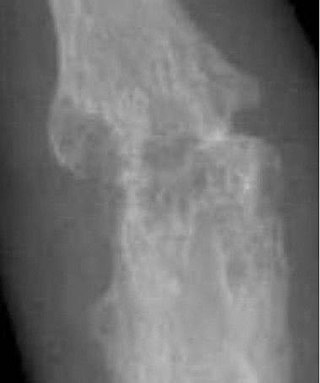
A diabetic foot disease is any condition that results directly from peripheral artery disease (PAD) or sensory neuropathy affecting the feet of people living with diabetes. Diabetic foot conditions can be acute or chronic complications of diabetes. Presence of several characteristic diabetic foot pathologies such as infection, diabetic foot ulcer and neuropathic osteoarthropathy is called diabetic foot syndrome. The resulting bone deformity is known as Charcot foot.

<span class="mw-page-title-main">Malum perforans</span> Medical condition

Malum perforans is a long-lasting, usually painless ulcer that penetrates deep into or through the skin, usually on the sole of the foot. It is often a complication in diabetes mellitus and other conditions affecting the nerves.

<span class="mw-page-title-main">Diabetic foot ulcer</span> Medical condition

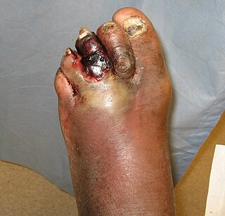
Diabetic foot ulcer is a breakdown of the skin and sometimes deeper tissues of the foot that leads to sore formation. It may occur due to a variety of mechanisms. It is thought to occur due to abnormal pressure or mechanical stress chronically applied to the foot, usually with concomitant predisposing conditions such as peripheral sensory neuropathy, peripheral motor neuropathy, autonomic neuropathy or peripheral arterial disease. It is a major complication of diabetes mellitus, and it is a type of diabetic foot disease. Secondary complications to the ulcer, such as infection of the skin or subcutaneous tissue, bone infection, gangrene or sepsis are possible, often leading to amputation.

Total contact casting (TCC) is a specially designed cast designed to take weight off of the foot (off-loading) in patients with diabetic foot ulcers (DFUs). Reducing pressure on the wound by taking weight off the foot has proven to be very effective in DFU treatment. DFUs are a major factor leading to lower leg amputations among the diabetic population in the US with 85% of amputations in diabetics being preceded by a DFU. Furthermore, the five-year post-amputation mortality rate among diabetics is estimated at 45% for those with neuropathic DFUs.

<span class="mw-page-title-main">Diabetic foot infection</span> Medical condition

Diabetic foot infection is any infection of the foot in a diabetic person. The most frequent cause of hospitalization for diabetic patients is due to foot infections. Symptoms may include pus from a wound, redness, swelling, pain, warmth, tachycardia, or tachypnea. Complications can include infection of the bone, tissue death, amputation, or sepsis. They are common and occur equally frequently in males and females. Older people are more commonly affected.

Nachiappan Chockalingam, professionally known as Nachi Chockalingam is a British scientist, academic practitioner and expert in Clinical Biomechanics. He is a professor at Staffordshire University and a Fellow of the Institute of Physics and Engineering in Medicine and has been appointed to a panel of experts for the Research Excellence Framework. He was elected as a Fellow of the International Society of Biomechanics in 2023. Between 2016 and 2022, he has contributed to the NIHR Research for Patient Benefit Panel and serves in multiple other review panels of global grant awarding bodies including the EPSRC, MRC and the European Commission. He contributed to development of podiatric biomechanics in the UK and played a pivotal role in the establishment of the journals such as the Footwear Science and is on the editorial panel for a number of scientific and clinical journals.

References

  1. Charcot Arthropathy at eMedicine
  2. Hirsch M et al., "Neuropathic osteoarthropathy of the shoulder secondary to syringomyelia". https://doi.org/10.1016/j.diii.2020.09.010
  3. 1 2 Snyder, Robert J.; et al. (1 November 2014). "The Management of Diabetic Foot Ulcers Through Optimal Off-Loading". Journal of the American Podiatric Medical Association . 104 (6): 555–567. doi:10.7547/8750-7315-104.6.555. ISSN   8750-7315.
  4. Raspovic, A. and K.B. Landorf, "A survey of offloading practices for diabetes-related plantar neuropathic foot ulcers". Journal of Foot and Ankle Research, 2014. 7: p. 35.
  5. Snyder, R.J., et al., "The management of diabetic foot ulcers through optimal off-loading building consensus guidelines and practical recommendations to improve outcomes". Journal of the American Podiatric Medical Association, 2014. 104(6): p. 555-567.
  6. Farid K, Farid M, Andrews CM. "Total contact casting as part of an adaptive care approach: a case study". Ostomy Wound Management , 2008. 54(6): 50–65.
  7. AOFAS. Foot ulcers and the total contact cast. Accessed 29.07.2015 at: https://www.aofas.org/footcaremd/conditions/diabetic-foot/Pages/Foot-Ulcers-and-the-Total-Contact-Cast.aspx
  8. Snyder, R.J. et al., "The management of diabetic foot ulcers through optimal off-loading building consensus guidelines and practical recommendations to improve outcomes". Journal of the American Podiatric Medical Association, 2014. 104(6): p. 555-567.
  9. Rogers LC et al. "The Charcot foot in diabetes". Diabetes Care . 2011;34(9):2123–9.