Related Research Articles
David Jerome Bloom was an American television journalist until his sudden death in 2003 after a deep vein thrombosis (DVT) became a pulmonary embolism.
Venous thrombosis is thrombosis in a vein, caused by a thrombus. A common form of venous thrombosis is deep vein thrombosis (DVT), when a blood clot forms in the deep veins. If a thrombus breaks off (embolizes) and flows towards the lungs, it can become a pulmonary embolism (PE), a blood clot in the lungs. The conditions of DVT only, DVT with PE, and PE only are captured by the term venous thromboembolism (VTE).
Factor V Leiden is a variant of human factor V, which causes an increase in blood clotting (hypercoagulability). Due to this mutation, protein C, an anticoagulant protein that normally inhibits the pro-clotting activity of factor V, is not able to bind normally to factor V, leading to a hypercoagulable state, i.e., an increased tendency for the patient to form abnormal and potentially harmful blood clots. Factor V Leiden is the most common hereditary hypercoagulability disorder amongst ethnic Europeans. It is named after the Dutch city of Leiden, where it was first identified in 1994 by Rogier Maria Bertina under the direction of Pieter Hendrick Reitsma. Despite the increased risk of venous thromboembolisms, people with one copy of this gene have not been found to have shorter lives than the general population.
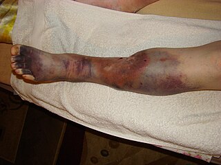
Deep vein thrombosis (DVT) is the formation of a blood clot in a deep vein, most commonly in the legs or pelvis. A minority of DVTs occur in the arms. Symptoms can include pain, swelling, redness, and enlarged veins in the affected area, but some DVTs have no symptoms. The most common life-threatening concern with DVT is the potential for a clot to embolize, travel as an embolus through the right side of the heart, and become lodged in a pulmonary artery that supplies blood to the lungs. This is called a pulmonary embolism (PE). DVT and PE comprise the cardiovascular disease of venous thromboembolism (VTE). About two-thirds of VTE manifests as DVT only, with one-third manifesting as PE with or without DVT. The most frequent long-term DVT complication is post-thrombotic syndrome, which can cause pain, swelling, a sensation of heaviness, itching, and in severe cases, ulcers. Recurrent VTE occurs in about 30% of those in the ten years following an initial VTE.
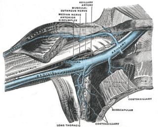
Paget–Schroetter disease, is a form of upper extremity deep vein thrombosis (DVT), a medical condition in which blood clots form in the deep veins of the arms. These DVTs typically occur in the axillary and/or subclavian veins.
In medicine, Homans' sign is considered by some physicians to be a sign of deep vein thrombosis (DVT). It was defined by John Homans in 1941 as discomfort behind the knee upon forced dorsiflexion of the foot. After many examples of false-positive Homans' signs were reported, Homans redefined it in 1944, stating that "discomfort need have no part in the reaction", and that increased resistance, involuntary flexure of the knee or pain in the calf upon forced dorsiflexion should be considered positive responses.
DVT is deep vein thrombosis, the formation of a blood clot in a deep vein, most commonly the legs.

Nadroparin is an anticoagulant belonging to a class of drugs called low molecular weight heparins (LMWHs). Nadroparin was developed by Sanofi-Synthélabo.
Post-thrombotic syndrome (PTS), also called postphlebitic syndrome and venous stress disorder is a medical condition that may occur as a long-term complication of deep vein thrombosis (DVT).
Venography is a procedure in which an x-ray of the veins, a venogram, is taken after a special dye is injected into the bone marrow or veins. The dye has to be injected constantly via a catheter, making it an invasive procedure. Normally the catheter is inserted by the groin and moved to the appropriate site by navigating through the vascular system.

Chronic venous insufficiency (CVI) is a medical condition in which blood pools in the veins, straining the walls of the vein. The most common cause of CVI is superficial venous reflux which is a treatable condition. As functional venous valves are required to provide for efficient blood return from the lower extremities, this condition typically affects the legs. If the impaired vein function causes significant symptoms, such as swelling and ulcer formation, it is referred to as chronic venous disease. It is sometimes called chronic peripheral venous insufficiency and should not be confused with post-thrombotic syndrome in which the deep veins have been damaged by previous deep vein thrombosis.
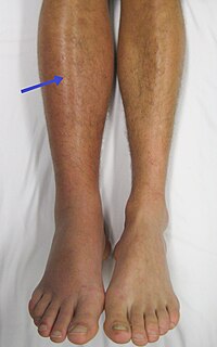
Pratt's sign is an indication of femoral deep vein thrombosis. It is seen as the presence of dilated pretibial veins in the affected leg, which remain dilated on raising the leg.

Venostasis, or venous stasis, is a condition of slow blood flow in the veins, usually of the legs.
Superficial thrombophlebitis is a thrombosis and inflammation of superficial veins which presents as a painful induration with erythema, often in a linear or branching configuration forming cords.
Pseudothrombophlebitis is a clinical condition where there are signs and symptoms of phlebitis in the absence of a thrombophlebitis lesion. Symptoms include pain, swelling, erythema and tenderness evolving over hours or days. It is often associated with the rupture or dissection of a popliteal cyst otherwise known as a Baker's cyst, although it can be associated with other disorders such as the arthritides. It may also occur as an orthopaedic surgical complication, secondary to trauma or as a presentation of septic arthritis. It is crucial to differentiate this condition from deep vein thrombosis as the treatment for DVT can cause adverse effects in patients with pseudothrombophlebitis.
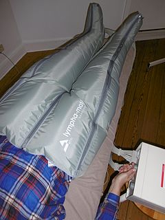
Intermittent pneumatic compression is a therapeutic technique used in medical devices that include an air pump and inflatable auxiliary sleeves, gloves or boots in a system designed to improve venous circulation in the limbs of patients who suffer edema or the risk of deep vein thrombosis (DVT) or pulmonary embolism (PE).
{{Multiple issues|

Apixaban, sold under the brand name Eliquis, is an anticoagulant medication used to treat and prevent blood clots and to prevent stroke in people with nonvalvular atrial fibrillation through directly inhibiting factor Xa. Specifically it is used to prevent blood clots following hip or knee replacement and in those with a history of prior clots. It is used as an alternative to warfarin and does not require monitoring by blood tests or dietary restrictions. It is taken by mouth.

Superficial vein thrombosis (SVT) is a blood clot formed in a superficial vein, a vein near the surface of the body. Usually there is thrombophlebitis, which is an inflammatory reaction around a thrombosed vein, presenting as a painful induration with redness. SVT itself has limited significance when compared to a deep vein thrombosis (DVT), which occurs deeper in the body at the deep venous system level. However, SVT can lead to serious complications, and is therefore no longer regarded as a benign condition. If the blood clot is too near the saphenofemoral junction there is a higher risk of pulmonary embolism, a potentially life-threatening complication.
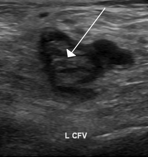
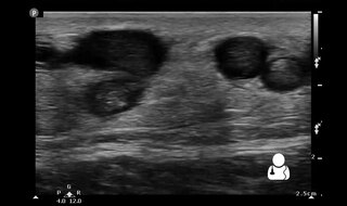
Ultrasonography in suspected deep vein thrombosis focuses primarily on the femoral vein and the popliteal vein, because thrombi in these veins are associated with the greatest risk of harmful pulmonary embolism.
References
- ↑ van Beek, Edwin J. R.; Büller, Harry R.; Oudkerk, Matthijs (2009). Deep Vein Thrombosis and Pulmonary Embolism. John Wiley and Sons. p. 57. ISBN 978-0-470-51717-8.
- ↑ PEABODY CN (October 1964). "An Objective Sign of Thrombophlebitis". Angiology. 15 (10): 434–5. doi:10.1177/000331976401501002. PMID 14229525.
| | This medical sign article is a stub. You can help Wikipedia by expanding it. |