8-Anilinonaphthalene-1-sulfonic acid

Last updated
8-Anilinonaphthalene-1-sulfonic acid
Phenylperi acid.png
Names
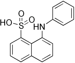
Preferred IUPAC name
8-Anilinonaphthalene-1-sulfonic acid
Other names
8-(Phenylamino)naphthalene-1-sulfonic acid
Identifiers
3D model (JSmol)
ChEBI
ChEMBL
ChemSpider
DrugBank
ECHA InfoCard 100.001.308 OOjs UI icon edit-ltr-progressive.svg
KEGG
PubChem CID
UNII
  • InChI=1S/C16H13NO3S/c18-21(19,20)15-11-5-7-12-6-4-10-14(16(12)15)17-13-8-2-1-3-9-13/h1-11,17H,(H,18,19,20) Yes check.svgY
    Key: FWEOQOXTVHGIFQ-UHFFFAOYSA-N Yes check.svgY
  • InChI=1/C16H13NO3S/c18-21(19,20)15-11-5-7-12-6-4-10-14(16(12)15)17-13-8-2-1-3-9-13/h1-11,17H,(H,18,19,20)
    Key: FWEOQOXTVHGIFQ-UHFFFAOYAB
  • O=S(=O)(O)c2c1c(cccc1ccc2)Nc3ccccc3
  • c1ccc(cc1)Nc2cccc3c2c(ccc3)S(=O)(=O)O
Properties
C16H13NO3S
Molar mass 299.34 g·mol−1
Except where otherwise noted, data are given for materials in their standard state (at 25 °C [77 °F], 100 kPa).
Yes check.svgY  verify  (what is  Yes check.svgYX mark.svgN ?)

8-Anilinonaphthalene-1-sulfonic acid (ANS), also called 1-anilino-8-naphthalenesulfonate, is an organic compound containing both a sulfonic acid and an amine group. This compound is used as a fluorescent molecular probe. [1] For example, ANS can be used to study conformational changes induced by ligand binding in proteins, as ANS's fluorescent properties will change as it binds to hydrophobic regions on the protein surface. Comparison of the fluorescence in the presence and absence of a particular ligand can thus give information about how the binding of the ligand changes the surface of the protein. Its permeability to mitochondrial membranes makes it particularly useful. [2]

Related Research Articles

<span class="mw-page-title-main">Fluorescent tag</span>

In molecular biology and biotechnology, a fluorescent tag, also known as a fluorescent label or fluorescent probe, is a molecule that is attached chemically to aid in the detection of a biomolecule such as a protein, antibody, or amino acid. Generally, fluorescent tagging, or labeling, uses a reactive derivative of a fluorescent molecule known as a fluorophore. The fluorophore selectively binds to a specific region or functional group on the target molecule and can be attached chemically or biologically. Various labeling techniques such as enzymatic labeling, protein labeling, and genetic labeling are widely utilized. Ethidium bromide, fluorescein and green fluorescent protein are common tags. The most commonly labelled molecules are antibodies, proteins, amino acids and peptides which are then used as specific probes for detection of a particular target.

A biosensor is an analytical device, used for the detection of a chemical substance, that combines a biological component with a physicochemical detector. The sensitive biological element, e.g. tissue, microorganisms, organelles, cell receptors, enzymes, antibodies, nucleic acids, etc., is a biologically derived material or biomimetic component that interacts with, binds with, or recognizes the analyte under study. The biologically sensitive elements can also be created by biological engineering. The transducer or the detector element, which transforms one signal into another one, works in a physicochemical way: optical, piezoelectric, electrochemical, electrochemiluminescence etc., resulting from the interaction of the analyte with the biological element, to easily measure and quantify. The biosensor reader device connects with the associated electronics or signal processors that are primarily responsible for the display of the results in a user-friendly way. This sometimes accounts for the most expensive part of the sensor device, however it is possible to generate a user friendly display that includes transducer and sensitive element. The readers are usually custom-designed and manufactured to suit the different working principles of biosensors.

<span class="mw-page-title-main">Fluorescence recovery after photobleaching</span>

Fluorescence recovery after photobleaching (FRAP) is a method for determining the kinetics of diffusion through tissue or cells. It is capable of quantifying the two-dimensional lateral diffusion of a molecularly thin film containing fluorescently labeled probes, or to examine single cells. This technique is very useful in biological studies of cell membrane diffusion and protein binding. In addition, surface deposition of a fluorescing phospholipid bilayer allows the characterization of hydrophilic surfaces in terms of surface structure and free energy.

<span class="mw-page-title-main">Förster resonance energy transfer</span> Photochemical energy transfer mechanism

Förster resonance energy transfer (FRET), fluorescence resonance energy transfer, resonance energy transfer (RET) or electronic energy transfer (EET) is a mechanism describing energy transfer between two light-sensitive molecules (chromophores). A donor chromophore, initially in its electronic excited state, may transfer energy to an acceptor chromophore through nonradiative dipole–dipole coupling. The efficiency of this energy transfer is inversely proportional to the sixth power of the distance between donor and acceptor, making FRET extremely sensitive to small changes in distance.

<span class="mw-page-title-main">DAPI</span> Fluorescent stain

DAPI, or 4′,6-diamidino-2-phenylindole, is a fluorescent stain that binds strongly to adenine–thymine-rich regions in DNA. It is used extensively in fluorescence microscopy. As DAPI can pass through an intact cell membrane, it can be used to stain both live and fixed cells, though it passes through the membrane less efficiently in live cells and therefore provides a marker for membrane viability.

A protein microarray is a high-throughput method used to track the interactions and activities of proteins, and to determine their function, and determining function on a large scale. Its main advantage lies in the fact that large numbers of proteins can be tracked in parallel. The chip consists of a support surface such as a glass slide, nitrocellulose membrane, bead, or microtitre plate, to which an array of capture proteins is bound. Probe molecules, typically labeled with a fluorescent dye, are added to the array. Any reaction between the probe and the immobilised protein emits a fluorescent signal that is read by a laser scanner. Protein microarrays are rapid, automated, economical, and highly sensitive, consuming small quantities of samples and reagents. The concept and methodology of protein microarrays was first introduced and illustrated in antibody microarrays in 1983 in a scientific publication and a series of patents. The high-throughput technology behind the protein microarray was relatively easy to develop since it is based on the technology developed for DNA microarrays, which have become the most widely used microarrays.

<span class="mw-page-title-main">Ligand (biochemistry)</span> Substance that forms a complex with a biomolecule

In biochemistry and pharmacology, a ligand is a substance that forms a complex with a biomolecule to serve a biological purpose. The etymology stems from Latin ligare, which means 'to bind'. In protein-ligand binding, the ligand is usually a molecule which produces a signal by binding to a site on a target protein. The binding typically results in a change of conformational isomerism (conformation) of the target protein. In DNA-ligand binding studies, the ligand can be a small molecule, ion, or protein which binds to the DNA double helix. The relationship between ligand and binding partner is a function of charge, hydrophobicity, and molecular structure.

<span class="mw-page-title-main">Thioflavin</span> Chemical compound

Thioflavins are fluorescent dyes that are available as at least two compounds, namely Thioflavin T and Thioflavin S. Both are used for histology staining and biophysical studies of protein aggregation. In particular, these dyes have been used since 1989 to investigate amyloid formation. They are also used in biophysical studies of the electrophysiology of bacteria. Thioflavins are corrosive, irritants, and are acutely toxic, causing serious eye damage. Thioflavin T has been used in research into Alzheimer's disease and other neurodegenerative diseases.

Bacterial display is a protein engineering technique used for in vitro protein evolution. Libraries of polypeptides displayed on the surface of bacteria can be screened using flow cytometry or iterative selection procedures (biopanning). This protein engineering technique allows us to link the function of a protein with the gene that encodes it. Bacterial display can be used to find target proteins with desired properties and can be used to make affinity ligands which are cell-specific. This system can be used in many applications including the creation of novel vaccines, the identification of enzyme substrates and finding the affinity of a ligand for its target protein.

<span class="mw-page-title-main">Conformational change</span> Change in the shape of a macromolecule, often induced by environmental factors

In biochemistry, a conformational change is a change in the shape of a macromolecule, often induced by environmental factors.

In biology, membrane fluidity refers to the viscosity of the lipid bilayer of a cell membrane or a synthetic lipid membrane. Lipid packing can influence the fluidity of the membrane. Viscosity of the membrane can affect the rotation and diffusion of proteins and other bio-molecules within the membrane, there-by affecting the functions of these things.

<i>alpha</i>-Parinaric acid Chemical compound

α-Parinaric acid is a conjugated polyunsaturated fatty acid. Discovered by Tsujimoto and Koyanagi in 1933, it contains 18 carbon atoms and 4 conjugated double bonds. The repeating single bond-double bond structure of α-parinaric acid distinguishes it structurally and chemically from the usual "methylene-interrupted" arrangement of polyunsaturated fatty acids that have double-bonds and single bonds separated by a methylene unit (−CH2−). Because of the fluorescent properties conferred by the alternating double bonds, α-parinaric acid is commonly used as a molecular probe in the study of biomembranes.

<span class="mw-page-title-main">Fluorescence in the life sciences</span> Scientific investigative technique

Fluorescence is used in the life sciences generally as a non-destructive way of tracking or analysing biological molecules. Some proteins or small molecules in cells are naturally fluorescent, which is called intrinsic fluorescence or autofluorescence. Alternatively, specific or general proteins, nucleic acids, lipids or small molecules can be "labelled" with an extrinsic fluorophore, a fluorescent dye which can be a small molecule, protein or quantum dot. Several techniques exist to exploit additional properties of fluorophores, such as fluorescence resonance energy transfer, where the energy is passed non-radiatively to a particular neighbouring dye, allowing proximity or protein activation to be detected; another is the change in properties, such as intensity, of certain dyes depending on their environment allowing their use in structural studies.

<span class="mw-page-title-main">Microscale thermophoresis</span> Biophysical technology for analyzing interactions

Microscale thermophoresis (MST) is a technology for the biophysical analysis of interactions between biomolecules. Microscale thermophoresis is based on the detection of a temperature-induced change in fluorescence of a target as a function of the concentration of a non-fluorescent ligand. The observed change in fluorescence is based on two distinct effects. On the one hand it is based on a temperature related intensity change (TRIC) of the fluorescent probe, which can be affected by binding events. On the other hand, it is based on thermophoresis, the directed movement of particles in a microscopic temperature gradient. Any change of the chemical microenvironment of the fluorescent probe, as well as changes in the hydration shell of biomolecules result in a relative change of the fluorescence detected when a temperature gradient is applied and can be used to determine binding affinities. MST allows measurement of interactions directly in solution without the need of immobilization to a surface.

<span class="mw-page-title-main">Quinaldine red</span> Chemical compound

Quinaldine red is a dark green–red or black solid that does not dissolve easily in water. In addition to being used as colored indicator, quinaldine red is also used as a fluorescence probe and an agent in bleaching.

A thermal shift assay (TSA) measures changes in the thermal denaturation temperature and hence stability of a protein under varying conditions such as variations in drug concentration, buffer pH or ionic strength, redox potential, or sequence mutation. The most common method for measuring protein thermal shifts is differential scanning fluorimetry (DSF) or thermofluor, which utilizes specialized fluorogenic dyes.

FlAsH-EDT<sub>2</sub> Chemical compound

FlAsH-EDT2 is an organoarsenic compound with molecular formula C24H18As2O5S4. Its structure is based around a fluorescein core with two 1,3,2-dithiarsolane substituents. It is used in bioanalytical research as a fluorescent label for visualising proteins in living cells. FlAsH-EDT2 is an abbreviation for fluorescin arsenical hairpin binder-ethanedithiol, and is a pale yellow or pinkish fluorogenic solid. It has a semi-structural formula (C2H4AsS2)2-(C13H5O3)-C6H4COOH, representing the dithiarsolane substituents bound to the hydroxyxanthone core, attached to an o-substituted molecule of benzoic acid.

A ligand binding assay (LBA) is an assay, or an analytic procedure, which relies on the binding of ligand molecules to receptors, antibodies or other macromolecules. A detection method is used to determine the presence and extent of the ligand-receptor complexes formed, and this is usually determined electrochemically or through a fluorescence detection method. This type of analytic test can be used to test for the presence of target molecules in a sample that are known to bind to the receptor.

Lanthanide probes are a non-invasive analytical tool commonly used for biological and chemical applications. Lanthanides are metal ions which have their 4f energy level filled and generally refer to elements cerium to lutetium in the periodic table. The fluorescence of lanthanide salts is weak because the energy absorption of the metallic ion is low; hence chelated complexes of lanthanides are most commonly used. The term chelate derives from the Greek word for “claw,” and is applied to name ligands, which attach to a metal ion with two or more donor atoms through dative bonds. The fluorescence is most intense when the metal ion has the oxidation state of 3+. Not all lanthanide metals can be used and the most common are: Sm(III), Eu(III), Tb(III), and Dy(III).

<span class="mw-page-title-main">TNP-ATP</span>

TNP-ATP is a fluorescent molecule that is able to determine whether a protein binds to ATP, and the constants associated with that binding. It is primarily used in fluorescence spectroscopy, but is also very useful as an acceptor molecule in FRET, and as a fluorescent probe in fluorescence microscopy and X-ray crystallography.

References

  1. Andras Malnasi-Csizmadia; György Hegyi; Ferenc Tölgyesi; Andrew G. Szent-Györgyi & László Nyitray (1999). "Fluorescence measurements detect changes in scallop myosin regulatory domain". European Journal of Biochemistry. 261 (2): 452–8. doi:10.1046/j.1432-1327.1999.00290.x. PMID   10215856.
  2. Gains N; Dawson AP (April 1975). "8-Anilinonaphthalene-1-sulphonate interaction with whole and disrupted mitochondria: a re-evaluation of the use of double-reciprocal plots in the derivation of binding parameters for fluorescent probes binding to mitochondrial membranes". Biochem. J. 148 (1): 157–60. doi:10.1042/bj1480157. PMC   1165518 . PMID   1156395.