APC/C activator protein CDH1

Last updated
SCF(Fbw7) ubiquitin ligase complex
SCF(Fbw7).PNG
Identifiers
SymbolCdh1, Hct1
PDB 2ovq
UniProt P53197
Search for
Structures Swiss-model
Domains InterPro

Cdh1 (cdc20 homolog 1) is one of the substrate adaptor protein of the anaphase-promoting complex (APC) in the budding yeast Saccharomyces cerevisiae . Functioning as an activator of the APC/C, Cdh1 regulates the activity and substrate specificity of this ubiquitin E3-ligase. The human homolog is encoded by the FZR1 gene, which is not to be confused with the CDH1 gene.

Contents

Introduction

Cdh1 plays a pivotal role in controlling cell division at the end of mitosis (telophase) and in the subsequent G1 phase of cell cycle: By recognizing and binding proteins (like mitotic cyclins) which contain a destruction box (D-box) and an additional degradation signal (KEN box), Cdh1 recruits them in a C-box-dependent mechanism to the APC for ubiquination and subsequent proteolysis. Cdh1 is required for the exit of mitosis. Furthermore, it is thought to be a possible target of a BUB2-dependent spindle checkpoint pathway. [1] [2]

Function

The anaphase-promoting complex/cyclosome (APC/c) is an ubiquitin E3-ligase complex. Once activated it attaches chains of ubiquitin molecules to its target substrates. These chains are recognised and the substrate is degraded by the Proteasome. Cdh1 is one of the co- activator proteins of APC/c and therefore contributes to the regulation of protein degradation, by providing substrate specificity to the E3-ligase in a cell cycle regulated manner.[ citation needed ]

Cdh1 can exist in several forms. It can be phosphorylated by CDKs, which inactivates it and it can be dephosphorylated by Cdc14. In the dephosphorylated form it can interact with APC/c and build the active ligase APCCdh1. [3]

Suppression of Cdh1 by RNA interference leads to an aberrant accumulation of APCCdh1 target proteins, such as cyclin A and B, the kinase AuroraB, PLK1, Skp2 and Cdc20, another APC/c co- activator. [4]

Stabilising G1-phase

The main function of Cdh1 is to suppress the re-accumulation of mitotic cyclins and other cell cycle determinants and therefore stabilising the G1-phase. It is inactive in early stages of mitosis and only becomes active in the transition from late mitosis to G1.[ citation needed ]

During the cell cycle Cdk gets activated through cyclins, this leads to the mitotic entry and promotes APCCdc20 activation. APCCdc20 degrades the cyclins, this and the activation of Cdc14 leads to the creation of APCCdh1. APCCdh1 keeps the cyclin concentration low and the Cdk inactive that maintains the G1-Phase. [5] [6]

G1/S transition

APCCdh1 is thought to prevent premature S-phase entry by degrading mitotic cyclins in G1 and regulate processes unrelated to the cell cycle. To enter S-phase APCCdh1 must be inactivated. This is made through degradation of the complex and through phosphorylation of Cdh1. [6]

Exit from mitosis

One characteristic of budding yeast cells exit from mitosis after chromosome segregation is the removal of the mitotic determinants. This requires the inactivation of mitotic CDKs which are inactivated through ubiquitin-dependent pathways. The protein phosphatase Cdc14 dephosphorylates Cdh1 and therefore activates APCCdh1. As a result the concentration of many APCCdh1 substrates (e.g. M-Cyclins) drops down at the cell exit from mitosis. [6]

Cdh1 functions as a tumour suppressor

Cdh1-deficient cells can proliferate but accumulate mitotic errors and have difficulties with cytokinesis.[ citation needed ]

It has been shown that APCCdh1-mediated degradation of PIk1 plays an important role in preventing mitosis in cells that have DNA-damage. In healthy cells Cdh1 stays inactive from late G1 to early mitosis. It stays inactive in early mitosis and only becomes active in the transition from late mitosis to G1. A cell that suffers from DNA-damage shows an active Cdh1 already in late G1 and therefore blocks the mitotic entry.[ citation needed ]

One substrate of APCCdh1 is the transcription factor Ets2, which is activated by the Ras-Raf-MAPK signalling pathway and induces the expression of cyclin D1. This pathway stimulates cell proliferation. It was shown that an increased expression of Ets2 can be associated with various cancer types, in the likes of cervical cancer or oesophageal squamous cell carcinoma. [6]

Function of Cdh1 in non-dividing cells

It was shown that APCCdh1 is active in adult brain and liver tissues. It seems that the complex has a function in axon growth, morphology and plasticity of synapses as well as in learning and memory. [6]

Structure

Fig. 1 There is no structure resolved for Cdh1 of Saccharomyces cerevisiae. There is a model based on template pdb2ovq, which shows the SCF(Fbw7) ubiquitin ligase complex. Fbw7 is also a WD repeat protein like Cdh1. (The model for Cdh1 can be found on the webpage of SWISS-MODEL Repository, see the external link at the end of this page) SCF(Fbw7).PNG
Fig. 1 There is no structure resolved for Cdh1 of Saccharomyces cerevisiae. There is a model based on template pdb2ovq, which shows the SCF(Fbw7) ubiquitin ligase complex. Fbw7 is also a WD repeat protein like Cdh1. (The model for Cdh1 can be found on the webpage of SWISS-MODEL Repository, see the external link at the end of this page)

The following structural informations are based on the cdh1 protein of Saccharomyces cerevisiae also named Hct1. Cdh1 is a cdc20 homolog and is Frizzy-related ( Drosophila ). The protein sequence of cdh1 consists of 566 amino acids and has a molecular weight of 62.8 kDa. Cdh1 comprises different domains important for its proper function, when it interacts with the APC/c complex and the various substrates. [9]

Activation and APC/c binding

In the N-terminal region at amino acid position 55-61 the cdh1 protein contains a C-Box motif, which is required for the association with the APC/c complex. Especially the residue R56 seems to be important for the binding to APC/c in vitro and Cdh1 function in vivo. [9] [10]

Cdh1 contains multiple phosphorylation sites for the kinase cdc28. When cdh1 is hyperphosphorytaled, the association of cdh1 to the APC/c is blocked, thus leading to the inactive form of cdh1. Activation can be induced by dephosphorylation through the phosphatase cdc14, which leads to the binding of cdh1 to the APC/c. [9] [11]

Cdh1 as well includes a poly-Ser in the N-terminal region from residue 32-38. In general serine, threonine and tyrosine side chains can act as phosphorylation sites for posttranslational modification. In the cdh1 protein amino acid modifications can be found at residue 156 being a phosphoserine and at residue 157 being a phosphothreonine. [9]

Cdh1 also contains a C-terminal Ile-Arg (IR) dipeptide motif at residue 565 and 566, which is suggested to bind to the Cdc27 subunit of APC. [9] [10] [12]

Substrate binding

Cdh1 has 7 WD repeats, which are located between the middle of the protein and the C-terminal end. They have a conserved core length of about 38 to 43 amino acids, which in general end with tryptophan-aspartic acid (WD). WD repeat proteins are assumed to form a circularized beta propeller structure, which is thought to be essential for the biological function. The WD repeats in cdh1 are suspected to be the binding sites for the APC/c substrates. Thus cdh1 seems to be a sort of linker between the APC/c complex and the substrates. The APC/c substrates contain a D-Box and/or a KEN-Box, which are important for the interaction with cdh1. [9] [13] [14] [15]

See also

Related Research Articles

<span class="mw-page-title-main">Telophase</span> Final stage of a cell division for eukaryotic cells both in mitosis and meiosis

Telophase is the final stage in both meiosis and mitosis in a eukaryotic cell. During telophase, the effects of prophase and prometaphase are reversed. As chromosomes reach the cell poles, a nuclear envelope is re-assembled around each set of chromatids, the nucleoli reappear, and chromosomes begin to decondense back into the expanded chromatin that is present during interphase. The mitotic spindle is disassembled and remaining spindle microtubules are depolymerized. Telophase accounts for approximately 2% of the cell cycle's duration.

<span class="mw-page-title-main">Anaphase-promoting complex</span> Cell-cycle regulatory complex

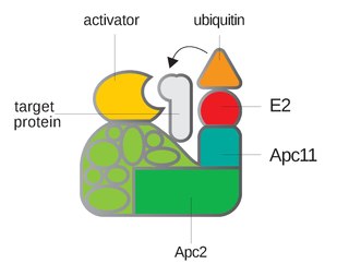
Anaphase-promoting complex is an E3 ubiquitin ligase that marks target cell cycle proteins for degradation by the 26S proteasome. The APC/C is a large complex of 11–13 subunit proteins, including a cullin (Apc2) and RING (Apc11) subunit much like SCF. Other parts of the APC/C have unknown functions but are highly conserved.

Maturation-promoting factor (abbreviated MPF, also called mitosis-promoting factor or M-Phase-promoting factor) is the cyclin-Cdk complex that was discovered first in frog eggs. It stimulates the mitotic and meiotic phases of the cell cycle. MPF promotes the entrance into mitosis (the M phase) from the G2 phase by phosphorylating multiple proteins needed during mitosis. MPF is activated at the end of G2 by a phosphatase, which removes an inhibitory phosphate group added earlier.

Endoreduplication is replication of the nuclear genome in the absence of mitosis, which leads to elevated nuclear gene content and polyploidy. Endoreplication can be understood simply as a variant form of the mitotic cell cycle (G1-S-G2-M) in which mitosis is circumvented entirely, due to modulation of cyclin-dependent kinase (CDK) activity. Examples of endoreplication characterized in arthropod, mammalian, and plant species suggest that it is a universal developmental mechanism responsible for the differentiation and morphogenesis of cell types that fulfill an array of biological functions. While endoreplication is often limited to specific cell types in animals, it is considerably more widespread in plants, such that polyploidy can be detected in the majority of plant tissues.

A spindle poison, also known as a spindle toxin, is a poison that disrupts cell division by affecting the protein threads that connect the centromere regions of chromosomes, known as spindles. Spindle poisons effectively cease the production of new cells by interrupting the mitosis phase of cell division at the spindle assembly checkpoint (SAC). However, as numerous and varied as they are, spindle poisons are not yet 100% effective at ending the formation of tumors (neoplasms). Although not 100% effective, substantive therapeutic efficacy has been found in these types of chemotherapeutic treatments. The mitotic spindle is composed of microtubules that aid, along with regulatory proteins, each other in the activity of appropriately segregating replicated chromosomes. Certain compounds affecting the mitotic spindle have proven highly effective against solid tumors and hematological malignancies.

<span class="mw-page-title-main">Deubiquitinating enzyme</span>

Deubiquitinating enzymes (DUBs), also known as deubiquitinating peptidases, deubiquitinating isopeptidases, deubiquitinases, ubiquitin proteases, ubiquitin hydrolases, ubiquitin isopeptidases, are a large group of proteases that cleave ubiquitin from proteins. Ubiquitin is attached to proteins in order to regulate the degradation of proteins via the proteasome and lysosome; coordinate the cellular localisation of proteins; activate and inactivate proteins; and modulate protein-protein interactions. DUBs can reverse these effects by cleaving the peptide or isopeptide bond between ubiquitin and its substrate protein. In humans there are nearly 100 DUB genes, which can be classified into two main classes: cysteine proteases and metalloproteases. The cysteine proteases comprise ubiquitin-specific proteases (USPs), ubiquitin C-terminal hydrolases (UCHs), Machado-Josephin domain proteases (MJDs) and ovarian tumour proteases (OTU). The metalloprotease group contains only the Jab1/Mov34/Mpr1 Pad1 N-terminal+ (MPN+) (JAMM) domain proteases.

<span class="mw-page-title-main">Cell cycle checkpoint</span> Control mechanism in the eukaryotic cell cycle

Cell cycle checkpoints are control mechanisms in the eukaryotic cell cycle which ensure its proper progression. Each checkpoint serves as a potential termination point along the cell cycle, during which the conditions of the cell are assessed, with progression through the various phases of the cell cycle occurring only when favorable conditions are met. There are many checkpoints in the cell cycle, but the three major ones are: the G1 checkpoint, also known as the Start or restriction checkpoint or Major Checkpoint; the G2/M checkpoint; and the metaphase-to-anaphase transition, also known as the spindle checkpoint. Progression through these checkpoints is largely determined by the activation of cyclin-dependent kinases by regulatory protein subunits called cyclins, different forms of which are produced at each stage of the cell cycle to control the specific events that occur therein.

<span class="mw-page-title-main">SCF complex</span>

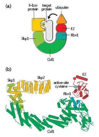
Skp, Cullin, F-box containing complex is a multi-protein E3 ubiquitin ligase complex that catalyzes the ubiquitination of proteins destined for 26S proteasomal degradation. Along with the anaphase-promoting complex, SCF has important roles in the ubiquitination of proteins involved in the cell cycle. The SCF complex also marks various other cellular proteins for destruction.

Polo-like kinases (Plks) are regulatory serine/threonin kinases of the cell cycle involved in mitotic entry, mitotic exit, spindle formation, cytokinesis, and meiosis. Only one Plk is found in the genomes of the fly Drosophila melanogaster (Polo), budding yeast (Cdc5) and fission yeast (Plo1). Vertebrates and other animals, however, have many Plk family members including Plk1, Plk2/Snk, Plk3/Prk/FnK, Plk4/Sak and Plk5. Of the vertebrate Plk family members, the mammalian Plk1 has been most extensively studied. During mitosis and cytokinesis, Plks associate with several structures including the centrosome, kinetochores, and the central spindle.

<span class="mw-page-title-main">CDC20</span> Protein-coding gene in the species Homo sapiens

The cell division cycle protein 20 homolog is an essential regulator of cell division that is encoded by the CDC20 gene in humans. To the best of current knowledge its most important function is to activate the anaphase promoting complex (APC/C), a large 11-13 subunit complex that initiates chromatid separation and entrance into anaphase. The APC/CCdc20 protein complex has two main downstream targets. Firstly, it targets securin for destruction, enabling the eventual destruction of cohesin and thus sister chromatid separation. It also targets S and M-phase (S/M) cyclins for destruction, which inactivates S/M cyclin-dependent kinases (Cdks) and allows the cell to exit from mitosis. A closely related protein, Cdc20homologue-1 (Cdh1) plays a complementary role in the cell cycle.

<span class="mw-page-title-main">CDC27</span> Protein-coding gene in the species Homo sapiens

Cell division cycle protein 27 homolog is a protein that in humans is encoded by the CDC27 gene.

<span class="mw-page-title-main">FBXO5</span>

F-box only protein 5 is a protein that in humans is encoded by the FBXO5 gene.

<span class="mw-page-title-main">CDC16</span> Protein-coding gene in the species Homo sapiens

Cell division cycle protein 16 homolog is a protein that in humans is encoded by the CDC16 gene.

<span class="mw-page-title-main">Wee1</span>

Wee1 is a nuclear kinase belonging to the Ser/Thr family of protein kinases in the fission yeast Schizosaccharomyces pombe. Wee1 has a molecular mass of 96 kDa and is a key regulator of cell cycle progression. It influences cell size by inhibiting the entry into mitosis, through inhibiting Cdk1. Wee1 has homologues in many other organisms, including mammals.

A series of biochemical switches control transitions between and within the various phases of the cell cycle. The cell cycle is a series of complex, ordered, sequential events that control how a single cell divides into two cells, and involves several different phases. The phases include the G1 and G2 phases, DNA replication or S phase, and the actual process of cell division, mitosis or M phase. During the M phase, the chromosomes separate and cytokinesis occurs.

<span class="mw-page-title-main">Cell division control protein 4</span>

Cdc4 is a substrate recognition component of the SCF ubiquitin ligase complex, which acts as a mediator of ubiquitin transfer to target proteins, leading to their subsequent degradation via the ubiquitin-proteasome pathway. Cdc4 targets primarily cell cycle regulators for proteolysis. It serves the function of an adaptor that brings target molecules to the core SCF complex. Cdc4 was originally identified in the model organism Saccharomyces cerevisiae. CDC4 gene function is required at G1/S and G2/M transitions during mitosis and at various stages during meiosis.

Cdc14 and Cdc14 are a gene and its protein product respectively. Cdc14 is found in most of the eukaryotes. Cdc14 was defined by Hartwell in his famous screen for loci that control the cell cycle of Saccharomyces cerevisiae. Cdc14 was later shown to encode a protein phosphatase. Cdc14 is dual-specificity, which means it has serine/threonine and tyrosine-directed activity. A preference for serines next to proline is reported. Many early studies, especially in the budding yeast Saccharomyces cerevisiae, demonstrated that the protein plays a key role in regulating late mitotic processes. However, more recent work in a range of systems suggests that its cellular function is more complex.

Clb5 and Clb6 are B-type, S-phase cyclins in yeast that assist in cell cycle regulation. Clb5 and Clb6 bind and activate Cdk1, and high levels of these cyclins are required for entering S-phase. S-phase cyclin binding to Cdk1 directly stimulates DNA replication as well as progression to the next phase of the cell cycle.

Mitotic exit is an important transition point that signifies the end of mitosis and the onset of new G1 phase for a cell, and the cell needs to rely on specific control mechanisms to ensure that once it exits mitosis, it never returns to mitosis until it has gone through G1, S, and G2 phases and passed all the necessary checkpoints. Many factors including cyclins, cyclin-dependent kinases (CDKs), ubiquitin ligases, inhibitors of cyclin-dependent kinases, and reversible phosphorylations regulate mitotic exit to ensure that cell cycle events occur in correct order with fewest errors. The end of mitosis is characterized by spindle breakdown, shortened kinetochore microtubules, and pronounced outgrowth of astral (non-kinetochore) microtubules. For a normal eukaryotic cell, mitotic exit is irreversible.

<span class="mw-page-title-main">Motifs targeted by APC/C</span>

The anaphase- promoting complex or cyclosome (APC/C) is a highly specific ubiquitin protein ligase responsible for triggering events of late mitosis. In early mitosis, Cdc20 levels rise and APC/C binds to form active APC/CCdc20. This then leads to the destruction of mitotic cyclins, securin, and other proteins to trigger chromosome separation in anaphase. In early anaphase, Cdk1 is inactivated, leading to the activation of Cdh1, the other activator subunit of APC/C. This then triggers the degradation of Cdc20 and leads to the activation of APC/CCdh1 through G1 to suppress S- phase cyclin-Cdk activity. At the end of G1, APC/CCdh1 is inactivated and S- phase and mitotic cyclins gets reaccumulate as the cell progresses to S phase.

References

  1. "Uniprot: P53197/Cdh1" . Retrieved 2010-04-13.
  2. Li M, Shin YH, Hou L, Huang X, Wei Z, Klann E, Zhang P (2008). "The adaptor protein of the anaphase promoting complex Cdh1 is essential in maintaining replicative lifespan and in learning and memory". Nat. Cell Biol. 10 (9): 1083–9. doi:10.1038/ncb1768. PMC   2914158 . PMID   19160489.
  3. Peters JM (2006). "The anaphase promoting complex/cyclosome: a machine designed to destroy". Nat. Rev. Mol. Cell Biol. 7 (9): 644–56. doi:10.1038/nrm1988. PMID   16896351. S2CID   31962403.
  4. Engelbert D (2007). Die Bedeutung des Anaphase-Promoting Komplex-Aktivators Cdh1 für die Erhaltung der genetischen Integrität humaner Zellen. Inauguraldissertation zur Erlangung der Doktorwürde (Thesis) (in German). Albert-Ludwigs-Universität Freiburg.
  5. Reis A, Chang HY, Levasseur M, Jones KT (2006). "APCcdh1 activity in mouse oocytes prevents entry into the first meiotic division". Nat. Cell Biol. 8 (5): 539–40. doi:10.1038/ncb1406. PMC   2435241 . PMID   16715549.
  6. 1 2 3 4 5 Li M, Zhang P (2009). "The function of APC/CCdh1 in cell cycle and beyond". Cell Div. 4: 2. doi:10.1186/1747-1028-4-2. PMC   2635356 . PMID   19152694.
  7. Kiefer F, Arnold K, Künzli M, Bordoli L, Schwede T (2009). "The SWISS-MODEL Repository and associated resources". Nucleic Acids Res. 37 (Database issue): D387–92. doi:10.1093/nar/gkn750. PMC   2686475 . PMID   18931379.
  8. Kopp J, Schwede T (January 2004). "The SWISS-MODEL Repository of annotated three-dimensional protein structure homology models". Nucleic Acids Res. 32 (Database issue): D230–4. doi:10.1093/nar/gkh008. PMC   308743 . PMID   14681401.
  9. 1 2 3 4 5 6 "Uniprot: P53197/Cdh1" . Retrieved 2010-04-10.
  10. 1 2 Thornton BR, Ng TM, Matyskiela ME, Carroll CW, Morgan DO, Toczyski DP (2006). "An architectural map of the anaphase-promoting complex". Genes Dev. 20 (4): 449–60. doi:10.1101/gad.1396906. PMC   1369047 . PMID   16481473.
  11. Manchado E, Eguren M, Malumbres M (2010). "The anaphase-promoting complex/cyclosome (APC/C): cell-cycle-dependent and -independent functions". Biochem. Soc. Trans. 38 (Pt 1): 65–71. doi:10.1042/BST0380065. PMID   20074037. S2CID   1889798.
  12. Burton JL, Tsakraklides V, Solomon MJ (May 2005). "Assembly of an APC-Cdh1-substrate complex is stimulated by engagement of a destruction box". Mol. Cell. 18 (5): 533–42. doi: 10.1016/j.molcel.2005.04.022 . PMID   15916960.
  13. Li D, Roberts R (2001). "WD-repeat proteins: structure characteristics, biological function, and their involvement in human diseases". Cell. Mol. Life Sci. 58 (14): 2085–97. doi:10.1007/PL00000838. PMID   11814058. S2CID   20646422.
  14. Kraft C, Vodermaier HC, Maurer-Stroh S, Eisenhaber F, Peters JM (2005). "The WD40 propeller domain of Cdh1 functions as a destruction box receptor for APC/C substrates". Mol. Cell. 18 (5): 543–53. doi: 10.1016/j.molcel.2005.04.023 . PMID   15916961.
  15. Pfleger CM, Kirschner MW (2000). "The KEN box: an APC recognition signal distinct from the D box targeted by Cdh1". Genes Dev. 14 (6): 655–65. doi:10.1101/gad.14.6.655. PMC   316466 . PMID   10733526.