Auranticordis | |
---|---|
![]() | |
Scientific classification ![]() | |
Domain: | Eukaryota |
Clade: | Diaphoretickes |
Clade: | SAR |
Phylum: | Cercozoa |
Class: | Imbricatea |
Order: | Marimonadida |
Family: | Auranticordidae |
Genus: | Auranticordis Chantangsi, Esson & Leander, 2008 |
Species: | A. quadriverberis |
Binomial name | |
Auranticordis quadriverberis Chantangsi, Esson & Leander, 2008 | |
Auranticordis is a genus of rare tetraflagellate eukaryotic organisms within the phylum Cercozoa. [1] Currently there is only one characterized species within this genus: Auranticordis quadriverberis.Auranticordis cells are heart shaped and can range from 35 - 75 μm long and 25 - 70 μm wide. [1] These cells move in a forward gliding motion, and are predators in interstitial marine sand habitats. Auranticordis cells contain several features that are unique and dissimilar from other cercozoans, such as black bodies, reduced acristate mitochondria (both features that may indicate a preference to a semi-anoxic habitat), putative primary endosymbionts, many orange pigmented extrusomes and the absence of permanently condensed chromosomes. [1]
The heart-like cell shape and orange colouration of Auranticordis quadriverberis are the basis for the genus name. [1] The latin words aurantium and cordis mean "orange" and "heart", respectively.
Auranticordis quadriverberis [1]
Currently, there is only one defined species with the genus Auranticordis: Auranticordis quadriverberis. It was first discovered and characterized in 2008 by Chantangsi, Esson & Leander [1] while completing a project focused on characterizing cercozoan diversity in British Columbia, Canada, and has since never been further investigated. This is likely due to the fact that Auranticordis quadriverberis is low in abundance and difficult to find, with only 65 cells isolated thus far. Additional species within this genus have not yet been discovered. [1]
Auranticordis has only been isolated and characterized from marine sand samples collected at Spanish Banks, British Columbia, a sandy tidal flat located on the southern shore of English Bay, BC. [1] Metabarcoding and environmental sequencing data from a variety of studies (data provided by the Global Biodiversity Information Facility) indicates that Auranticordis may be a cosmopolitan species however no further Auranticordis individuals have been isolated. [2] The current known habitat of Auranticordis, based on the type species, are benthic environments, specifically interstitial marine sand. Evidence of this is both provided by its discovery in marine sand samples, and the presence of muciferous bodies (a type of extrusome) on the external surface of the cell which may aid in allowing Auranticordis cells to adhere to sand by producing a sticky substance. Seasonality of Auranticordis is currently unknown due to limited research available. Auranticordis (Auranticordis quadriverberis) has only been observed and isolated in the months of March and May 2007.
It is currently unknown whether or not Auranticordis is a purely heterotrophic species, consuming other microorganisms as food, or whether or not it may photosynthesize as well. [1] Auranticordis is likely predatory and feeds on bacteria based on observed ingested bacteria within the cell, however the exact method of feeding is unknown. Predators of Auranticordis are currently unknown and it is likely that Auranticordis plays a minimal role in the food web and nutrient cycling within marine sand environments given the low abundance of this genus.
Auranticordis cells are orange in colour and large, with Auranticordis quadriverberis cells observed to be 35 - 75 μm long and 25 - 70 μm wide. [1] Auranticordis cells do not contain a wall, are heart shaped and consist of four lobes. From the anterior to posterior end, these cells are covered in longitudinal ridges supported by microtubules. Auranticordis cells have four hair-covered flagella arranged in pairs that originate from the anterior end of the cell, lay along the ventral groove and emerge at the posterior end of the cell, allowing the cell to glide forwards. [1] This trait is dissimilar from most cercozoans, which are typically biflagellated, with the exception of Cholamonas cyrtodiopsidis, which is another tetraflagellated cercozoan. Cells are consistently observed to be flagellated, with pseudopodia and amoeboid stages not observed. As well, Auranticordis cells are not able to significantly alter their shape.
The cytoplasm of Auranticordis contains many vacuoles, Golgi bodies and lipid globules. There are a large number of small orange muciferous bodies (a type of extrusome) arranged in linear rows beneath the cell membrane visible on the surface of the cell which result in the orange colouration of Auranticordis cells. [1] These extrusomes secrete a sticky substance called mucilage through small pores present in the grooves between the ridges on the cell surface. Similar extrusomes have been observed in Cryothecomonas armigera, however other extrusome types found commonly in cercozoans are not present in Auranticordis. The cytoplasmic components and organization of Auranticordis are similar to those of the Protaspa genus, another cercozoan lineage. [1]
Within the cytoplasm, black inclusions are found near the anterior end of the cell. [1] These black inclusions may be associated with a semi-anoxic lifestyle, as similar black inclusions have been noted in semi-anoxic ciliate and euglenid lineages. As well, while most cercozoans contain mitochondria with tubular cristae, no distinct mitochondria are observed within Auranticordis cells. Instead, near the edges of the cell within the cytoplasm, structures similar to acristate mitochondria are observed. These putative mitochondria-like structures are smaller in contrast to the distinct mitochondria present among other cercozoans, and possibly could be reduced as an adaptation to a semi-anoxic environment.
Pale orange bodies are observed throughout the cytoplasm of Auranticordis cells; found more frequently near the anterior end of the cell and in variable but high numbers: 2 - 30 bodies per cell. [1] The pale orange bodies are small, with each pale body having a diameter between 4 - 5 μm. Bound by two inner membranes, these cells bodies contain sack-like vesicles surrounding the outer membrane. The edge regions of the bodies are abundant with unstacked thylakoids, formed from the innermost membrane. The central region of each pale orange body does not contain any thylakoids, is electron dense and contains viral particles.
Auranticordis cells are uninucleate with a single large nucleus located at the anterior end of each cell. [1] The position of the nucleus of Auranticordis is found to be associated with the basal bodies within each cell. Several nucleoli are present within the nucleus, and the chromosomes within the nucleus are not permanently condensed.
Auranticordis cells are found to contain many pale orange bodies which are double inner-membrane bound. [1] Based on the ultrastructure of these endosymbionts, specifically the presence of two inner membranes with one forming unstacked thylakoids, it is possible that these bodies are primary endosymbionts and could indicate that an independent primary endosymbiosis occurred within the Auranticordis lineage. This could be similar to the endosymbiotic event which occurred in Paulinella chromatophora, another cercozoan. These pale orange bodies are likely cyanobacterial in origin, based on their ultrastructure and the existence of free living cyanobacteria which are similar in colouration. The origin of these bodies has yet to be confirmed, however several possibilities have been theorized. The first possibility is that they are in fact cyanobacteria or eukaryotic cells with similar plastids which were engulfed and are early in the process of digestion within Auranticordis cells, however no evidence of digestion has been observed. [1] A second possibility for the origin of these pale orange bodies is that they are secondary endosymbionts which are not permanent fixtures within Auranticordis cells but in fact, are continuously restored within the cell via kleptoplasty of a different eukaryotic cell. The third possibility for the origin and function of the pale orange bodies is that they are permanent plastids acquired via primary endosymbiosis of a free-living cyanobacterial cell.
The genetics of Auranticordis are currently not well understood, however the placement of Auranticordis within Cercozoa is well supported through molecular phylogenetic analysis of small-subunit rDNA. [1]
Currently, Auranticordis has no known practical importance. If the pale orange bodies are in fact integrated plastids acquired via primary endosymbiosis, then Auranticordis cells would represent one of only three occurrences of primary endosymbiosis within eukaryotic lineages; the other two being the primary endosymbiosis that resulted in the plastids within the archaeplastids and the plastids present in Paulinella chromatophora. [1]
The cell is the basic structural and functional unit of all forms of life. Every cell consists of cytoplasm enclosed within a membrane; many cells contain organelles, each with a specific function. The term comes from the Latin word cellula meaning 'small room'. Most cells are only visible under a microscope. Cells emerged on Earth about 4 billion years ago. All cells are capable of replication, protein synthesis, and motility.
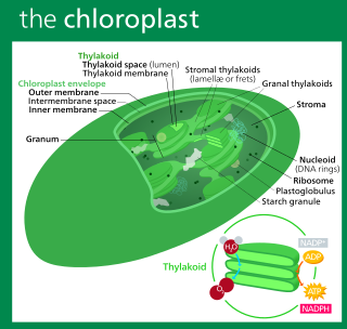
A chloroplast is a type of membrane-bound organelle known as a plastid that conducts photosynthesis mostly in plant and algal cells. The photosynthetic pigment chlorophyll captures the energy from sunlight, converts it, and stores it in the energy-storage molecules ATP and NADPH while freeing oxygen from water in the cells. The ATP and NADPH is then used to make organic molecules from carbon dioxide in a process known as the Calvin cycle. Chloroplasts carry out a number of other functions, including fatty acid synthesis, amino acid synthesis, and the immune response in plants. The number of chloroplasts per cell varies from one, in unicellular algae, up to 100 in plants like Arabidopsis and wheat.
In cell biology, an organelle is a specialized subunit, usually within a cell, that has a specific function. The name organelle comes from the idea that these structures are parts of cells, as organs are to the body, hence organelle, the suffix -elle being a diminutive. Organelles are either separately enclosed within their own lipid bilayers or are spatially distinct functional units without a surrounding lipid bilayer. Although most organelles are functional units within cells, some function units that extend outside of cells are often termed organelles, such as cilia, the flagellum and archaellum, and the trichocyst.
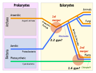
Symbiogenesis is the leading evolutionary theory of the origin of eukaryotic cells from prokaryotic organisms. The theory holds that mitochondria, plastids such as chloroplasts, and possibly other organelles of eukaryotic cells are descended from formerly free-living prokaryotes taken one inside the other in endosymbiosis. Mitochondria appear to be phylogenetically related to Rickettsiales bacteria, while chloroplasts are thought to be related to cyanobacteria.
A plastid, pl. plastids, is a membrane-bound organelle found in the cells of plants, algae, and some other eukaryotic organisms. They are considered to be intracellular endosymbiotic cyanobacteria.
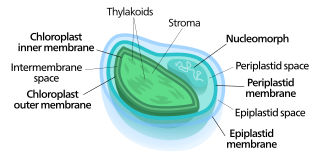
Nucleomorphs are small, vestigial eukaryotic nuclei found between the inner and outer pairs of membranes in certain plastids. They are thought to be vestiges of primitive red and green algal nuclei that were engulfed by a larger eukaryote. Because the nucleomorph lies between two sets of membranes, nucleomorphs support the endosymbiotic theory and are evidence that the plastids containing them are complex plastids. Having two sets of membranes indicate that the plastid, a prokaryote, was engulfed by a eukaryote, an alga, which was then engulfed by another eukaryote, the host cell, making the plastid an example of secondary endosymbiosis.
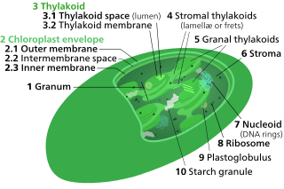
Chloroplasts contain several important membranes, vital for their function. Like mitochondria, chloroplasts have a double-membrane envelope, called the chloroplast envelope, but unlike mitochondria, chloroplasts also have internal membrane structures called thylakoids. Furthermore, one or two additional membranes may enclose chloroplasts in organisms that underwent secondary endosymbiosis, such as the euglenids and chlorarachniophytes.
Gymnophryidae is a small family of amoeboids that lack shells and produce thin, reticulose pseudopods. These contain microtubules and have a granular appearance, owing to the presence of extrusomes, but are distinct from the pseudopods of Foraminifera. They are included among the Cercozoa, but differ from other cercozoans in having mitochondria with flat cristae, rather than tubular cristae.
An apicoplast is a derived non-photosynthetic plastid found in most Apicomplexa, including Toxoplasma gondii, and Plasmodium falciparum and other Plasmodium spp., but not in others such as Cryptosporidium. It originated from algae through secondary endosymbiosis; there is debate as to whether this was a green or red alga. The apicoplast is surrounded by four membranes within the outermost part of the endomembrane system. The apicoplast hosts important metabolic pathways like fatty acid synthesis, isoprenoid precursor synthesis and parts of the heme biosynthetic pathway.
Paulinella is a genus of at least eleven species including both freshwater and marine amoeboids. Like many members of euglyphids it is covered by rows of siliceous scales, and use filose pseudopods to crawl over the substrate of the benthic zone.
Rhodomonas is a genus of cryptomonads. It is characterized by its red colour, the square-shaped plates of its inner periplast, its short furrow ending in a gullet, and a distinctly shaped chloroplast closely associated with its nucleomorph. Historically, Rhodomonas was characterized by its red chloroplast alone, but this no longer occurs as its taxonomy has become increasingly based on molecular and cellular data. Currently, there is some debate about the taxonomic validity of Rhodomonas as a genus and further research is needed to verify its taxonomic status. Rhodomonas is typically found in marine environments, although freshwater reports exist. It is commonly used as a live feed for various aquaculture species.
Geminigera /ˌdʒɛmɪnɪˈdʒɛɹə/ is a genus of cryptophyte from the family Geminigeraceae. Named for its unique pyrenoids, Geminigera is a genus with a single mixotrophic species. It was discovered in 1968 and is known for living in very cold temperatures such as under the Antarctic ice. While originally considered to be part of the genus Cryptomonas, the genus Geminigera was officially described in 1991 by D. R. A. Hill.
Guillardia is a genus of marine biflagellate cryptomonad algae with a plastid obtained through secondary endosymbiosis of a red alga.
Mesodinium rubrum is a species of ciliates. It constitutes a plankton community and is found throughout the year, most abundantly in spring and fall, in coastal areas. Although discovered in 1908, its scientific importance came into light in the late 1960s when it attracted scientists by the recurrent red colouration it caused by forming massive blooms, that cause red tides in the oceans.
Kraken is a genus of amoebae within the Cercozoa, containing the sole species Kraken carinae. These amoebae are characterized by a small round cell body and a network of thin and very long filopodia that can reach up to a mm in diameter. Kraken amoebae feed on bacteria and live in freshwater and soil systems.
Platysulcus tardus is an eukaryotic microorganism that was recently discovered to be the earliest diverging lineage of the Heterokont phylogenetic tree. It is the only member of the family Platysulcidae, order Platysulcida and class Platysulcea.
Durinskia is a genus of dinoflagellates that can be found in freshwater and marine environments. This genus was created to accommodate its type species, Durinskia baltica, after major classification discrepancies were found. While Durinskia species appear to be typical dinoflagellates that are armored with cellulose plates called theca, the presence of a pennate diatom-derived tertiary endosymbiont is their most defining characteristic. This genus is significant to the study of endosymbiotic events and organelle integration since structures and organelle genomes in the tertiary plastids are not reduced. Like some dinoflagellates, species in Durinskia may cause blooms.
Viridiraptoridae, previously known as clade X, is a clade of heterotrophic protists in the phylum Cercozoa. They're a family of glissomonads, a group containing a vast, mostly undescribed diversity of soil and freshwater organisms.
Orciraptor is a genus of heterotrophic protists, containing the single species Orciraptor agilis. It belongs to the family Viridiraptoridae, in the phylum Cercozoa.
Bihospites is a genus of symbiontid euglenozoans characterized by the presence of two species of epibiotic bacteria on the cell surface. Bihospites cells are clear, biflagellated, and uninucleated, that range between 40–120 μm long and 15–30 μm wide. Bihospites, as well as other members of the symbiontids, are found in semi-anoxic to anoxic sediments in benthic marine environments. Each cell surface is covered by both rod-shaped and spherical-shaped epibiotic bacteria that may share a commensalistic or mutualistic relationship with Bihospites host cells. Bihospites cells are highly contractile and contain several morphological synapomorphies which are present in euglenozoans, however they also contain several unique morphological traits including a unique C-shaped feeding apparatus.
{{cite journal}}
: Cite journal requires |journal=
(help)