Ball-and-socket joint

Last updated
Ball and socket joint
Gelenke Zeichnung01.jpg
1: Ball and socket joint; 2: Condyloid joint (Ellipsoid); 3: Saddle joint; 4 Hinge joint; 5: Pivot joint;
Gray327.png
Capsule of shoulder-joint (distended). Anterior aspect.
Identifiers
TA98 A03.0.00.050
TA2 1562
FMA 75301
Anatomical terminology

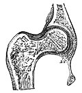
The ball-and-socket joint (or spheroid joint) is a type of synovial joint in which the ball-shaped surface of one rounded bone fits into the cup-like depression of another bone. The distal bone is capable of motion around an indefinite number of axes, which have one common center. This enables the joint to move in many directions.

Contents

An enarthrosis is a special kind of spheroidal joint in which the socket covers the sphere beyond its equator. [1]

Examples of joints

Examples of this form of articulation are found in the hip, where the round head of the femur (ball) rests in the cup-like acetabulum (socket) of the pelvis; and in the shoulder joint, where the rounded upper extremity of the humerus (ball) rests in the cup-like glenoid fossa (socket) of the shoulder blade. [2] (The shoulder also includes a sternoclavicular joint.)

Related Research Articles

<span class="mw-page-title-main">Femur</span> Thigh bone

The femur, or thigh bone is the only bone in the thigh. The thigh is the region of the lower limb between the hip and the knee. In many four-legged animals the femur is the upper bone of the hindleg.

<span class="mw-page-title-main">Acetabulum</span> Cavity where the thigh bone (femur) articulates with the pelvis

The acetabulum also called the cotyloid cavity, is a concave surface of the pelvis. The head of the femur meets with the pelvis at the acetabulum, forming the hip joint.

<span class="mw-page-title-main">Thigh</span> The part of the body between the pelvis and the knee; upper leg

In anatomy, the thigh is the area between the hip (pelvis) and the knee. Anatomically, it is part of the lower limb.

<span class="mw-page-title-main">Hip dysplasia (canine)</span> Joint abnormality in dogs

In dogs, hip dysplasia is an abnormal formation of the hip socket that, in its more severe form, can eventually cause lameness and arthritis of the joints. It is a genetic (polygenic) trait that is affected by environmental factors. It is common in many dog breeds, particularly the larger breeds, and is the most common single cause of arthritis of the hips.

<span class="mw-page-title-main">Hip</span> Anatomical region between the torso and the legs, holding the buttocks and genital region

In vertebrate anatomy, the hip, or coxa(pl.: coxae) in medical terminology, refers to either an anatomical region or a joint on the outer (lateral) side of the pelvis.

<span class="mw-page-title-main">Proximal femoral focal deficiency</span> Medical condition

Proximal femoral focal deficiency (PFFD), also known as Congenital Femoral Deficiency (CFD), is a rare, non-hereditary birth defect that affects the pelvis, particularly the hip bone, and the proximal femur. The disorder may affect one side or both, with the hip being deformed and the leg shortened.

<span class="mw-page-title-main">Hip dislocation</span> Orthopedic injury

A hip dislocation is when the thighbone (femur) separates from the hip bone (pelvis). Specifically it is when the ball–shaped head of the femur separates from its cup–shaped socket in the hip bone, known as the acetabulum. The joint of the femur and pelvis is very stable, secured by both bony and soft-tissue constraints. With that, dislocation would require significant force which typically results from significant trauma such as from a motor vehicle collision or from a fall from elevation. Hip dislocations can also occur following a hip replacement or from a developmental abnormality known as hip dysplasia.

<span class="mw-page-title-main">Ischium</span> Lower and back region of the hip bone

The ischium forms the lower and back region of the hip bone.

<span class="mw-page-title-main">Pubis (bone)</span> Most forward-facing of the three main regions making up the os coxa

In vertebrates, the pubis or pubic bone forms the lower and anterior part of each side of the hip bone. The pubis is the most forward-facing of the three bones that make up the hip bone. The left and right pubic bones are each made up of three sections, a superior ramus, inferior ramus, and a body.

<span class="mw-page-title-main">Upper extremity of femur</span>

The upper extremity, proximal extremity or superior epiphysis of the femur is the part of the femur closest to the pelvic bone and the trunk. It contains the following structures:

<span class="mw-page-title-main">Glenoid fossa</span> Part of the shoulder

The glenoid fossa of the scapula or the glenoid cavity is a bone part of the shoulder. The word glenoid is pronounced or and is from Greek: gléne, "socket", reflecting the shoulder joint's ball-and-socket form. It is a shallow, pyriform articular surface, which is located on the lateral angle of the scapula. It is directed laterally and forward and articulates with the head of the humerus; it is broader below than above and its vertical diameter is the longest.

<span class="mw-page-title-main">Femoral head ostectomy</span> Surgical removal of the head and neck of the femur

A femoral head ostectomy is a surgical operation to remove the head and neck from the femur. It is performed to alleviate pain, and is a salvage procedure, reserved for condition where pain can not be alleviated in any other way. It is common in veterinary surgery. Other names are excision arthroplasty of the femoral head and neck, Girdlestone's operation, Girdlestone procedure, and femoral head and neck ostectomy.

<span class="mw-page-title-main">Acetabular labrum</span> Ring of cartilage that surrounds the acetabulum of the hip

The acetabular labrum is a fibrocartilaginous ring which surrounds the circumference of the acetabulum of the hip, deepening the acetabulum. The labrum is attached onto the bony rim and transverse acetabular ligament. It is triangular in cross-section.

<span class="mw-page-title-main">Femoral head</span>

The femoral head is the highest part of the thigh bone (femur). It is supported by the femoral neck.

<span class="mw-page-title-main">Hip resurfacing</span>

Hip resurfacing has been developed as a surgical alternative to total hip replacement (THR). The procedure consists of placing a cap, which is hollow and shaped like a mushroom, over the head of the femur while a matching metal cup is placed in the acetabulum, replacing the articulating surfaces of the person's hip joint and removing very little bone compared to a THR. When the person moves the hip, the movement of the joint induces synovial fluid to flow between the hard metal bearing surfaces lubricating them when the components are placed in the correct position. The surgeon's level of experience with hip resurfacing is most important; therefore, the selection of the right surgeon is crucial for a successful outcome. Health-related quality of life measures are markedly improved and the person's satisfaction is favorable after hip resurfacing arthroplasty.

<span class="mw-page-title-main">Skeletal system of the horse</span> The skeletal system is made of many interconnected tissues including bone, cartilage, and tendons

The skeletal system of the horse is a skeletal system of a horse that has three major functions in the body. It protects vital organs, provides framework, and supports soft parts of the body. Horses typically have 205 bones. The pelvic limb typically contains 19 bones, while the thoracic limb contains 20 bones.

<span class="mw-page-title-main">Hip bone</span> Bone of the pelvis

The hip bone is a large flat bone, constricted in the center and expanded above and below. In some vertebrates it is composed of three parts: the ilium, ischium, and the pubis.

<span class="mw-page-title-main">Hip arthroscopy</span>

Hip arthroscopy refers to the viewing of the interior of the acetabulofemoral (hip) joint through an arthroscope and the treatment of hip pathology through a minimally invasive approach. This technique is sometimes used to help in the treatment of various joint disorders and has gained popularity because of the small incisions used and shorter recovery times when compared with conventional surgical techniques. Hip arthroscopy was not feasible until recently, new technology in both the tools used and the ability to distract the hip joint has led to a recent surge in the ability to do hip arthroscopy and the popularity of it.

<span class="mw-page-title-main">Pelvis</span> Lower torso of the human body

The pelvis is the lower part of the trunk, between the abdomen and the thighs, together with its embedded skeleton.

<span class="mw-page-title-main">Hip pain</span>

Pain in the hip is the experience of pain in the muscles or joints in the hip/ pelvic region, a condition commonly arising from any of a number of factors. Sometimes it is closely associated with lower back pain.

References

PD-icon.svgThis article incorporates text in the public domain from page 287 of the 20th edition of Gray's Anatomy (1918)

  1. Platzer, Werner (2008) Color Atlas of Human Anatomy, Volume 1, p.28
  2. And the phalanges (toes, fingers). Introduction to Joints: Synovial Joints - Ball and Socket Joints Archived 2011-07-20 at the Wayback Machine