
A melanocytic nevus is a type of melanocytic tumor that contains nevus cells. Some sources equate the term mole with "melanocytic nevus", but there are also sources that equate the term mole with any nevus form.

Nevus is a nonspecific medical term for a visible, circumscribed, chronic lesion of the skin or mucosa. The term originates from nævus, which is Latin for "birthmark"; however, a nevus can be either congenital or acquired. Common terms, including mole, birthmark, and beauty mark, are used to describe nevi, but these terms do not distinguish specific types of nevi from one another.

Melanoma is a group of serious skin cancers that arise from pigment cells (melanocytes); acral lentiginous melanoma is a kind of lentiginous skin melanoma. Acral lentiginous melanoma is the most common subtype in people with darker skins and is rare in people with lighter skin types. Acral lentiginous melanoma is observed on the palms, soles, under the nails and in the oral mucosa. It occurs on non-hair-bearing surfaces of the body, which have not necessarily been exposed to sunlight. It is also found on mucous membranes. It is the most common form of melanoma diagnosed amongst Asian and sub-Saharan African ethnic groups. The average age at diagnosis is between sixty and seventy years.

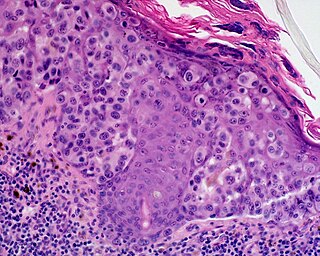
A dysplastic nevus or atypical mole is a nevus (mole) whose appearance is different from that of common moles. In 1992, the NIH recommended that the term "dysplastic nevus" be avoided in favor of the term "atypical mole". An atypical mole may also be referred to as an atypical melanocytic nevus, atypical nevus, B-K mole, Clark's nevus, dysplastic melanocytic nevus, or nevus with architectural disorder.

Dysplastic nevus syndrome, also known as familial atypical multiple mole–melanoma (FAMMM) syndrome, is an inherited cutaneous condition described in certain families, and characterized by unusual nevi and multiple inherited melanomas. First described in 1820, the condition is inherited in an autosomal dominant pattern, and caused by mutations in the CDKN2A gene. In addition to melanoma, individuals with the condition are at increased risk for pancreatic cancer.
Ocular melanosis (OM) is a blue-gray and/or brown lesion of the conjunctiva that can be separated into benign conjunctival epithelial melanosis (BCEM) and primary acquired melanosis (PAM), of which the latter is considered a risk factor for uveal melanoma. The disease is caused by an increase of melanocytes in the iris, choroid, and surrounding structures. Overproduction of pigment by these cells can block the trabecular meshwork through which fluid drains from the eye. The increased fluid in the eye leads to increased pressure, which can lead to glaucoma. In humans, this is sometimes known as pigment dispersion syndrome.
Lentigo maligna melanoma is a melanoma that has evolved from a lentigo maligna, as seen as a lentigo maligna with melanoma cells invading below the boundaries of the epidermis. They are usually found on chronically sun damaged skin such as the face and the forearms of the elderly.

The congenital melanocytic nevus is a type of melanocytic nevus found in infants at birth. This type of birthmark occurs in an estimated 1% of infants worldwide; it is located in the area of the head and neck 15% of the time.

Nevus of Ota is a hyperpigmentation that occurs on the face, most often appearing on the white of the eye. It also occurs on the forehead, nose, cheek, periorbital region, and temple.

Blue nevus is a type of melanocytic nevus. The blue colour is caused by the pigment being deeper in the skin than in ordinary nevi. In principle they are harmless but they can sometimes be mimicked by malignant lesions, i.e. some melanomas can look like a blue nevus.
Roy G. Geronemus is an American dermatologist in the field of laser treatment of skin disorders.
Halo nevus is a mole that is surrounded by a depigmented ring or 'halo'.
A Spitz nevus is a benign skin lesion. A type of melanocytic nevus, it affects the epidermis and dermis.
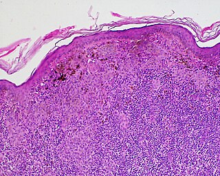
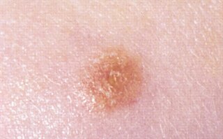
A benign melanocytic nevus is a cutaneous condition characterised by well-circumscribed, pigmented, round or ovoid lesions, generally measuring from 2 to 6 mm in diameter. A benign melanocytic nevus may feature hair or pigmentation as well.
Pseudomelanoma is a cutaneous condition in which melanotic skin lesions clinically resemble a superficial spreading melanoma at the site of a recent shave removal of a melanocytic nevus.
Oral pigmentation is asymptomatic and does not usually cause any alteration to the texture or thickness of the affected area. The colour can be uniform or speckled and can appear solitary or as multiple lesions. Depending on the site, depth, and quantity of pigment, the appearance can vary considerably.

An acral nevus is a cutaneous condition of the palms, soles, fingers, or toes, characterized by a skin lesion that is usually macular or only slightly elevated, and may display a uniform brown or dark brown color, often with linear striations.

Nevus depigmentosus is a loss of pigment in the skin which can be easily differentiated from vitiligo. Although age factor has not much involvement in the nevus depigmentosus but in about 19% of the cases these are noted at birth. Their size may however grow in proportion to growth of the body. The distribution is also fairly stable and are nonprogressive hypopigmented patches. The exact cause of nevus depigmentosus is still not clearly understood. A sporadic defect in the embryonic development has been suggested to be a causative factor. It has been described as "localised albinism", though this is incorrect.

Neurocutaneous melanosis is a congenital disorder characterized by the presence of congenital melanocytic nevi on the skin and melanocytic tumors in the leptomeninges of the central nervous system. These lesions may occur in the amygdala, cerebellum, cerebrum, pons and spinal cord of patients. Although typically asymptomatic, malignancy occurs in the form of leptomeningeal melanoma in over half of patients. Regardless of the presence of malignancy, patients with symptomatic neurocutaneous melanosis generally have a poor prognosis with few treatment options. The pathogenesis of neurocutaneous melanosis is believed to be related to the abnormal postzygotic development of melanoblasts and mutations of the NRAS gene.
