
The rib cage is an endoskeletal enclosure in the thorax of most vertebrate animals that comprises the ribs, vertebral column and sternum, which protects vital organs such as the heart, lungs and great vessels. The circumferential enclosure formed by left and right rib cages, together known as the thoracic cage, is a semi-rigid bony and cartilaginous structure which surrounds the thoracic cavity and supports the shoulder girdles to form the core part of the axial skeleton.

A joint or articulation is the connection made between bones, ossicles, or other hard structures in the body which link an animal's skeletal system into a functional whole. They are constructed to allow for different degrees and types of movement. Some joints, such as the knee, elbow, and shoulder, are self-lubricating, almost frictionless, and are able to withstand compression and maintain heavy loads while still executing smooth and precise movements. Other joints such as sutures between the bones of the skull permit very little movement in order to protect the brain and the sense organs. The connection between a tooth and the jawbone is also called a joint, and is described as a fibrous joint known as a gomphosis. Joints are classified both structurally and functionally.

In anatomy, the extrapyramidal system is a part of the motor system network causing involuntary actions. The system is called extrapyramidal to distinguish it from the tracts of the motor cortex that reach their targets by traveling through the pyramids of the medulla. The pyramidal tracts may directly innervate motor neurons of the spinal cord or brainstem, whereas the extrapyramidal system centers on the modulation and regulation of anterior (ventral) horn cells.

The axial skeleton is the part of the skeleton that consists of the bones of the head and trunk of a vertebrate. In the human skeleton, it consists of 80 bones and is composed of six parts; the skull, also the ossicles of the middle ear, the hyoid bone, the rib cage, sternum and the vertebral column. The axial skeleton together with the appendicular skeleton form the complete skeleton. Another definition of axial skeleton is the bones including the vertebrae, sacrum, coccyx, skull, ribs, and sternum.

The four classical muscles of mastication elevate the mandible and move it forward/backward and laterally, facilitating biting and chewing. Other muscles are responsible for opening the jaw, namely the geniohyoid, mylohyoid, and digastric muscles.
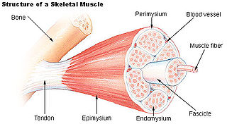
Epimysium is the fibrous tissue envelope that surrounds skeletal muscle. It is a layer of dense irregular connective tissue which ensheaths the entire muscle and protects muscles from friction against other muscles and bones. It also allows a muscle to contract and move powerfully while maintaining its structural integrity.
A synovial bursa, usually simply bursa, is a small fluid-filled sac lined by synovial membrane with an inner capillary layer of viscous synovial fluid. It provides a cushion between bones and tendons and/or muscles around a joint. This helps to reduce friction between the bones and allows free movement. Bursae are found around most major joints of the body.

A synarthrosis is a type of joint which allows no movement under normal conditions. Sutures and gomphoses are both synarthroses. Joints which allow more movement are called amphiarthroses or diarthroses. Syndesmoses are considered to be amphiarthrotic, because they allow a small amount of movement.

A hinge joint is a bone joint in which the articular surfaces are molded to each other in such a manner as to permit motion only in one plane. According to one classification system they are said to be uniaxial. The direction which the distal bone takes in this motion is seldom in the same plane as that of the axis of the proximal bone; there is usually a certain amount of deviation from the straight line during flexion.

Motion, the process of movement, is described using specific anatomical terms. Motion includes movement of organs, joints, limbs, and specific sections of the body. The terminology used describes this motion according to its direction relative to the anatomical position of the body parts involved. Anatomists and others use a unified set of terms to describe most of the movements, although other, more specialized terms are necessary for describing unique movements such as those of the hands, feet, and eyes.

Amphiarthrosis is a type of continuous, slightly movable joint. Most amphiarthroses are held together by cartilage, as a result of which limited movements between the bones is made possible. An example is the joints of the vertebral column only allow for small movements between adjacent vertebrae, but when added together, these movements provide the flexibility that allows the body to twist, or bend to the front, back, or side.
A condyloid joint is an ovoid articular surface, or condyle that is received into an elliptical cavity. This permits movement in two planes, allowing flexion, extension, adduction, abduction, and circumduction.
A saddle joint is a type of synovial joint in which the opposing surfaces are reciprocally concave and convex. It is found in the thumb, the thorax, the middle ear, and the heel.

A plane joint is a synovial joint which, under physiological conditions, allows only gliding movement.
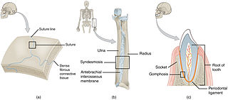
In anatomy, fibrous joints are joints connected by fibrous tissue, consisting mainly of collagen. These are fixed joints where bones are united by a layer of white fibrous tissue of varying thickness. In the skull, the joints between the bones are called sutures. Such immovable joints are also referred to as synarthroses.

Cartilaginous joints are connected entirely by cartilage. Cartilaginous joints allow more movement between bones than a fibrous joint but less than the highly mobile synovial joint. Cartilaginous joints also forms the growth regions of immature long bones and the intervertebral discs of the spinal column.
A tendon sheath is a layer of synovial membrane around a tendon. It permits the tendon to stretch and not adhere to the surrounding fascia. It contains a lubricating fluid that allows for smooth motions of the tendon during muscle contraction and joint movements.
The term gaze is frequently used in physiology to describe coordinated motion of the eyes and neck. The lateral gaze is controlled by the paramedian pontine reticular formation (PPRF). The vertical gaze is controlled by the rostral interstitial nucleus of medial longitudinal fasciculus and the interstitial nucleus of Cajal.

Anatomical terminology is a form of scientific terminology used by anatomists, zoologists, and health professionals such as doctors, physicians, and pharmacists.

A multiaxial joint is a diarthrosis that allows for several directions of movement.