Bioreporter

Last updated
Anatomy of a bioreporter organism. Upon exposure to a specific analyte, the promoter/reporter gene complex is transcribed into messenger RNA (mRNA) and then translated into a reporter protein that is ultimately responsible for generating a signal. Bioreporter.jpg
Anatomy of a bioreporter organism. Upon exposure to a specific analyte, the promoter/reporter gene complex is transcribed into messenger RNA (mRNA) and then translated into a reporter protein that is ultimately responsible for generating a signal.

Bioreporters are intact, living microbial cells that have been genetically engineered to produce a measurable signal in response to a specific chemical or physical agent in their environment. Bioreporters contain two essential genetic elements, a promoter gene and a reporter gene. The promoter gene is turned on (transcribed) when the target agent is present in the cell’s environment. The promoter gene in a normal bacterial cell is linked to other genes that are then likewise transcribed and then translated into proteins that help the cell in either combating or adapting to the agent to which it has been exposed. In the case of a bioreporter, these genes, or portions thereof, have been removed and replaced with a reporter gene. As a result, turning on the promoter gene also turns on the reporter gene, leading to the production of reporter proteins that output a detectable signal. The presence of a signal indicates that the bioreporter has sensed a particular agent in its environment.

Contents

Originally developed for fundamental analysis of factors affecting gene expression, bioreporters were early on applied for the detection of environmental contaminants [1] and have since evolved into fields as diverse as medical diagnostics, precision agriculture, food safety assurance, process monitoring and control, and bio-microelectronic computing. Their versatility stems from the fact that there exist a large number of reporter gene systems that are capable of generating a variety of signals. Additionally, reporter genes can be genetically inserted into bacterial, yeast, plant, and mammalian cells, thereby providing considerable functionality over a wide range of host vectors.

Reporter gene systems

Several types of reporter genes are available for use in the construction of bioreporter organisms, and the signals they generate can usually be categorized as either colorimetric, fluorescent, luminescent, chemiluminescent or electrochemical. Although each functions differently, their end product always remains the same – a measurable signal that is proportional to the concentration of the unique chemical or physical agent to which they have been exposed. In some instances, the signal only occurs when a secondary substrate is added to the bioassay (luxAB, Luc, and aequorin). For other bioreporters, the signal must be activated by an external light source (GFP and UMT), and for a select few bioreporters, the signal is completely self-induced, with no exogenous substrate or external activation being required (luxCDABE). The following sections outline in brief some of the reporter gene systems available and their existing applications.

Bacterial luciferase (Lux)

Bioluminescence emitted from colonies of microbial cells containing the genes for bacterial luciferase. Biolumplate.jpg
Bioluminescence emitted from colonies of microbial cells containing the genes for bacterial luciferase.

Luciferase is a generic name for an enzyme that catalyzes a light-emitting reaction. Luciferases can be found in bacteria, algae, fungi, jellyfish, insects, shrimp, and squid, and the resulting light that these organisms produce is termed bioluminescence. In bacteria, the genes responsible for the light-emitting reaction (the lux genes) have been isolated and used extensively in the construction of bioreporters that emit a blue-green light with a maximum intensity at 490 nm. [2] Three variants of lux are available, one that functions at < 30°C, another at < 37°C, and a third at < 45°C. The lux genetic system consists of five genes, luxA, luxB, luxC, luxD, and luxE. Depending on the combination of these genes used, several different types of bioluminescent bioreporters can be constructed.

luxAB Bioreporters

luxAB bioreporters contain only the luxA and luxB genes, which together are responsible for generating the light signal. However, to fully complete the light-emitting reaction, a substrate must be supplied to the cell. Typically, this occurs through the addition of the chemical decanal at some point during the bioassay procedure. Numerous luxAB bioreporters have been constructed within bacterial, yeast, insect, nematode, plant, and mammalian cell systems.

luxCDABE Bioreporters

Instead of containing only the luxA and luxB genes, bioreporters can contain all five genes of the lux cassette, thereby allowing for a completely independent light generating system that requires no extraneous additions of substrate nor any excitation by an external light source. So in this bioassay, the bioreporter is simply exposed to a target analyte and a quantitative increase in bioluminescence results, often within less than one hour. Due to their rapidity and ease of use, along with the ability to perform the bioassay repetitively in real time and on-line, makes luxCDABE bioreporters extremely attractive. Consequently, they have been incorporated into a diverse array of detection methodologies ranging from the sensing of environmental contaminants to the real-time monitoring of pathogen infections in living mice.

Nonspecific lux Bioreporters

Nonspecific lux bioreporters are typically used for the detection of chemical toxins. They are usually designed to continuously bioluminesce. Upon exposure to a chemical toxin, either the cell dies or its metabolic activity is retarded, leading to a decrease in bioluminescent light levels. Their most familiar application is in the Microtox assay where, following a short exposure to several concentrations of the sample, the decreased bioluminescence can be correlated to relative levels of toxicity. [3] [4]

Firefly luciferase (Luc)

Firefly luciferase catalyzes a reaction that produces visible light in the 550 to 575 nm range. A click-beetle luciferase is also available that produces light at a peak closer to 595 nm. Both luciferases require the addition of an exogenous substrate (luciferin) for the light reaction to occur. Numerous luc-based bioreporters have been constructed for the detection of a wide array of inorganic and organic compounds of environmental concern. The most promising applications, however, probably rely on introducing the genetic code of the firefly luciferase into other eukaryotic cells and tissues. [5]

Medical diagnostics

Insertion of the luc genes into a human cervical carcinoma cell line (HeLa) illustrated that tumor-cell clearance could be visualized within a living mouse by simply scanning with a charge-coupled device camera, allowing for chemotherapy treatment to rapidly be monitored on-line and in real-time. [6] In another example, the luc genes were inserted into human breast cancer cell lines to develop a bioassay for the detection and measurement of substances with potential estrogenic and antiestrogenic activity. [7]

Research on gene regulation

Particular promoters can be placed upstream of the luc gene, that is, the luc sequence can be fused to the promoter sequence at DNA level. If such construct is not too large in size, it can simply be introduced into eukaryotic cells using plasmids. This approach is widely used to study the activity of a given promoter in a given cell/tissue type, since the amount of light produced by the luciferase is directly proportional to the promoter activity. [8] In addition to studying the promoters, firefly luciferase assays offer the option of studying transcriptional activators: in these experiments typically the GAL4/UAS_system is used and its Gal4 upstream activating DNA sequence (UAS) is placed upstream the luc gene while the different activators or the different variants/fragments of the same activator are fused to the GAL4 DNA binding module at protein level. This way the transcriptional activity of the different GAL4 fusion proteins can be directly compared using light as a readout. [9]

Aequorin

Aequorin is a photoprotein isolated from the bioluminescent jellyfish Aequorea victoria . Upon addition of calcium ions (Ca2+) and coelenterazine, a reaction occurs whose result is the generation of blue light in the 460 to 470 nm range. Aequorin has been incorporated into human B cell lines for the detection of pathogenic bacteria and viruses in what is referred to as the Cell CANARY assay (Cellular Analysis and Notification of Antigen Risks and Yields). [10] The B cells are genetically engineered to produce aequorin. Upon exposure to antigens of different pathogens, the recombinant B cells emit light as a result of activation of an intracellular signaling cascade that releases calcium ions inside the cell.

Green fluorescent protein (GFP)

Green fluorescent protein (GFP) is also a photoprotein isolated and cloned from the jellyfish Aequorea victoria . [11] Variants have also been isolated from the sea pansy Renilla reniformis . GFP, like aequorin, produces a blue fluorescent signal, but without the required addition of an exogenous substrate. All that is required is an ultraviolet light source to activate the fluorescent properties of the photoprotein. This ability to autofluoresce makes GFP highly desirable in biosensing assays since it can be used on-line and in to monitor intact, living cells. Additionally, the ability to alter GFP to produce light emissions besides blue (i.e., cyan, red, and yellow) allows it to be used as a multianalyte detector. Consequently, GFP has been used extensively in bioreporter constructs within bacterial, yeast, nematode, plant, and mammalian hosts.

Uroporphyrinogen (urogen) III methyltransferase (UMT)

Uroporphyrinogen (urogen) III methyltransferase (UMT) catalyzes a reaction that yields two fluorescent products which produce a red-orange fluorescence in the 590 to 770 nm range when illuminated with ultraviolet light. [12] So as with GFP, no addition of exogenous substrates is required. UMT has been used as a bioreporter for the selection of recombinant plasmids, as a marker for gene transcription in bacterial, yeast, and mammalian cells, and for the detection of toxic salts such as arsenite and antimonite.

Related Research Articles

<span class="mw-page-title-main">Green fluorescent protein</span> Protein that exhibits bright green fluorescence when exposed to ultraviolet light

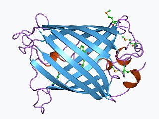
The green fluorescent protein (GFP) is a protein that exhibits green fluorescence when exposed to light in the blue to ultraviolet range. The label GFP traditionally refers to the protein first isolated from the jellyfish Aequorea victoria and is sometimes called avGFP. However, GFPs have been found in other organisms including corals, sea anemones, zoanithids, copepods and lancelets.

<span class="mw-page-title-main">Bioluminescence</span> Emission of light by a living organism

Bioluminescence is the production and emission of light by living organisms. It is a form of chemiluminescence. Bioluminescence occurs widely in marine vertebrates and invertebrates, as well as in some fungi, microorganisms including some bioluminescent bacteria, and terrestrial arthropods such as fireflies. In some animals, the light is bacteriogenic, produced by symbiotic bacteria such as those from the genus Vibrio; in others, it is autogenic, produced by the animals themselves.

<span class="mw-page-title-main">Chemiluminescence</span> Emission of light as a result of a chemical reaction

Chemiluminescence is the emission of light (luminescence) as the result of a chemical reaction, i.e. a chemical reaction results in a flash or glow of light. A standard example of chemiluminescence in the laboratory setting is the luminol test. Here, blood is indicated by luminescence upon contact with iron in hemoglobin. When chemiluminescence takes place in living organisms, the phenomenon is called bioluminescence. A light stick emits light by chemiluminescence.

<span class="mw-page-title-main">Luciferase</span> Enzyme family

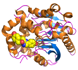
Luciferase is a generic term for the class of oxidative enzymes that produce bioluminescence, and is usually distinguished from a photoprotein. The name was first used by Raphaël Dubois who invented the words luciferin and luciferase, for the substrate and enzyme, respectively. Both words are derived from the Latin word lucifer, meaning "lightbearer", which in turn is derived from the Latin words for "light" (lux) and "to bring or carry" (ferre).

<span class="mw-page-title-main">Reporter gene</span> Technique in molecular biology

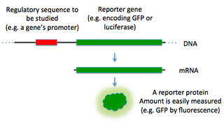
In molecular biology, a reporter gene is a gene that researchers attach to a regulatory sequence of another gene of interest in bacteria, cell culture, animals or plants. Such genes are called reporters because the characteristics they confer on organisms expressing them are easily identified and measured, or because they are selectable markers. Reporter genes are often used as an indication of whether a certain gene has been taken up by or expressed in the cell or organism population.

<i>Aliivibrio fischeri</i> Species of bacterium

Aliivibrio fischeri is a Gram-negative, rod-shaped bacterium found globally in marine environments. This species has bioluminescent properties, and is found predominantly in symbiosis with various marine animals, such as the Hawaiian bobtail squid. It is heterotrophic, oxidase-positive, and motile by means of a single polar flagella. Free-living A. fischeri cells survive on decaying organic matter. The bacterium is a key research organism for examination of microbial bioluminescence, quorum sensing, and bacterial-animal symbiosis. It is named after Bernhard Fischer, a German microbiologist.

pGLO

The pGLO plasmid is an engineered plasmid used in biotechnology as a vector for creating genetically modified organisms. The plasmid contains several reporter genes, most notably the green fluorescent protein (GFP) and the ampicillin resistance gene. GFP was isolated from the jelly fish Aequorea victoria. Because it shares a bidirectional promoter with a gene for metabolizing arabinose, the GFP gene is expressed in the presence of arabinose, which makes the transgenic organism express its fluorescence under UV light. GFP can be induced in bacteria containing the pGLO plasmid by growing them on +arabinose plates. pGLO is made by Bio-Rad Laboratories.

<span class="mw-page-title-main">Two-hybrid screening</span> Molecular biology technique

Two-hybrid screening is a molecular biology technique used to discover protein–protein interactions (PPIs) and protein–DNA interactions by testing for physical interactions between two proteins or a single protein and a DNA molecule, respectively.

<span class="mw-page-title-main">Aequorin</span> Calcium-activated photoprotein

Aequorin is a calcium-activated photoprotein isolated from the hydrozoan Aequorea victoria. Its bioluminescence was studied decades before the protein was isolated from the animal by Osamu Shimomura in 1962. In the animal, the protein occurs together with the green fluorescent protein to produce green light by resonant energy transfer, while aequorin by itself generates blue light.

<span class="mw-page-title-main">Bimolecular fluorescence complementation</span>

Bimolecular fluorescence complementation is a technology typically used to validate protein interactions. It is based on the association of fluorescent protein fragments that are attached to components of the same macromolecular complex. Proteins that are postulated to interact are fused to unfolded complementary fragments of a fluorescent reporter protein and expressed in live cells. Interaction of these proteins will bring the fluorescent fragments within proximity, allowing the reporter protein to reform in its native three-dimensional structure and emit its fluorescent signal. This fluorescent signal can be detected and located within the cell using an inverted fluorescence microscope that allows imaging of fluorescence in cells. In addition, the intensity of the fluorescence emitted is proportional to the strength of the interaction, with stronger levels of fluorescence indicating close or direct interactions and lower fluorescence levels suggesting interaction within a complex. Therefore, through the visualisation and analysis of the intensity and distribution of fluorescence in these cells, one can identify both the location and interaction partners of proteins of interest.

Within the field of molecular biology, a protein-fragment complementation assay, or PCA, is a method for the identification and quantification of protein–protein interactions. In the PCA, the proteins of interest are each covalently linked to fragments of a third protein. Interaction between the bait and the prey proteins brings the fragments of the reporter protein in close proximity to allow them to form a functional reporter protein whose activity can be measured. This principle can be applied to many different reporter proteins and is also the basis for the yeast two-hybrid system, an archetypical PCA assay.

<span class="mw-page-title-main">GUS reporter system</span>

The GUS reporter system is a reporter gene system, particularly useful in plant molecular biology and microbiology. Several kinds of GUS reporter gene assay are available, depending on the substrate used. The term GUS staining refers to the most common of these, a histochemical technique.

<span class="mw-page-title-main">Bioluminescence imaging</span>

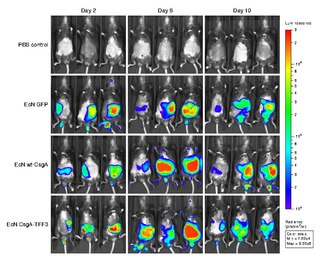
Bioluminescence imaging (BLI) is a technology developed over the past decades (1990's and onward). that allows for the noninvasive study of ongoing biological processes Recently, bioluminescence tomography (BLT) has become possible and several systems have become commercially available. In 2011, PerkinElmer acquired one of the most popular lines of optical imaging systems with bioluminescence from Caliper Life Sciences.

<span class="mw-page-title-main">GAL4/UAS system</span> Biochemical method

The GAL4-UAS system is a biochemical method used to study gene expression and function in organisms such as the fruit fly. It is based on the finding by Hitoshi Kakidani and Mark Ptashne, and Nicholas Webster and Pierre Chambon in 1988 that Gal4 binding to UAS sequences activates gene expression. The method was introduced into flies by Andrea Brand and Norbert Perrimon in 1993 and is considered a powerful technique for studying the expression of genes. The system has two parts: the Gal4 gene, encoding the yeast transcription activator protein Gal4, and the UAS, an enhancer to which GAL4 specifically binds to activate gene transcription.

<span class="mw-page-title-main">Renilla-luciferin 2-monooxygenase</span>

Renilla-luciferin 2-monooxygenase, Renilla luciferase, or RLuc, is a bioluminescent enzyme found in Renilla reniformis, belonging to a group of coelenterazine luciferases. Of this group of enzymes, the luciferase from Renilla reniformis has been the most extensively studied, and due to its bioluminescence requiring only molecular oxygen, has a wide range of applications, with uses as a reporter gene probe in cell culture, in vivo imaging, and various other areas of biological research. Recently, chimeras of RLuc have been developed and demonstrated to be the brightest luminescent proteins to date, and have proved effective in both noninvasive single-cell and whole body imaging.

Autoinducers are signaling molecules that are produced in response to changes in cell-population density. As the density of quorum sensing bacterial cells increases so does the concentration of the autoinducer. Detection of signal molecules by bacteria acts as stimulation which leads to altered gene expression once the minimal threshold is reached. Quorum sensing is a phenomenon that allows both Gram-negative and Gram-positive bacteria to sense one another and to regulate a wide variety of physiological activities. Such activities include symbiosis, virulence, motility, antibiotic production, and biofilm formation. Autoinducers come in a number of different forms depending on the species, but the effect that they have is similar in many cases. Autoinducers allow bacteria to communicate both within and between different species. This communication alters gene expression and allows bacteria to mount coordinated responses to their environments, in a manner that is comparable to behavior and signaling in higher organisms. Not surprisingly, it has been suggested that quorum sensing may have been an important evolutionary milestone that ultimately gave rise to multicellular life forms.

Douglas C. Prasher is an American molecular biologist. He is known for his work to clone and sequence the genes for the photoprotein aequorin and green fluorescent protein (GFP) and for his proposal to use GFP as a tracer molecule. He communicated his pioneering work to Martin Chalfie and Roger Y. Tsien, but by 1991 he was unable to obtain further research funding, and left academia. Eventually, he had to abandon science. Chalfie and Tsien were awarded the 2008 Nobel Prize in Chemistry for work that they publicly acknowledged was substantially based on Prasher's work; through their efforts and those of others, he returned to scientific research in June 2010.

Photoproteins are a type of enzyme produced by bioluminescent organisms. They add to the function of the luciferins whose usual light-producing reaction is catalyzed by the enzyme luciferase.

<span class="mw-page-title-main">John Woodland Hastings</span>

John Woodland "Woody" Hastings, was a leader in the field of photobiology, especially bioluminescence, and was one of the founders of the field of circadian biology. He was the Paul C. Mangelsdorf Professor of Natural Sciences and Professor of Molecular and Cellular Biology at Harvard University. He published over 400 papers and co-edited three books.

<span class="mw-page-title-main">Bioluminescent bacteria</span>

Bioluminescent bacteria are light-producing bacteria that are predominantly present in sea water, marine sediments, the surface of decomposing fish and in the gut of marine animals. While not as common, bacterial bioluminescence is also found in terrestrial and freshwater bacteria. These bacteria may be free living or in symbiosis with animals such as the Hawaiian Bobtail squid or terrestrial nematodes. The host organisms provide these bacteria a safe home and sufficient nutrition. In exchange, the hosts use the light produced by the bacteria for camouflage, prey and/or mate attraction. Bioluminescent bacteria have evolved symbiotic relationships with other organisms in which both participants benefit close to equally. Another possible reason bacteria use luminescence reaction is for quorum sensing, an ability to regulate gene expression in response to bacterial cell density.

References

  1. King, J.M.H. et al. (1990) Rapid, sensitive bioluminescence reporter technology for naphthalene exposure and biodegradation. Science 249, 778–781.http://www.sciencemag.org/cgi/content/abstract/249/4970/778?ck=nck
  2. Meighen, E.A. (1994) Genetics of bacterial bioluminescence. Annu. Rev. Genet. 28, 117-139. http://arjournals.annualreviews.org/doi/abs/10.1146/annurev.ge.28.120194.001001
  3. Hermens, J. et al. (1985) Quantitative structure-activity relationships and mixture toxicity of organic chemicals in Photobacterium phosphoreum: the Microtox test. Ecotoxicol. Environ. Saf. 9, 17-25. https://doi.org/10.1007%2Fs11434-006-2168-z
  4. Taghavi K, Fath PM, Hosseinkhani S, Mirzaei M, Behrooj H, Kiani A, Abedini A, Razavi F. Construction and genetic improvement of copper bioreporter Escherichia Coli. Biomedical and Biotechnology Research Journal (BBRJ). 2018 Jan 1;2(1):26-30. https://journals.lww.com/BBRJ/fulltext/2018/02010/Construction_and_Genetic_Improvement_of_Copper.5.aspx
  5. Contag, C.H. et al. (2000) Use of reporter genes for optical measurements of neoplastic disease in vivo. Neoplasia 2 (1-2), 41-52. http://neoreviews.aappublications.org/cgi/content/extract/1/12/e225
  6. Legler, J. et al. (1999) Development of a stably transfected estrogen receptor-mediated luciferase reporter gene assay in the human T47D breast cancer cell line. Toxicol. Sci. 48 (1), 55-66. http://toxsci.oxfordjournals.org/cgi/content/abstract/48/1/55
  7. Kovács KA, Steinmann M, Halfon O, Magistretti PJ, Cardinaux JR (September 2006). "C/EBPbeta couples dopamine signalling to substance P precursor gene expression in striatal neurones". Journal of Neurochemistry. 98 (5): 1390–9. doi:10.1111/j.1471-4159.2006.03957.x. PMID   16771829. S2CID   36225447.
  8. Kovács KA, Steinmann M, Halfon O, Magistretti PJ, Cardinaux JR (November 2015). "Complex regulation of CREB-binding protein by homeodomain-interacting protein kinase 2" (PDF). Cellular Signalling. 27 (11): 2252–60. doi:10.1016/j.cellsig.2015.08.001. PMID   26247811.
  9. Rider, T. et al. (1999) In NASA/NCI Biomedical Imaging Symposium.
  10. Misteli, T. and Spector, D.L. (1997) Application of the green fluorescent protein in cell biology and biotechnology. Nat. Biotechnol. 15, 961-964.
  11. Sattler, I. et al. (1995) Cloning, sequencing, and expression of the uroporphyrinogen-III methyltransferase cobA gene of Propionibacterium freudenreichii (shermanii). J. Bacteriol. 177, 1564–1569.http://jb.asm.org/cgi/content/abstract/177/6/1564