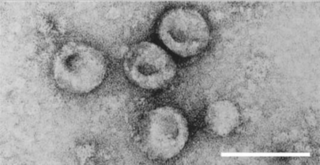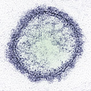
Bluetongue disease is a noncontagious, insect-borne, viral disease of ruminants, mainly sheep and less frequently cattle, yaks, goats, buffalo, deer, dromedaries, and antelope. It is caused by Bluetongue virus (BTV). The virus is transmitted by the midges Culicoides imicola, Culicoides variipennis, and other culicoids.

Rift Valley fever (RVF) is a viral disease of humans and livestock that can cause mild to severe symptoms. The mild symptoms may include: fever, muscle pains, and headaches which often last for up to a week. The severe symptoms may include: loss of sight beginning three weeks after the infection, infections of the brain causing severe headaches and confusion, and bleeding together with liver problems which may occur within the first few days. Those who have bleeding have a chance of death as high as 50%.

Orf is a farmyard pox, a type of zoonosis. It causes small pustules in the skin of primarily sheep and goats, but can also occur on the hands of humans. A pale halo forms around a red centre. It may persist for several weeks before crusting and then either resolves or leaves a hard lump. There is usually only one lesion, but there may be many, and they are not painful. Sometimes there are swollen lymph glands.

Pestivirus is a genus of viruses, in the family Flaviviridae. Viruses in the genus Pestivirus infect mammals, including members of the family Bovidae and the family Suidae. There are 11 species in this genus. Diseases associated with this genus include: hemorrhagic syndromes, abortion, and fatal mucosal disease.
SMEDI is a reproductive disease of swine caused by Porcine parvovirus (PPV) and Porcine enterovirus. The term SMEDI usually indicates Porcine enterovirus, but it also can indicate Porcine parvovirus, which is a more important cause of the syndrome. SMEDI also causes abortion, neonatal death, and decreased male fertility.
Porcine parvovirus (PPV), a virus in the species Ungulate protoparvovirus 1 of genus Protoparvovirus in the virus family Parvoviridae, causes reproductive failure of swine characterized by embryonic and fetal infection and death, usually in the absence of outward maternal clinical signs. The disease develops mainly when seronegative dams are exposed oronasally to the virus anytime during about the first half of gestation, and conceptuses are subsequently infected transplacentally before they become immunocompetent. There is no definitive evidence that infection of swine other than during gestation is of any clinical or economic significance. The virus is ubiquitous among swine throughout the world and is enzootic in most herds that have been tested. Diagnostic surveys have indicated that PPV is the major infectious cause of embryonic and fetal death. In addition to its direct causal role in reproductive failure, PPV can potentiate the effects of porcine circovirus type II (PCV2) infection in the clinical course of postweaning multisystemic wasting syndrome (PMWS).
Bovine alphaherpesvirus 1 (BoHV-1) is a virus of the family Herpesviridae and the subfamily Alphaherpesvirinae, known to cause several diseases worldwide in cattle, including rhinotracheitis, vaginitis, balanoposthitis, abortion, conjunctivitis, and enteritis. BoHV-1 is also a contributing factor in shipping fever, also known as bovine respiratory disease (BRD). It is spread horizontally through sexual contact, artificial insemination, and aerosol transmission and it may also be transmitted vertically across the placenta. BoHV-1 can cause both clinical and subclinical infections, depending on the virulence of the strain. Although these symptoms are mainly non-life-threatening it is an economically important disease as infection may cause a drop in production and affect trade restrictions. Like other herpesviruses, BoHV-1 causes a lifelong latent infection and sporadic shedding of the virus. The sciatic nerve and trigeminal nerve are the sites of latency. A reactivated latent carrier is normally the source of infection in a herd. The clinical signs displayed are dependent on the virulence of the strain. There is a vaccine available which reduces the severity and incidence of disease. Some countries in Europe have successfully eradicated the disease by applying a strict culling policy.

Feline viral rhinotracheitis (FVR) is an upper respiratory or pulmonary infection of cats caused by Felid alphaherpesvirus 1 (FeHV-1), of the family Herpesviridae. It is also commonly referred to as feline influenza, feline coryza, and feline pneumonia but, as these terms describe other very distinct collections of respiratory symptoms, they are misnomers for the condition. Viral respiratory diseases in cats can be serious, especially in catteries and kennels. Causing one-half of the respiratory diseases in cats, FVR is the most important of these diseases and is found worldwide. The other important cause of feline respiratory disease is feline calicivirus.

Feline calicivirus (FCV) is a virus of the family Caliciviridae that causes disease in cats. It is one of the two important viral causes of respiratory infection in cats, the other being Felid alphaherpesvirus 1. FCV can be isolated from about 50% of cats with upper respiratory infections. Cheetahs are the other species of the family Felidae known to become infected naturally.

Bovine malignant catarrhal fever (BMCF) is a fatal lymphoproliferative disease caused by a group of ruminant gamma herpes viruses including Alcelaphine gammaherpesvirus 1 (AlHV-1) and Ovine gammaherpesvirus 2 (OvHV-2) These viruses cause unapparent infection in their reservoir hosts, but are usually fatal in cattle and other ungulates such as deer, antelope, and buffalo. In Southern Africa the disease is known as snotsiekte, from the Afrikaans.

Neospora is a single celled parasite of livestock and companion animals. It was not discovered until 1984 in Norway, where it was found in dogs. Neosporosis, the disease that affects cattle and companion animals, has a worldwide distribution. Neosporosis causes abortions in cattle and paralysis in companion animals. It is highly transmissible and some herds can have up to a 90% prevalence. Up to 33% of pregnancies can result in aborted fetuses on one dairy farm. In many countries this organism is the main cause of abortion in cattle. Neosporosis is now considered as a major cause of abortion in cattle worldwide. Many reliable diagnostic tests are commercially available. Neospora caninum does not appear to be infectious to humans. In dogs, Neospora caninum can cause neurological signs, especially in congenitally infected puppies, where it can form cysts in the central nervous system.

Sheeppox is a highly contagious disease of sheep caused by a poxvirus different from the benign orf. This virus is in the family Poxviridae and genus Capripoxvirus. Sheeppox virus (SPV) is the most severe of all the animal pox diseases and can result in some of the most significant economic consequences due to poor wool and leather quality.

Bovine viral diarrhea (BVD), bovine viral diarrhoea or mucosal disease, previously referred to as bovine virus diarrhea (BVD), is an economically significant disease of cattle that is found in the majority of countries throughout the world. Worldwide reviews of the economically assessed production losses and intervention programs incurred by BVD infection have been published. The causative agent, bovine viral diarrhea virus (BVDV), is a member of the genus Pestivirus of the family Flaviviridae.
Visna-maedi virus from the genus Lentivirus and subfamily Orthoretrovirinae, is a retrovirus that causes encephalitis and chronic pneumonitis in sheep. It is known as visna when found in the brain, and maedi when infecting the lungs. Lifelong, persistent infections in sheep occur in the lungs, lymph nodes, spleen, joints, central nervous system, and mammary glands; The condition is sometimes known as ovine progressive pneumonia (OPP), particularly in the United States, or Montana sheep disease. White blood cells of the monocyte/macrophage lineage are the main target of the virus.

Foot-and-mouth disease (FMD) or hoof-and-mouth disease (HMD) is an infectious and sometimes fatal viral disease that affects cloven-hoofed animals, including domestic and wild bovids. The virus causes a high fever lasting two to six days, followed by blisters inside the mouth and near the hoof that may rupture and cause lameness.
Bovine gammaherpesvirus 4 (BoHV-4) is a member of the Herpesviridae family. It is part of the subfamily Gammaherpesvirinae and genus Rhadinovirus. Infection is normally sub-clinical but can cause reproductive disease in cattle such as endometritis, vulvovaginitis and mastitis. Transmission is both vertical and horizontal. It can also be indirectly spread by fomites. Distribution is worldwide and the virus infects a range of ruminants, including bison, buffalo, sheep and goats.

Schmallenberg orthobunyavirus, also called Schmallenberg virus, abbreviated SBV, is a virus that causes congenital malformations and stillbirths in cattle, sheep, goats, and possibly alpaca. It appears to be transmitted by midges, which are likely to have been most active in causing the infection in the Northern Hemisphere summer and autumn of 2011, with animals subsequently giving birth from late 2011. Schmallenberg virus falls in the Simbu serogroup of orthobunyaviruses. It is considered to be most closely related to the Sathuperi and Douglas viruses.
Capripoxvirus is a genus of viruses in the subfamily Chordopoxvirinae and the family Poxviridae. Capripoxviruses are among the most serious of all animal poxviruses. All CaPV are notifiable diseases to the OIE. Sheep, goat, and cattle serve as natural hosts. These viruses cause negative economic consequences by damaging hides and wool and forcing the establishment of trade restrictions in response to an outbreak. The genus consists of three species: sheeppox virus (SPPV), goatpox virus (GTPV), and lumpy skin disease virus (LSDV). They share no serological relationship with camel pox, horse pox, or avian poxes. Capripoxviruses for sheeppox and goatpox infect only sheep and goat respectively. However, it is probable that North American relatives, the mountain goat and mountain sheep, may be susceptible to the strains but has not been experimentally proven. Lumpy skin disease virus affects primarily cattle, but studies have been shown that giraffes and impala are also susceptible to LSDV. Humans cannot be infected by Capripoxviruses.
Bovine respiratory disease (BRD) is the most common and costly disease affecting beef cattle in the world. It is a complex, bacterial or viral infection that causes pneumonia in calves which can be fatal. The infection is usually a sum of three codependent factors: stress, an underlying viral infection, and a new bacterial infection. The diagnosis of the disease is complex since there are multiple possible causes.
Cache Valley orthobunyavirus (CVV) is a member of the order Bunyavirales, genus Orthobunyavirus, and serogroup Bunyamwera, which was first isolated in 1956 from Culiseta inornata mosquitos collected in Utah's Cache Valley. CVV is an enveloped arbovirus, nominally 80–120 nm in diameter, whose genome is composed of three single-stranded, negative-sense RNA segments. The large segment of related bunyaviruses is approximately 6800 bases in length and encodes a probable viral polymerase. The middle CVV segment has a 4463-nucleotide sequence and the smallest segment encodes for the nucleocapsid, and a second non-structural protein. CVV has been known to cause outbreaks of spontaneous abortion and congenital malformations in ruminants such as sheep and cattle. CVV rarely infects humans, but when they are infected it has caused encephalitis and multiorgan failure.


