
Microscopy is the technical field of using microscopes to view objects and areas of objects that cannot be seen with the naked eye. There are three well-known branches of microscopy: optical, electron, and scanning probe microscopy, along with the emerging field of X-ray microscopy.

In optics, the refractive index of an optical medium is a dimensionless number that gives the indication of the light bending ability of that medium.
In optics, an index-matching material is a substance, usually a liquid, cement (adhesive), or gel, which has an index of refraction that closely approximates that of another object.
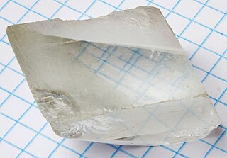
Birefringence is the optical property of a material having a refractive index that depends on the polarization and propagation direction of light. These optically anisotropic materials are described as birefringent or birefractive. The birefringence is often quantified as the maximum difference between refractive indices exhibited by the material. Crystals with non-cubic crystal structures are often birefringent, as are plastics under mechanical stress.

A microscope slide is a thin flat piece of glass, typically 75 by 26 mm and about 1 mm thick, used to hold objects for examination under a microscope. Typically the object is mounted (secured) on the slide, and then both are inserted together in the microscope for viewing. This arrangement allows several slide-mounted objects to be quickly inserted and removed from the microscope, labeled, transported, and stored in appropriate slide cases or folders etc.
Petrography is a branch of petrology that focuses on detailed descriptions of rocks. Someone who studies petrography is called a petrographer. The mineral content and the textural relationships within the rock are described in detail. The classification of rocks is based on the information acquired during the petrographic analysis. Petrographic descriptions start with the field notes at the outcrop and include macroscopic description of hand-sized specimens. The most important petrographer's tool is the petrographic microscope. The detailed analysis of minerals by optical mineralogy in thin section and the micro-texture and structure are critical to understanding the origin of the rock.
A total internal reflection fluorescence microscope (TIRFM) is a type of microscope with which a thin region of a specimen, usually less than 200 nanometers can be observed.

A Wollaston prism is an optical device, invented by William Hyde Wollaston, that manipulates polarized light. It separates light into two separate linearly polarized outgoing beams with orthogonal polarization. The two beams will be polarized according to the optical axis of the two right angle prisms.

A Nicol prism is a type of polarizer. It is an optical device made from calcite crystal used to convert ordinary light into plane polarized light. It is made in such a way that it eliminates one of the rays by total internal reflection, i.e. the ordinary ray is eliminated and only the extraordinary ray is transmitted through the prism.

William Nicol FRSE FCS was a Scottish geologist and physicist who invented the Nicol prism, the first device for obtaining plane-polarized light, in 1828.

A polarizer or polariser is an optical filter that lets light waves of a specific polarization pass through while blocking light waves of other polarizations. It can filter a beam of light of undefined or mixed polarization into a beam of well-defined polarization, known as polarized light. Polarizers are used in many optical techniques and instruments. Polarizers find applications in photography and LCD technology. In photography, a polarizing filter can be used to filter out reflections.

A Glan–Thompson prism is a type of polarizing prism similar to the Nicol prism and Glan–Foucault prism.

A polarimeter is a scientific instrument used to measure optical rotation: the angle of rotation caused by passing linearly polarized light through an optically active substance.

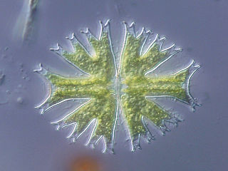
Optical mineralogy is the study of minerals and rocks by measuring their optical properties. Most commonly, rock and mineral samples are prepared as thin sections or grain mounts for study in the laboratory with a petrographic microscope. Optical mineralogy is used to identify the mineralogical composition of geological materials in order to help reveal their origin and evolution.
Differential interference contrast (DIC) microscopy, also known as Nomarski interference contrast (NIC) or Nomarski microscopy, is an optical microscopy technique used to enhance the contrast in unstained, transparent samples. DIC works on the principle of interferometry to gain information about the optical path length of the sample, to see otherwise invisible features. A relatively complex optical system produces an image with the object appearing black to white on a grey background. This image is similar to that obtained by phase contrast microscopy but without the bright diffraction halo. The technique was invented by Francis Hughes Smith. The "Smith DIK" was produced by Ernst Leitz Wetzlar in Germany and was difficult to manufacture. DIC was then developed further by Polish physicist Georges Nomarski in 1952.

A Nomarski prism is a modification of the Wollaston prism that is used in differential interference contrast microscopy. It is named after its inventor, Polish and naturalized-French physicist Georges Nomarski. Like the Wollaston prism, the Nomarski prism consists of two birefringent crystal wedges cemented together at the hypotenuse. One of the wedges is identical to a conventional Wollaston wedge and has the optical axis oriented parallel to the surface of the prism. The second wedge of the prism is modified by cutting the crystal so that the optical axis is oriented obliquely with respect to the flat surface of the prism. The Nomarski modification causes the light rays to come to a focal point outside the body of the prism, and allows greater flexibility so that when setting up the microscope the prism can be actively focused.

A petrographic microscope is a type of optical microscope used to identify rocks and minerals in thin sections. The microscope is used in optical mineralogy and petrography, a branch of petrology which focuses on detailed descriptions of rocks. The method includes aspects of polarized light microscopy (PLM).

In light microscopy, oil immersion is a technique used to increase the resolving power of a microscope. This is achieved by immersing both the objective lens and the specimen in a transparent oil of high refractive index, thereby increasing the numerical aperture of the objective lens.

The Sénarmont prism is a type of polariser. It is made from two prisms of a birefringent material such as calcite, usually cemented together. The Sénarmont prism is named after Henri Hureau de Sénarmont. It is similar to the Rochon and Wollaston prisms.
The operation of a photon scanning tunneling microscope (PSTM) is analogous to the operation of an electron scanning tunneling microscope, with the primary distinction being that PSTM involves tunneling of photons instead of electrons from the sample surface to the probe tip. A beam of light is focused on a prism at an angle greater than the critical angle of the refractive medium in order to induce total internal reflection within the prism. Although the beam of light is not propagated through the surface of the refractive prism under total internal reflection, an evanescent field of light is still present at the surface.

