Choledochoduodenostomy | |
---|---|
![]() | |
Specialty | Gastroenterology |
Choledochoduodenostomy (CDD) is a surgical procedure to create an anastomosis, a surgical connection, between the common bile duct (CBD) and an alternative portion of the duodenum. [1] In healthy individuals, the CBD meets the pancreatic duct at the ampulla of Vater, which drains via the major duodenal papilla to the second part of duodenum. [2] In cases of benign conditions such as narrowing of the distal CBD or recurrent CBD stones, performing a CDD provides the diseased patient with CBD drainage and decompression. [3] A side-to-side anastomosis is usually performed. [4]
Bile from the gallbladder is carried to the CBD and emptied into the duodenum. CBD drainage might be obstructed due to distal CBD stricture, which is narrowing of the CBD due to the presence of scar tissue within the duct, and choledocholithiasis, the presence of gallstones. Obstruction can occur when gallstones may be too large to pass through the CBD into the duodenum.[ medical citation needed ]
Liver tests are performed before and after the operation. [5] During surgery, the duodenum should be repositioned in close proximity with the CBD to ensure a tension-free anastomosis. 8 incisions are made, with one in the CBD and one in the duodenum. Sutures are performed between the incisions to create a new pathway. [6] Postoperative complications include inflammation and narrowing within the surgical site and sump syndrome. [5] Given that the duodenum is in a diseased state, or a tension-free anastomosis cannot be created, a CDD should not be performed and alternative bypass procedures could be considered. [5] Currently, CDD accounts for approximately 1% of all biliary operations to provide CBD drainage. 38% of the patients undergo CDD as a primary operation (first treatment given for a disease) and 60% of the patients undergo CDD as a secondary procedure (a surgical procedure which is performed to improve conditions found to exist during the primary surgery). CDD is more often performed in the elderly with the mean age of the patients being around 61 years. [7]
CDD creates an anastomosis to allow free flow of bile from the CBD into the duodenum. [1] Side-to-side anastomosis and end-to-side anastomosis are two procedures that can be done. Side-to-side anastomosis is preferred as the distal CBD blood supply is poor and more suitable to the laparoscopic approach, which requires limited anterior CBD dissection. Performing an end-to-side anastomosis risks ischemia and recurrent stenosis. [8]
CDD is an alternative to choledochojejunostomy, a procedure which involves creating an anastomosis of the CBD to the jejunum. [9] CDD is less technically challenging as the procedure is fast and simple with less anastomotic sites and does not require the creation of a Roux-en-Y anastomosis. [5] It also has better endoscopic access to the biliary tree and more physiologic bile drainage. [8]
CDD is used in the treatment of biliary obstruction caused by various factors shown below or when alternative treatment options were not accessible. Other less common indications include operative injuries, or chronic dilation of the CBD. [6]
Gallstone disease is considered to be one of the most common digestive diseases. It is prevalent regardless of ethnicity with rates in the United States and Europe reported to be approximately 10-15%. [10] It affects about 25 million people in the US and nearly 1 million people diagnosed annually will need to be treated. Studies have shown that 58-72% of patients with symptomatic gallstones will have ongoing symptoms and complications. [11]
With multiple stones present, the difficulty in clearance of the CBD can lead to biliary blockage. A CDD allows for the passage of any retained gallstones, [6] such as impacted stones, or stones where endoscopic treatments of extraction are not possible. [5]
The narrowing of the Ampulla of Vater is either verified by a 3mm instrumental probe being unable to pass through the ampulla, or the lack of dye flowing into the duodenum when a cholangiogram is carried out, indicating obstructed bile flow. Any attempts to enlarge the stenosis can lead to perforation of the duodenum wall or CBD, or damage to the pancreas, further limiting bile flow. [6] This is present in approximately 10% of patients with persistent or recurrent biliary colic after cholecystectomy. [12]
In addition to disrupted pancreatic function and physiology, long-term pancreatic inflammation can lead to distal CBD blockage. Chronic pancreatitis poses a high risk for developing pancreatic cancer. Creating an alternative passage from the CBD to the duodenum is done when surgical resection of the tumour is not available. [6] The prevalence of this disease is 0.05% in industrialised countries. This disease often develops in patients between the ages of 30 and 40, and is more common in men. [13]
Cancerous tumours arising from the CBD, the ampulla, or the portion of the duodenum near the ampulla can result in distal CBD obstruction. When the masses are incapable of being surgically removed, CDD can be performed. In some cases where the growing tumour occludes the new pathway, CDD will only provide palliative relief. It is recommended to use alternative procedures that allow for relief farther away from the tumor. [6] In the United States, an estimated 6000-7000 new cases of gallbladder carcinoma and 3000-4000 new cases of bile ducts carcinoma are diagnosed annually. [14]
Contraindications for the procedure are based on the patient’s physiologic condition of the CBD and the duodenum.
Duodenal ulceration can lead to inflammation or fibrosis of the duodenum. [3] Duodenal scarring or blockage makes it subpar for an anastomosis to be performed. As an alternative, a choledochojejunostomy can be performed. [15]
Malignant cell growth, such as a pancreatic head tumor, [3] can prevent proper repositioning of the duodenum to be in close contact with the bile duct. [5] Performing a CDD may lead to a tension-filled surgical anastomosis, leading to bile leakage and jaundice. [6]
There is also the possibility of active tumour growth obstructing the CBD. Alternative procedures could be considered, such as a Roux-en-Y hepaticojejunostomy (a connection made between the hepatic duct and the jejunum). [3]
Before surgery, liver function tests are carried out. An ultrasound is conducted to establish the location of biliary tract obstruction as well as the diameter of the CBD. Antibiotic prophylaxis are provided to the patient. Pre-surgical biliary stenting (a tube used to keep the biliary duct open) should be avoided, as it increases the likelihood of contracting infections and makes performing surgical anastomosis more difficult. [5]
A closed suction drain is placed posterior to the CDD and left in place for usually a week. [3] If the output is less than 40 cm3/day, the drain is removed. [16] It can be sent for a bilirubin and alkaline phosphatase test if there are concerns regarding the output. [5] After the surgery, nasogastric suction is usually maintained for 2–3 days and the tube is removed when there is low output. Once the tube is in place, it can be used to give the patient food and medicine.[ medical citation needed ] A fluid diet is started after surgery and the diet is advanced as tolerated by the patient. [16] Liver function tests should be restudied as needed during the postoperative recovery period. The average hospital stay ranges from 3–8 days but patients can be discharged as early as postoperative day 4. [17]
A CT scan or a fistulogram (an x-ray procedure used to investigate any abnormalities in the anastomosis) can be used to determine whether there is an increased bilirubin concentration within the intra-abdominal fluid. Given there is no blockage downstream of the anastomosis, most bile leakages will heal on their own. [5]
Anastomotic stricture can lead to cholangitis, or inflammation of the bile duct. [1] Narrowing of the bile duct leads to a buildup of pressure, causing the intercellular gaps to widen, exposing the cells to microorganisms found within the bile, resulting in inflammation. [18] Symptoms include jaundice and pain in the upper right quadrant. A CT scan or an endoscopic retrograde cholangiopancreatography (ECRP) could be performed to determine the site of inflammation. Cholangiocarcinoma commonly occurs in patients with repeated episodes of cholangitis. [5]
Performing an anastomosis alters the structural composition of bile ducts, which can result in bile stricture obstruction. [19] It leads to slowing of the biliary flow and dilation of the CBD, causing more progressive symptoms of jaundice. [6] An anastomotic stricture is more likely to occur if the choledoctomy incision was less than 2 cm in length. The anastomosis could be enlarged by using endoscopic balloon dilation, which consists of inflating a balloon after it is placed in a stenosed region, as a means of widening the stenosed area. [5]
Sump syndrome occurs when the nonfunctional portion of the distal CBD (between the site of anastomosis and the ampulla of Vater) acts as a site for stones or debris to gather. [20]
The reported incidence of the sump syndrome is between 0.4% and 3.3%. [8] Possible symptoms include pain, cholangitis, jaundice, and pancreatitis. [5] Diagnosis and treatment can be carried out by an ERCP, where the accumulated debris identified in the blind end of the CBD is then extracted. [5] [21] Alternative treatment procedures include making an incision in the major duodenal papilla to enlarge the bile duct opening (biliary sphincterotomy). [21]
In 1888, a German surgeon named Bernhard Riedel performed the first CDD. He intended to cut across the CBD and implant the severed end into the duodenum; however, this idea was scrapped and he performed lateral anastomosis of the dilated CBD to the duodenum. The patient died due to infected bile leakage into the peritoneal cavity. [22]
A German surgeon named Otto Sprengel reported the first recovery following CDD in 1891. [22]
Following cholecystectomy and choledochotomy (a surgical incision of CBD), an American surgeon named W.J. Mayo reported successful treatment of CBD stricture. He sutured the end of the dilated portion of the CBD to the duodenum in 1905. [22]
In vertebrates, the gallbladder, also known as the cholecyst, is a small hollow organ where bile is stored and concentrated before it is released into the small intestine. In humans, the pear-shaped gallbladder lies beneath the liver, although the structure and position of the gallbladder can vary significantly among animal species. It receives and stores bile, produced by the liver, via the common hepatic duct, and releases it via the common bile duct into the duodenum, where the bile helps in the digestion of fats.
A bile duct is any of a number of long tube-like structures that carry bile, and is present in most vertebrates.
A gallstone is a stone formed within the gallbladder from precipitated bile components. The term cholelithiasis may refer to the presence of gallstones or to any disease caused by gallstones, and choledocholithiasis refers to the presence of migrated gallstones within bile ducts.
Cholecystectomy is the surgical removal of the gallbladder. Cholecystectomy is a common treatment of symptomatic gallstones and other gallbladder conditions. In 2011, cholecystectomy was the eighth most common operating room procedure performed in hospitals in the United States. Cholecystectomy can be performed either laparoscopically, or via an open surgical technique.
Endoscopic retrograde cholangiopancreatography (ERCP) is a technique that combines the use of endoscopy and fluoroscopy to diagnose and treat certain problems of the biliary or pancreatic ductal systems. It is primarily performed by highly skilled and specialty trained gastroenterologists. Through the endoscope, the physician can see the inside of the stomach and duodenum, and inject a contrast medium into the ducts in the biliary tree and pancreas so they can be seen on radiographs.
The bile duct is a part of the biliary tract. It is formed by the union of the common hepatic duct and cystic duct. It ends by uniting with the pancreatic duct to form the hepatopancreatic ampulla. It possesses its own sphincter to enable regulation of bile flow.
Courvoisier's principle states that a painless palpably enlarged gallbladder accompanied with mild jaundice is unlikely to be caused by gallstones. Usually, the term is used to describe the physical examination finding of the right-upper quadrant of the abdomen. This sign implicates possible malignancy of the gallbladder or pancreas and the swelling is unlikely due to gallstones.
A pancreaticoduodenectomy, also known as a Whipple procedure, is a major surgical operation most often performed to remove cancerous tumours from the head of the pancreas. It is also used for the treatment of pancreatic or duodenal trauma, or chronic pancreatitis. Due to the shared blood supply of organs in the proximal gastrointestinal system, surgical removal of the head of the pancreas also necessitates removal of the duodenum, proximal jejunum, gallbladder, and, occasionally, part of the stomach.
Common bile duct stone, also known as choledocholithiasis, is the presence of gallstones in the common bile duct (CBD). This condition can cause jaundice and liver cell damage. Treatments include choledocholithotomy and endoscopic retrograde cholangiopancreatography (ERCP).
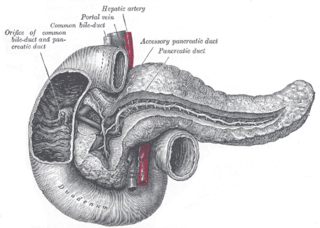
The pancreatic duct, or duct of Wirsung, is a duct joining the pancreas to the common bile duct. This supplies it with pancreatic juice from the exocrine pancreas, which aids in digestion.
A biliary fistula is a type of fistula in which bile flows along an abnormal connection from the bile ducts into nearby hollow structure. Types of biliary fistula include:
Ascending cholangitis, also known as acute cholangitis or simply cholangitis, is inflammation of the bile duct, usually caused by bacteria ascending from its junction with the duodenum. It tends to occur if the bile duct is already partially obstructed by gallstones.
The biliary tract refers to the liver, gallbladder and bile ducts, and how they work together to make, store and secrete bile. Bile consists of water, electrolytes, bile acids, cholesterol, phospholipids and conjugated bilirubin. Some components are synthesized by hepatocytes ; the rest are extracted from the blood by the liver.
Percutaneous transhepatic cholangiography, percutaneous hepatic cholangiogram (PTHC) is a radiological technique used to visualize the anatomy of the biliary tract. A contrast medium is injected into a bile duct in the liver, after which X-rays are taken. It allows access to the biliary tree in cases where endoscopic retrograde cholangiopancreatography has been unsuccessful. Initially reported in 1937, the procedure became popular in 1952.
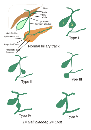
Choledochal cysts are congenital conditions involving cystic dilatation of bile ducts. They are uncommon in western countries but not as rare in East Asian nations like Japan and China.
Periampullary cancer is a cancer that forms near the ampulla of Vater, an enlargement of the ducts from the liver and pancreas where they join and enter the small intestine. It consists of:
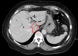
Pneumobilia is the presence of gas in the biliary system. It is typically detected by ultrasound or a radiographic imaging exam, such as CT, or MRI. It is a common finding in patients that have recently undergone biliary surgery or endoscopic biliary procedure. While the presence of air within biliary system is not harmful, this finding may alternatively suggest a pathological process, such as a biliary-enteric anastomosis, an infection of the biliary system, an incompetent sphincter of Oddi, or spontaneous biliary-enteric fistula.
Sphincter of Oddi dysfunction refers to a group of functional disorders leading to abdominal pain due to dysfunction of the Sphincter of Oddi: functional biliary sphincter of Oddi and functional pancreatic sphincter of Oddi disorder. The sphincter of Oddi is a sphincter muscle, a circular band of muscle at the bottom of the biliary tree which controls the flow of pancreatic juices and bile into the second part of the duodenum. The pathogenesis of this condition is recognized to encompass stenosis or dyskinesia of the sphincter of Oddi ; consequently the terms biliary dyskinesia, papillary stenosis, and postcholecystectomy syndrome have all been used to describe this condition. Both stenosis and dyskinesia can obstruct flow through the sphincter of Oddi and can therefore cause retention of bile in the biliary tree and pancreatic juice in the pancreatic duct.
Biliary endoscopic sphincterotomy is a procedure where the sphincter of Oddi and the segment of the common bile duct where it enters the duodenum are cannulated and then cut with a sphincterotome, a device that includes a wire which cuts with an electric current (electrocautery).
Antrectomy, also called distal gastrectomy, is a type of gastric resection surgery that involves the removal of the stomach antrum to treat gastric diseases causing the damage, bleeding, or blockage of the stomach. This is performed using either the Billroth I (BI) or Billroth II (BII) reconstruction method. Quite often, antrectomy is used alongside vagotomy to maximise its safety and effectiveness. Modern antrectomies typically have a high success rate and low mortality rate, but the exact numbers depend on the specific conditions being treated.
{{cite journal}}
: Cite journal requires |journal=
(help)